. 2012 Dec;1821(12):1501-7.
doi: 10.1016/j.bbalip.2012.08.017. Epub 2012 Aug 30.

Phosphatidylethanolamine is externalized at the surface of microparticles

Affiliations

Phosphatidylethanolamine is externalized at the surface of microparticles

Michael C Larson et al. Biochim Biophys Acta. 2012 Dec.

Abstract

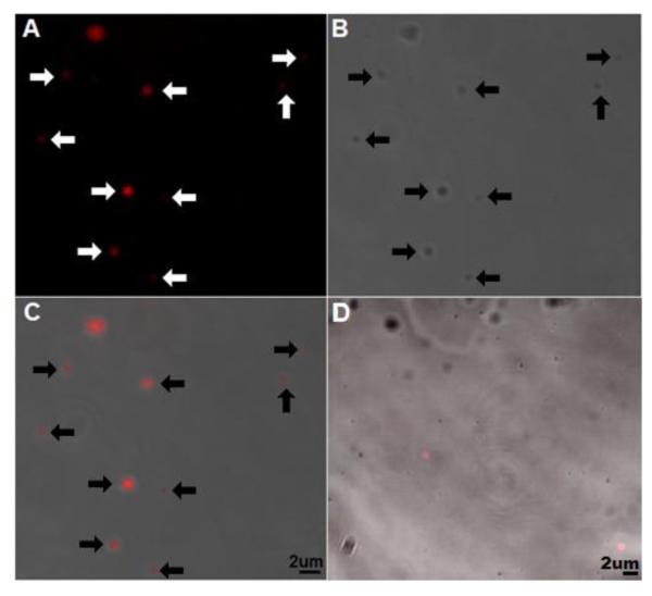
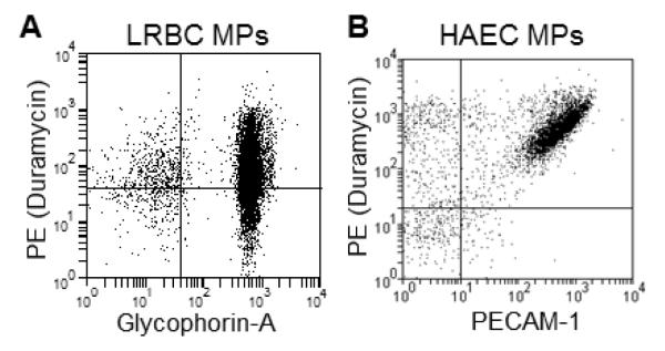
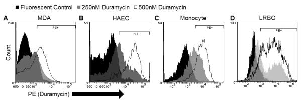
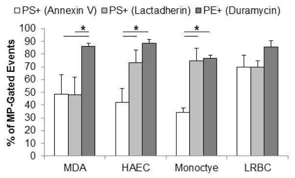
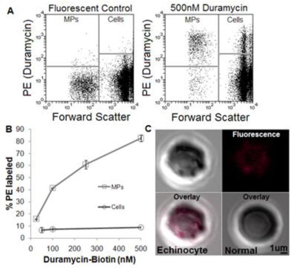
Microparticles (MPs) are membrane-bound vesicles shed normally or as a result of various (pathological) stimuli. MPs contain a wealth of bio-active macromolecules. Aminophospholipid phosphatidylserine (PS) is present on the surface of many MPs. As PS and phosphatidylethanolamine (PE) are related, yet distinct aminophospholipids, the purpose of this study was to systematically and directly assess PE exposure on MPs. We examined MPs from various human cellular sources (human breast cancer, endothelial, red and white blood cells) by flow cytometry using a PE-specific probe, duramycin, and two PS-specific probes, annexin V and lactadherin. PS and PE exposure percentage was comparable on vascular and blood cell-derived MPs (80-90% of MP-gated events). However, the percentage of malignant breast cancer MPs exposing PE (~90%) was significantly higher than PS (~50%). Thus, while PS and PE exposure can result from a general loss of membrane asymmetry, there may also be distinct mechanisms of PE and PS exposure on MPs that vary by cellular source.


Figures

Figure 1
Figure 1
Confocal imaging of duramcyin-labeled cancer MPs To label exposed phosphatidylethanolamine (PE) MDA MB-231 breast cancer cell (MDA) microparticles (MPs) were stained with 500nM duramycin-biotin and 250nM streptavidin Alexafluor-647, pelleted, and resuspended in 1:1 PBS:O.C.T. A) 647 signal, B) phase contrast, C) and overlay show fluorescence localizes to MPs (arrows added for emphasis). D) Fluorescent control (duramycin-biotin omitted) overlay. C-D) Lower right corner of overlays, bar indicates 2 μm. O.C.T., optimal cutting temperature embedding medium. Images representative of 2 separate experiments.
Figure 2
Figure 2
Duramycin binds MPs from various cellular sources MP-rich supernatants from aged donor units (LRBC) and human aortic endothelial cells stimulated with LPS (HAECs) were examined with flow cytometry after staining with 500 nM duramycin-biotin, 125 nM fluorescent streptavidin and 1:200 fluorescently-labeled antibodies. A) PE and glycophorin A staining. B) PE with PECAM-1 staining. Representative of N=3. Gating was based on isotype and fluorescent (streptavidin-only) controls.
Figure 3
Figure 3
Duramycin titration on MPs MP-rich supernatants from A) MDA, B) HAEC, C) monocyte, and D) LRBC were labeled with duramycin-biotin (or 125 nM fluorescent SA only) at various concentrations. Representative of 3-6 experiments.
Figure 4
Figure 4
Duramycin binds MPs from a variety of cells. MP-rich supernatants from A) MDA, B) HAEC, C) monocyte, and D) LRBC were labeled with duramycin-biotin (or 125 nM fluorescent SA only) at or near saturating conditions (1:50 annexin V, 32nM lactadherin, and 500 nM duramycin). Annexin V stained 48, 42, 34, and 70%; lactadherin stained 48, 73, 75, and 70%; duramycin stained 86, 88, 76, and 85% of MP-gated events of MP-rich supernatant from MDA, HAEC, monocyte, and LRBC, respectively. SA, streptavidin Alexafluor.
Figure 5
Figure 5
Duramycin-PE binding on RBCs and RBC MPs A) Fluorescent (SA only) control (left) and duramycin-stained donor blood (right). B) Duramycin bound a larger portion of LRBC-derived MPs compared with cells over a wide range of concentrations. C) Confocal imaging of duramycin-stained RBCs show PE exposure on echinocytes (upper half and lower left) but not normal cells (lower right).
Figure 6
Figure 6
Duramycin inhibits breast cancer microparticle-mediated coagulation A) Control plasma or MDA MP-containing media did not clot even after 10 minutes of adding calcium. Relative to MDA MPs in plasma, the addition of duramycin lengthened the time prior to clot formation. B) This resulted in a dose-dependent increase in the coagulation time (or decrease in the coagulability) of the breast cancer MPs. N>=4.

References

    1. Mause SF, Weber C. Microparticles: protagonists of a novel communication network for intercellular information exchange. Circ. Res. 2010;107:1047–1057. - PubMed
    1. Amabile N, Rautou P-E, Tedgui A, Boulanger CM. Microparticles: key protagonists in cardiovascular disorders. Semin. Thromb. Hemost. 2010;36:907–916. - PubMed
    1. Tushuizen ME, Diamant M, Sturk A, Nieuwland R. Cell-derived microparticles in the pathogenesis of cardiovascular disease: friend or foe? Arterioscler. Thromb. Vasc. Biol. 2011;31:4–9. - PubMed
    1. Rak J. Microparticles in cancer. Semin. Thromb. Hemost. 2010;36:888–906. - PubMed
    1. Zwicker JI. Predictive value of tissue factor bearing microparticles in cancer associated thrombosis. Thromb. Res. 2010;125(Suppl 2):S89–91. - PubMed

Publication types

MeSH terms