. 2012 Oct;33(5):1338-45.
doi: 10.1016/j.neuro.2012.08.010. Epub 2012 Aug 31.

Perinatal exposure to 50 ppb sodium arsenate induces hypothalamic-pituitary-adrenal axis dysregulation in male C57BL/6 mice

Affiliations

Perinatal exposure to 50 ppb sodium arsenate induces hypothalamic-pituitary-adrenal axis dysregulation in male C57BL/6 mice

Samantha L Goggin et al. Neurotoxicology. 2012 Oct.

Abstract

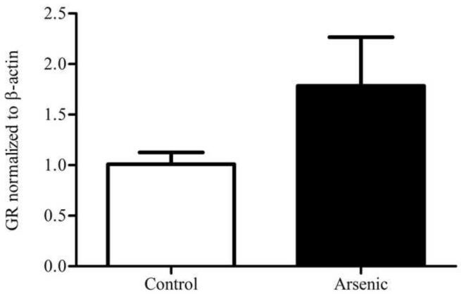
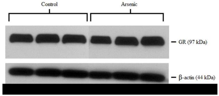
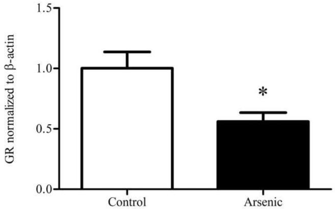
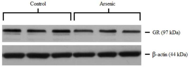
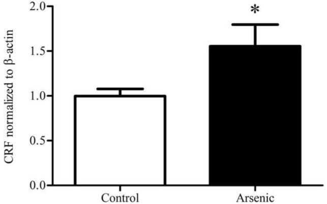
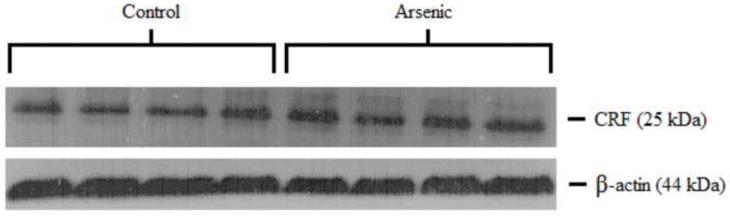
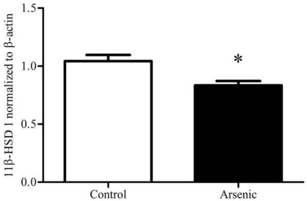
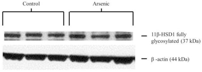
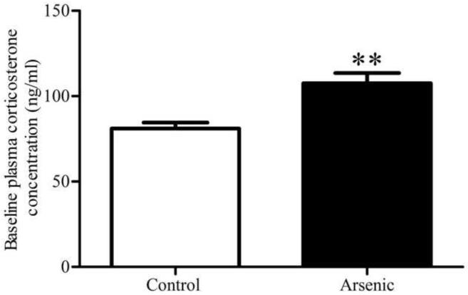
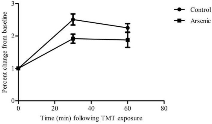
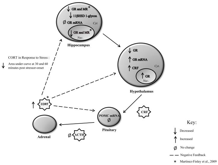
Over the past two decades, key advancements have been made in understanding the complex pathology that occurs following not only high levels of arsenic exposure (>1 ppm) but also levels previously considered to be low (<100 ppb). Past studies have characterized the deleterious effects of arsenic on the various functions of cardiovascular, pulmonary, immunological, respiratory, endocrine and neurological systems. Other research has demonstrated an elevated risk of a multitude of cancers and increased rates of psychopathology, even at very low levels of arsenic exposure. The hypothalamic-pituitary-adrenal (HPA) axis represents a multisite integration center that regulates a wide scope of biological and physiological processes: breakdown within this system can generate an array of far-reaching effects, making it an intriguing candidate for arsenic-mediated damage. Using a mouse model, we examined the effects of perinatal exposure to 50 ppb sodium arsenate on the functioning of the HPA axis through the assessment of corticotrophin-releasing factor (CRF), proopiomelanocortin (Pomc) mRNA, adrenocorticotrophin hormone (ACTH), corticosterone (CORT), 11β-hydroxysteroid dehydrogenase Type 1 (11β-HSD 1), and glucocorticoid receptor (GR) protein and mRNA. Compared to controls, we observed that the perinatal arsenic-exposed offspring exhibit an increase in hypothalamic CRF, altered CORT secretion both at baseline and in response to a stressor, decreased hippocampal 11β-HSD 1 and altered subcellular GR distribution in the hypothalamus. These data indicate significant HPA axis impairment at post-natal day 35 resulting from perinatal exposure to 50 ppb sodium arsenate. Our findings suggest that the dysregulation of this critical regulatory axis could underlie important molecular and cognitive pathology observed following exposure to arsenic.


Figures

Figure 1
Figure 1
Subcellular distribution of GR in the hypothalamus of control and arsenic exposed animals. (a) Levels of GR protein are elevated in the nuclear fraction of the hypothalamus (p=.118), though this was not of statistical significance. (b) Representative western blot of nuclear GR in control and perinatal arsenic-exposed offspring. (c) GR protein is decreased in the cytosolic fraction of the hypothalamus (p=.011). Data are presented as corrected to β-actin and normalized to controls. (d) Representative western blot of cytosolic GR in control and perinatal arsenic-exposed offspring. Data are presented as mean±SEM for n=8-9 litters.
Figure 1
Figure 1
Subcellular distribution of GR in the hypothalamus of control and arsenic exposed animals. (a) Levels of GR protein are elevated in the nuclear fraction of the hypothalamus (p=.118), though this was not of statistical significance. (b) Representative western blot of nuclear GR in control and perinatal arsenic-exposed offspring. (c) GR protein is decreased in the cytosolic fraction of the hypothalamus (p=.011). Data are presented as corrected to β-actin and normalized to controls. (d) Representative western blot of cytosolic GR in control and perinatal arsenic-exposed offspring. Data are presented as mean±SEM for n=8-9 litters.
Figure 2
Figure 2
CRF in the hypothalamus (a) CRF in the hypothalamus is significantly elevated in the arsenic-exposed offspring. (b) Representative western blot for hypothalamic CRF. Data are presented as normalized to β-actin and corrected to controls (p=.045). Results are presented as mean±SEM for n=8 litters.
Figure 2
Figure 2
CRF in the hypothalamus (a) CRF in the hypothalamus is significantly elevated in the arsenic-exposed offspring. (b) Representative western blot for hypothalamic CRF. Data are presented as normalized to β-actin and corrected to controls (p=.045). Results are presented as mean±SEM for n=8 litters.
Figure 3
Figure 3
11β-HSD 1 in the hippocampus control and perinatal arsenic-exposed offspring. (a) The fully glycosylated form of 11β-HSD 1 is significantly decreased in the arsenic-exposed group (p=.005). (b) Representative western blot for the glycosylated and un-glycosylated forms of 11β-HSD 1 in the arsenic-exposed group and controls. Data were normalized to β-actin and corrected to controls. Data are presented as mean±SEM for n=8 litters in control and n=10 litters in the arsenic group.
Figure 3
Figure 3
11β-HSD 1 in the hippocampus control and perinatal arsenic-exposed offspring. (a) The fully glycosylated form of 11β-HSD 1 is significantly decreased in the arsenic-exposed group (p=.005). (b) Representative western blot for the glycosylated and un-glycosylated forms of 11β-HSD 1 in the arsenic-exposed group and controls. Data were normalized to β-actin and corrected to controls. Data are presented as mean±SEM for n=8 litters in control and n=10 litters in the arsenic group.
Figure 4
Figure 4
Baseline and stress-induced plasma corticosterone concentration in arsenic and control groups. (a) Baseline plasma CORT concentration was significantly increased in the arsenic-exposed offspring (p=.003), a replication of our previous finding included in Martinez-Finley et al., 2008. Data are presented as mean±SEM for n=8 litters in control and n=10 litters in the arsenic group. (b) Stress-induced plasma corticosterone at 30 and 60 minutes post stressor-onset: area under curve was significantly decreased in the arsenic-exposed offspring (p=.02) in n=7 litters.
Figure 4
Figure 4
Baseline and stress-induced plasma corticosterone concentration in arsenic and control groups. (a) Baseline plasma CORT concentration was significantly increased in the arsenic-exposed offspring (p=.003), a replication of our previous finding included in Martinez-Finley et al., 2008. Data are presented as mean±SEM for n=8 litters in control and n=10 litters in the arsenic group. (b) Stress-induced plasma corticosterone at 30 and 60 minutes post stressor-onset: area under curve was significantly decreased in the arsenic-exposed offspring (p=.02) in n=7 litters.
Figure 5
Figure 5
Summary of alterations induced by perinatal exposure to 50 ppb sodium arsenate in male, ~PN35 mice.

References

    1. Allan AM, Liang X, Luo Y, Pak C, Li X, Szulwach KE, Chen D, Jin P, Zhao X. The loss of methyl-CpG binding protein 1 leads to autism-like behavioral deficits. Human Molecular Genetics. 2008;17(13):2047–57. - PMC - PubMed
    1. ATSDR . Toxicological Profile for Arsenic (Update) Agency for Toxic Substances and Disease Registry, SUDHHS, PHS; Washington, DC: 1999. - PubMed
    1. Blum A, Martin HJ, Maser E. Human 11beta-hydroxysteroid dehydrogenase type 1 is enzymatically active in its nonglycosylated form. Biochem Biophys Res Commun. 2000;276(2):428–34. - PubMed
    1. Buckley CT, Caldwell KK. Fear conditioning is associated with altered integration of PLC and ERK signaling in the hippocampus. Pharmacol Biochem Behav. 2004;79:633–40. - PubMed
    1. Bodwell JE, Kingsley LA, Hamilton JW. Arsenic at very low concentrations alters glucocorticoid receptor (GR) –mediated gene activation but not GR-mediated gene repression: complex dose-response effects are closely correlated with levels of activated GR and require a functional GR DNA binding domain. Chem Res Toxicol. 2004;17:1064–76. - PubMed

Publication types

MeSH terms

LinkOut - more resources