Review
. 2012 Nov 1;4(11):a012310.
doi: 10.1101/cshperspect.a012310.

Imaging translation in single cells using fluorescent microscopy

Affiliations
Review

Imaging translation in single cells using fluorescent microscopy

Jeffrey A Chao et al. Cold Spring Harb Perspect Biol. .

Abstract

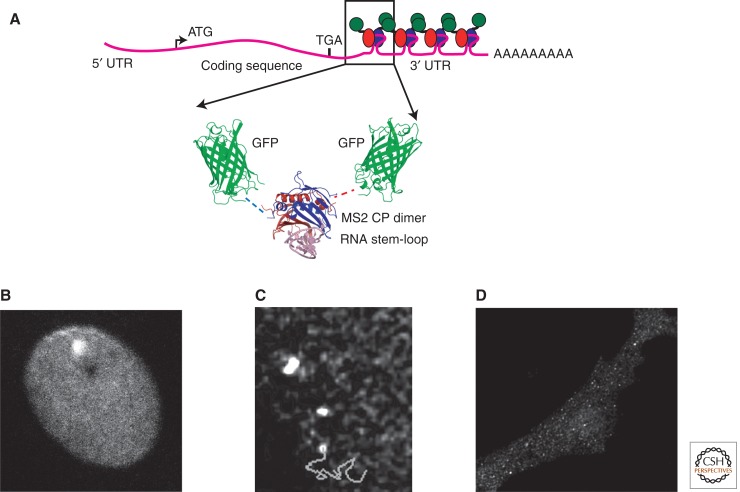
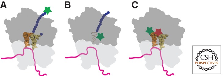
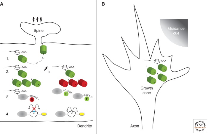
The regulation of translation provides a mechanism to control not only the abundance of proteins, but also the precise time and subcellular location that they are synthesized. Much of what is known concerning the molecular basis for translational control has been gleaned from experiments (e.g., luciferase assays and polysome analysis) that measure average changes in the protein synthesis of a population of cells, however, mechanistic insights can be obscured in ensemble measurements. The development of fluorescent microscopy techniques and reagents has allowed translation to be studied within its cellular context. Here we highlight recent methodologies that can be used to study global changes in protein synthesis or regulation of specific mRNAs in single cells. Imaging of translation has provided direct evidence for local translation of mRNAs at synapses in neurons and will become an important tool for studying translational control.


Figures

Figure 1.
Figure 1.
MS2 system for fluorescent labeling of mRNAs in living cells. (A) Schematic of a reporter mRNA that contains multiple copies of the MS2 stem-loop (boxed) that bind to the MS2 coat protein fused to GFP (arrows). The binding of many MCP-GFP molecules to a single transcript allows the RNA to be observed in living cells. (B) Transcription in the nucleus detected by binding of MCP-GFP to nascent mRNAs at the transcription site. (From Darzacq et al. 2007; reprinted, with permission, from the author.) (C) Tracking of single mRNAs (trajectory shown as line) in the nucleoplasm. (From Shav-tal et al. 2004; reprinted, with permission, from The American Association for the Advancement of Science © 2004.) (D) Single mRNAs detected in the cytoplasm. (From Fusco et al. 2003; reprinted, with permission, from Elsevier © 2003.)
Figure 2.
Figure 2.
Global measurements of translation in single cells. (A) The incorporation of noncanonical amino acids using FUNCAT allows newly synthesized proteins to be fluorescently labeled (green star). Methionine analogs are incorporated into the elongating polypeptide chain that can be conjugated to fluorescent dyes. (B) Fluorescent analogs of puromycin (green star) prematurely terminate translation and allow nascent polypetides to be fluorescently labeled. (C) FRET between adjacent fluorescent tRNAs (green and red stars are FRET pairs) allows the cellular location of active translation to be observed. Only when fluorescently labeled tRNAs are bound to the ribosome in adjacent sites, is the FRET signal observable.
Figure 3.
Figure 3.
Strategies for visualization of newly synthesized proteins in neurons. Polarized cellular architecture of neurons provides an ideal system to study localized translation in discrete subcellular domains. In dendrites (A), a small structure called the spine receives signals and transmits the information to the cell body. During this process, localized protein synthesis can be visualized using a variety of strategies depicted in the illustration. 1. Anchored GFP: Postsynaptic stimulation leads to synthesis of myristoylated GFP (green cylinder) that becomes anchored to the membrane thereby demarcating the site of active translation. 2. Photoconvertible GFP: Conversion of the preexisting population of fluorescent proteins (cylinders) from green to red by a brief exposure to ultraviolet light allows visualization of the newly synthesized population of fluorescent proteins. 3. Biarsenical dyes: Association of cell-permeable biarsenical dyes, FlAsH (F) and ReAsH (R) to the tetracysteine motif (CCXXCC) permits detection of translation using a pulse-chase approach. 4. TimeSTAMP: A self-cleaving protease (white) cassette that carries an epitope tag (yellow) becomes detectable on addition of the protease inhibitor (blue) by immunofluorescence methods. In axon growth cones (B), the directionality of guidance cues (upper right quadrant) can lead to asymmetric translation thereby leading to rapid growth cone turning and extension. Most, if not all, of the strategies employed in dendrites can be used in growth cones and other subcellular compartments to further characterize localized GFP translation (green cylinder) in neurons.

References

    1. Aakalu G, Smith WB, Nguyen N, Jiang C, Schuman EM 2001. Dynamic visualization of local protein synthesis in hippocampal neurons. Neuron 30: 489–502 - PubMed
    1. Adams SR, Campbell RE, Gross LA, Martin BR, Walkup GK, Yao Y, Llopis J, Tsien RY 2002. New biarsenical ligands and tetracysteine motifs for protein labeling in vitro and in vivo: Synthesis and biological applications. J Am Chem Soc 124: 6063–6076 - PubMed
    1. Ando R, Hama H, Yamamoto-Hino M, Mizuno H, Miyawaki A 2002. An optical marker based on the UV-induced green-to-red photoconversion of a fluorescent protein. Proc Natl Acad Sci 99: 12651–12656 - PMC - PubMed
    1. Barhoom S, Kaur J, Cooperman BS, Smorodinsky NI, Smilansky Z, Ehrlich M, Elroy-Stein O 2011. Quantitative single cell monitoring of protein synthesis at subcellular resolution using fluorescently labeled tRNA. Nucleic Acids Res 39: e129. - PMC - PubMed
    1. Bassell GJ, Zhang H, Byrd AL, Femino AM, Singer RH, Taneja KL, Lifshitz LM, Herman IM, Kosik KS 1998. Sorting of β-actin mRNA and protein to neurites and growth cones in culture. J Neurosci 18: 251–265 - PMC - PubMed

LinkOut - more resources