. 2012 Oct 17;31(20):4020-34.
doi: 10.1038/emboj.2012.251. Epub 2012 Sep 7.

Alternative 3'-end processing of long noncoding RNA initiates construction of nuclear paraspeckles

Affiliations

Alternative 3'-end processing of long noncoding RNA initiates construction of nuclear paraspeckles

Takao Naganuma et al. EMBO J. .

Abstract

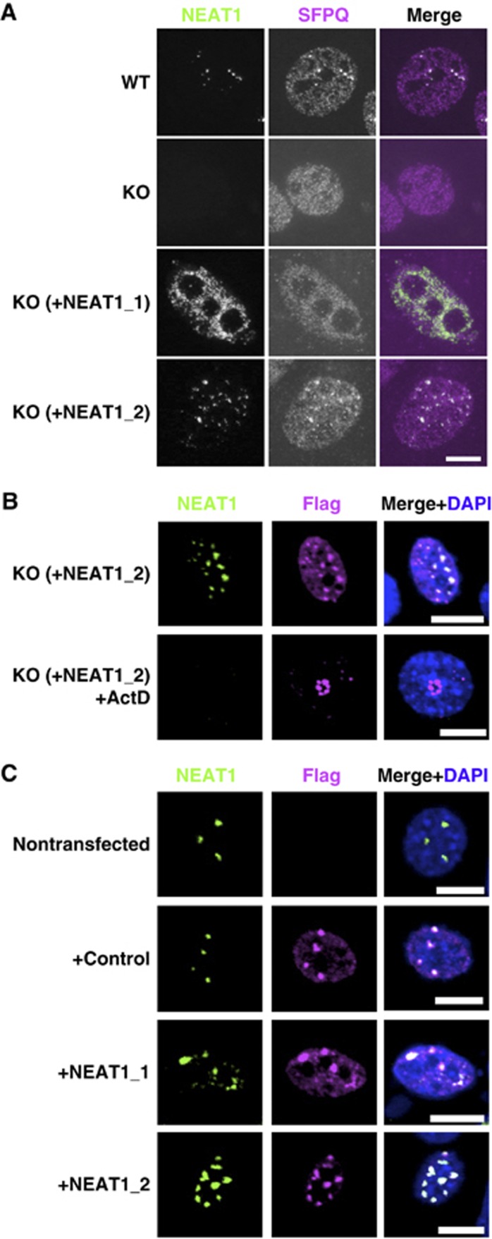
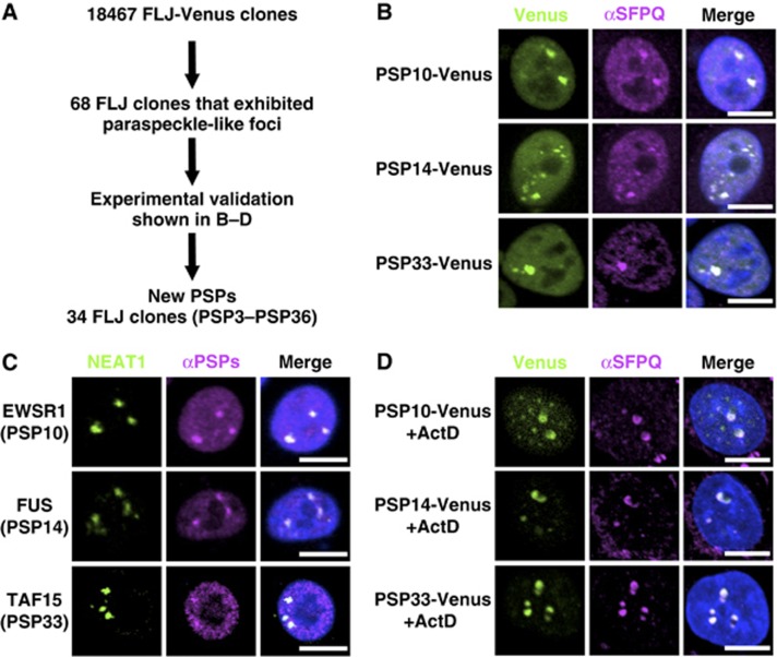
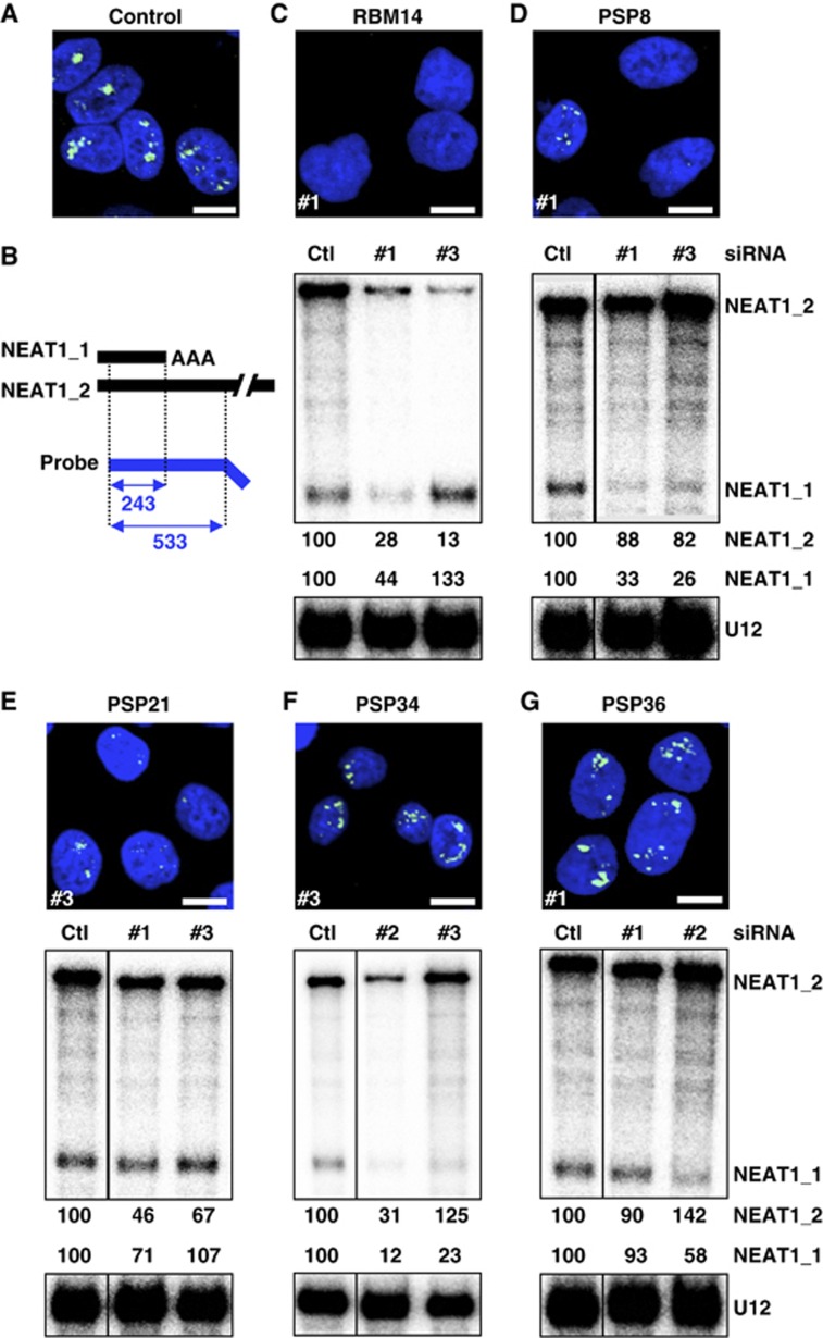
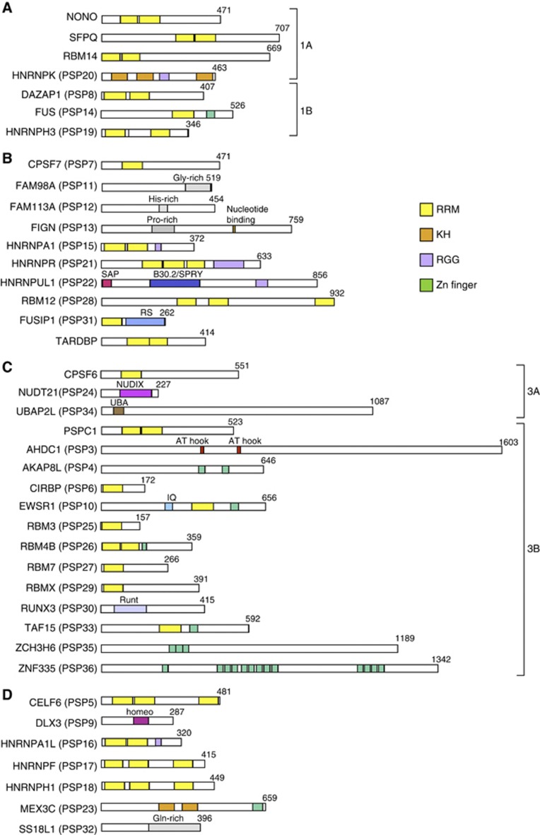
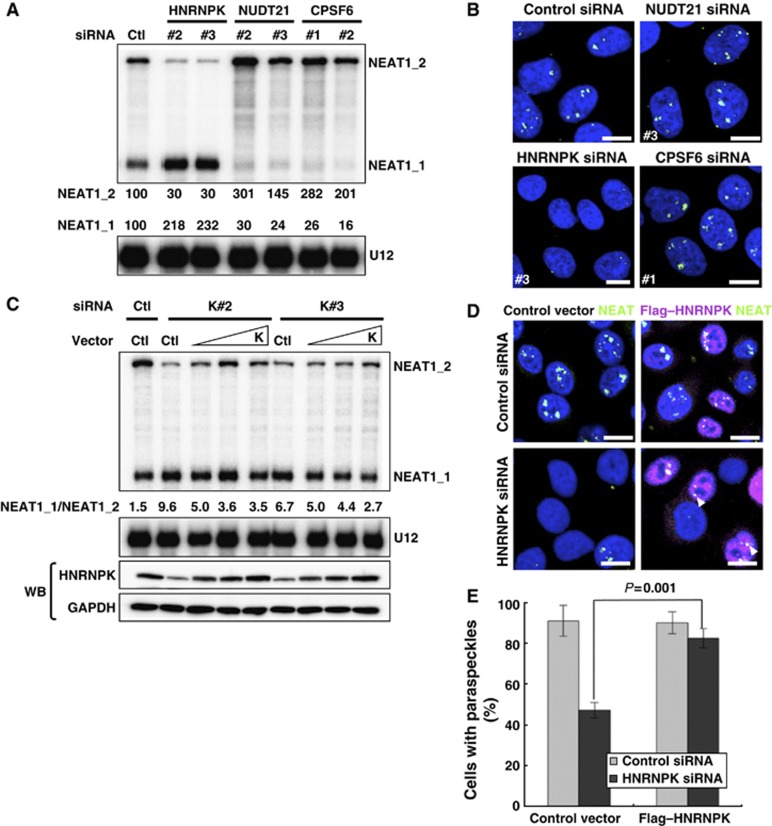
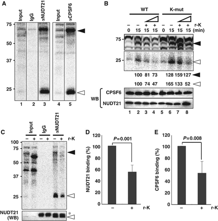
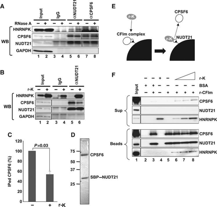
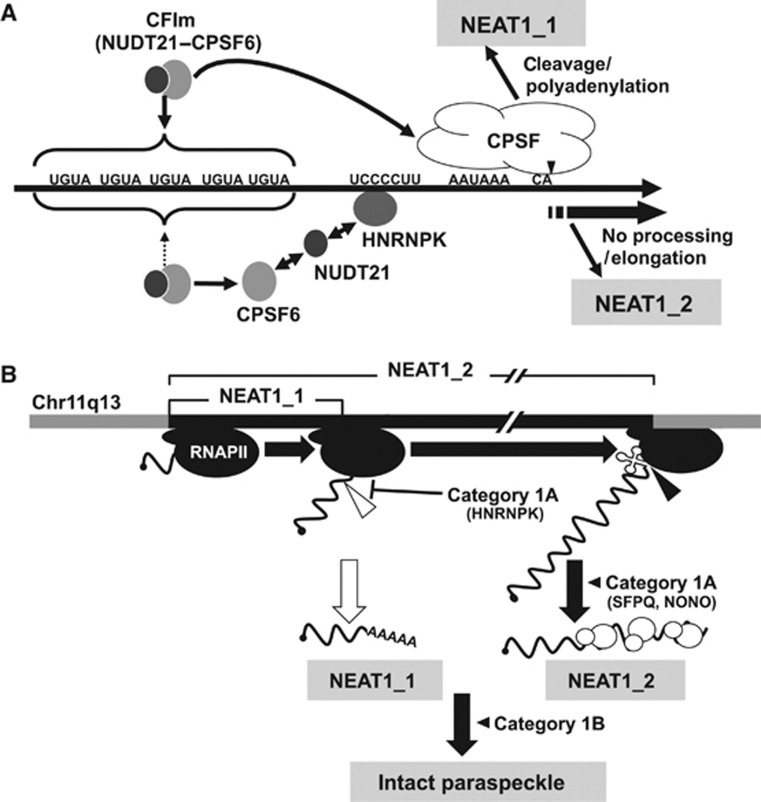
Paraspeckles are unique subnuclear structures built around a specific long noncoding RNA, NEAT1, which is comprised of two isoforms produced by alternative 3'-end processing (NEAT1_1 and NEAT1_2). To address the precise molecular processes that lead to paraspeckle formation, we identified 35 paraspeckle proteins (PSPs), mainly by colocalization screening with a fluorescent protein-tagged full-length cDNA library. Most of the newly identified PSPs possessed various putative RNA-binding domains. Subsequent RNAi analyses identified seven essential PSPs for paraspeckle formation. One of the essential PSPs, HNRNPK, appeared to affect the production of the essential NEAT1_2 isoform by negatively regulating the 3'-end polyadenylation of the NEAT1_1 isoform. An in vitro 3'-end processing assay revealed that HNRNPK arrested binding of the CPSF6-NUDT21 (CFIm) complex in the vicinity of the alternative polyadenylation site of NEAT1_1. In vitro binding assays showed that HNRNPK competed with CPSF6 for binding to NUDT21, which was the underlying mechanism to arrest CFIm binding by HNRNPK. This HNRNPK function led to the preferential accumulation of NEAT1_2 and initiated paraspeckle construction with multiple PSPs.


Conflict of interest statement

The authors declare that they have no conflict of interest.

Figures

Figure 1
Figure 1
NEAT1_2 is a potent RNA component for paraspeckle formation. (A) NEAT1_2 but not NEAT1_1 rescues paraspeckle formation. Intact paraspeckles were detected by RNA-FISH with the antisense probe of mouse NEAT1 ncRNA and coimmunostaining of endogenous SFPQ. Paraspeckles, which were observed in WT MEF cells, were undetectable in MEF cells prepared from NEAT1-knockout mice (KO). Paraspeckles were detected in KO-MEF cells transfected with a plasmid expressing NEAT1_1 (KO+NEAT1_1) or NEAT1_2 (KO+NEAT1_2). (B) Effect of actinomycin D treatment on the reformed paraspeckle-like foci. KO-MEF cells were cotransfected with plasmids expressing NEAT1_2 ncRNA and SFPQ–Flag. Transfected cells were treated with 0.3 μg/ml actinomycin D for 4 h. Reformed paraspeckle-like foci were visualized with RNA-FISH of NEAT1 and coimmunostained with anti-Flag M2 antibody. (C) NEAT1_2 ncRNA is more competent than NEAT1_1 ncRNA at elevating the number of paraspeckles. NIH3T3 cells were cotransfected with expression plasmids of control (+control), NEAT1_1 (+NEAT1_1), or NEAT1_2 (+NEAT1_2), together with SFPQ–Flag. The counted paraspeckle numbers are shown in Supplementary Figure S1D. Scale bar, 10 μm.
Figure 2
Figure 2
Identification of novel PSP components. (A) Experimental strategy to identify new PSPs. (B) Selection of FLJ-Venus clones that localize to paraspeckle-like nuclear foci. Paraspeckles were visualized by the immunostaining of SFPQ. Three representatives (PSP10, PSP14, and PSP33) of the new PSPs are shown. (C) Confirmation of the paraspeckle localization of endogenous PSP counterparts. Antibodies against each counterpart of PSP10, PSP14, and PSP33 (EWSR1, FUS and TAF15, respectively) were employed to monitor the localization (see Supplementary Table S4). NEAT1 ncRNA was used as a paraspeckle marker. (D) Effect of actinomycin D treatment on the localization patterns of selected PSPs. Localization of the Venus clones in B was monitored after actinomycin D treatment. SFPQ is an endogenous PSP control. Data regarding other PSPs are shown in Supplementary Figures S2 and S3. Scale bar, 10 μm.
Figure 3
Figure 3
Functional assignment of new PSPs in paraspeckle formation by extensive RNAi treatment. Paraspeckle appearance and NEAT1 levels were monitored by RNA-FISH and RPA to detect NEAT1_1 and NEAT1_2. (A) Paraspeckles in cells treated with control siRNA. (B) Schema for the RPA probe used and the protected fragments (with size, nt) of NEAT1_1 and NEAT1_2. Data for a representative from each category (1A: RBM14, 1B: PSP8/DAZAP1, 2: PSP21/HNRNPR, 3A: PSP34/UBAP2L, 3B: PSP36/ZNF335) are shown in (CG), respectively. The siRNA numbers (#) used in the RNA-FISH analysis are shown at the lower left of each photo. For RPA, the ratio of band intensities of the two isoforms, normalized by those of U12 snRNA, is shown below (Ctl was defined as 100%). RNAi data regarding all PSPs are compiled in Supplementary Figure S4. Their quantified data are shown in Supplementary Table S3. The siRNAs used are shown in Supplementary Table S6. Scale bar, 10 μm. Figure source data can be found with the Supplementary data.
Figure 4
Figure 4
Compilation of PSPs. Schematics of the major domains of the PSPs that belong to category 1 (A), 2 (B), and 3 (C) are grouped and shown. Uncategorized PSPs are shown in (D). Subcategories in categories 1 and 3 are shown as 1A and 1B in (A) and 3A and 3B in (C). The amino-acid length of each PSP is shown in the right corner. The colour codes of four putative RNA-binding domains are shown on the right.
Figure 5
Figure 5
Alternative 3′-end processing of NEAT1 is the initial essential step underlying paraspeckle formation. (A) PSPs required for the alternative 3′-end processing of NEAT1. RPA was performed as in Figure 3. Data are shown for NUDT21 and CPSF6, which are required for NEAT1_1 3′-end processing, and HNRNPK, which is required for NEAT1_2 synthesis by interfering with NEAT1_1 3′-end processing. (B) Paraspeckle appearance in HeLa cells treated with siRNAs against NUDT21, CPSF6, or HNRNPK. (C) Plasmid rescue of a defect of NEAT1_2 synthesis in HNRNPK-eliminated cells. Two siRNAs against HNRNPK (K#2 and K#3) were used to eliminate endogenous HNRNPK, and HNRNPK rescue plasmid (K) was introduced at three concentrations (1–5 μg). NEAT1 ncRNA levels were measured by RPA as in Figure 3. The ratios of NEAT1_1 to NEAT1_2 (NEAT1_1/NEAT1_2) are shown below the upper panel. GADPH and HNRNPK were detected by western blotting (WB). (D) Paraspeckle formation is rescued by plasmid expression of HNRNPK. The siRNAs and rescue plasmids used are shown on the left and top, respectively. Paraspeckles were detected by RNA-FISH of NEAT1. Transfected cells were visualized by immunostaining with αFlag. Arrowheads indicate paraspeckles that formed in the rescued cells. Scale bar, 10 μm. (E). Quantification of the results in (D). Cells possessing more than one paraspeckle-like focus were counted. Total cell numbers counted are the siRNAs used are shown in Supplementary Table S6. P-value was calculated by Student’s t-test. The cell numbers counted for control and HNRNPK-eliminated cells were 152 and 136, respectively.
Figure 6
Figure 6
Roles of CFIm and HNRNPK in 3′-end processing of NEAT1_1. (A) Schematic representation of the substrate RNAs for the in vitro processing reaction that contains the region spanning the 3′-end of NEAT1_1. Numbers indicate distance from the polyadenylation site of NEAT1_1. Middle scheme represents putative sequences around the NEAT1_1 3′-end processing site that are recognized by the CFIm complex (CFBS: red boxes) or HNRNPK (KBS: blue box). Mutated positions on the mutant substrates (CFIm-mut, K-mut, and PAS-mut) are indicated. (B) Recapitulation of CFIm-dependent 3′-end processing of NEAT1_1 in vitro. Incubation time is shown above each panel. Substrate RNAs are represented on the top. Unprocessed and processed bands are shown with closed and open triangles, respectively, on the right. Processing efficiencies (%) are shown below each panel. (C) Average values of the processing efficiencies obtained from three independent experiments. (D) Detection of sequence-specific RNA binding of HNRNPK. Gel mobility shift assay to detect binding of recombinant HNRNPK protein (r-K) with RNA fragments (30 nt) derived from WT and K-mut, WT oligo, and K-mut oligo, respectively, are shown. The RNA–protein complex and free RNA are shown with closed and open triangles, respectively. Amounts of supplemented r-K (μg) are shown above each panel.
Figure 7
Figure 7
Molecular mechanism of the alternative 3′-end processing of NEAT1. (A) Detection of RNA binding of CFIm during in vitro processing by UV-crosslinking. UV-crosslinked WT substrate RNA-binding proteins were detected as 32P-labelled proteins on SDS–PAGE. The 68- and 25-kDa UV-crosslinked RNA-binding proteins (closed and open arrows, respectively) were immunoprecipitated with antibodies against CPSF6 and NUDT21. (B) RNA bindings of CPSF6 and NUDT21 are affected by the addition of r-K. The two RNA substrates (WT and K-mut) employed are shown on the top. Addition of r-K (+) at two concentrations (10 × and 30 × excess of endogenous HNRNPK in HNE) and the incubation time (min) are shown above. Closed and open triangles are as in (A). Intensities of the 68- and 25-kDa bands were quantified and normalized by the levels of CPSF6 and NUDT21, respectively, which were detected by the western blot (WB) shown below. Molecular weight marker is shown on the left. (C) Confirmation of r-K-dependent inhibition of RNA binding of CPSF6 and NUDT21. Presence (+) or absence (−) of r-K (30 × excess) is indicated above the panel. UV-crosslinking of r-K (∼55 kDa) was detected in the Input lane (+r-K) and in Supplementary Figure S6B. Closed and open triangles are as in (A). Amounts of NUDT21 in the input samples and immunoprecipitated samples were detected by the WB shown below the panel. Asterisk represents IgG light chain. (D) Quantification of immunoprecipitated, UV-crosslinked NUDT21 in the presence (+) or absence (−) of r-K (open triangle in the upper panel in C). Data were normalized with the total amounts of NUDT21 in each immunoprecipitation sample (lower panel in C). Graph shows the average (with s.d.) of three independent experiments. P-value was calculated by Student’s t-test. (E) Quantification of immunoprecipitated CPSF6, as in (D). Antibodies are shown in Supplementary Table S4. Figure source data can be found with the Supplementary data.
Figure 8
Figure 8
HNRNPK captures NUDT21 from the functional CFIm complex. (A) Interaction between HNRNPK and NUDT21 in vivo. Immunoprecipitations with αNUDT21 and αCPSF6 were performed in the presence (+) or absence (−) of RNase A. CPSF6, NUDT21, and HNRNPK were detected by WB. (B) The CPSF6–NUDT21 interaction is affected by HNRNPK. HNE was incubated with r-K in the presence of WT RNA. Immunoprecipitation with αNUDT21 was performed in the presence (+) or absence (−) of r-K. CPSF6, NUDT21, HNRNPK, and GAPDH were detected by WB. Immunoprecipitation of CPSF6 was diminished in the presence of r-K. Asterisk represents nonspecific binding of excess r-K to IgG. (C) Quantification of immunoprecipitated CPSF6 in the presence or absence of r-K. Relative amount (%) of CPSF6 was normalized by the immunoprecipitated NUDT21 level (B). Graph shows the average (with SD) of three independent experiments. P-value was calculated by Student’s t-test. (D) Purified CFIm complex. The SDS–PAGE gel was stained with Coomassie Brilliant Blue. Both CPSF6 and SBP–NUDT21 possess Flag- and HA-tags. Size maker is shown on the left. (E) Schematics of the competitive binding assay carried out in F. The CFIm complex (30 pmol NUDT21–CPSF6 complex, white circles) was immobilized on streptavidin-conjugated beads (black quarter circle) through SBP-tag (small black circle). Recombinant HNRNPK (0–90 pmol r-K, grey circle) was mixed with the beads. Binding of r-K with NUDT21 is expected to lead to dissociation of CPSF6 from the beads. BSA (90 pmol) was used as a control. (F) Detection of CPSF6, NUDT21, and HNRNPK in bead (Beads) and supernatant (Sup) fractions. Proteins in the binding reaction are shown by+on the top of the panels. Each protein was detected by western blot, as shown on the right of the panels. Input lanes were loaded with 1/30 of the proteins used. Antibodies used are shown in Supplementary Table S4. Figure source data can be found with the Supplementary data.
Figure 9
Figure 9
(A) Models of NEAT1 isoform synthesis. NEAT1 border region is shown with the critical sequence elements. Pathways for NEAT1_1 and NEAT1_2 synthesis are shown above and below the NEAT1 scheme. For NEAT1_2 synthesis, HNRNPK binds with the UCCCCUU sequence, captures NUDT21 from the functional CFIm (CPSF6–NUDT21), and arrests CFIm binding to upstream UGUA sequences. (B) Current model of intact paraspeckle formation. The essential steps, including 1) ongoing transcription of NEAT1 by RNA polymerase II (RNAPII), 2) NEAT1_2 synthesis by alternative 3′-end processing, 3) NEAT1_2 stabilization by category 1A proteins, such as SFPQ and NONO, and 4) subsequent assembly step(s), are schematized and represented with bold black arrows. Category 1B proteins act in an essential step other than NEAT1_2 accumulation. NEAT1_1 synthesis is dispensable; therefore, it is shown with a white arrow. The 3′-ends of NEAT1_1 and NEAT1_2 are formed by distinct mechanisms: canonical polyadenylation (open triangle) and RNase P cleavage (closed triangle). The significance of the noncanonical 3′-end processing of NEAT1_2 remains uncertain.

References

    1. Arias-Palomo E, Yamashita A, Fernández IS, Núñez-Ramírez R, Bamba Y, Izumi N, Ohno S, Llorca O (2011) The nonsense-mediated mRNA decay SMG-1 kinase is regulated by large-scale conformational changes controlled by SMG-8. Genes Dev 25: 153–164 - PMC - PubMed
    1. Auboeuf D, Dowhan DH, Dutertre M, Martin N, Berget SM, O’Malley BW (2005) A subset of nuclear receptor coregulators act as coupling proteins during synthesis and maturation of RNA transcripts. Mol Cell Biol 25: 5307–5316 - PMC - PubMed
    1. Bomsztyk K, Denisenko O, Ostrowski J (2004) hnRNP K: one protein multiple processes. Bioessays 26: 629–638 - PubMed
    1. Bond CS, Fox AH (2009) Paraspeckles: nuclear bodies built on long noncoding RNA. J Cell Biol 186: 637–644 - PMC - PubMed
    1. Brown RS (2005) Zinc finger proteins: getting a grip on RNA. Curr Opin Struct Biol 15: 94–98 - PubMed

Publication types

MeSH terms