Review
. 2012 Jul-Aug;2(4):118-23.
doi: 10.4161/bioa.21131. Epub 2012 Jul 1.

Structural biology of the PCI-protein fold

Affiliations
Review

Structural biology of the PCI-protein fold

Andrew M Ellisdon et al. Bioarchitecture. 2012 Jul-Aug.

Abstract

The PCI fold is based on a stack of α-helices topped with a winged-helix domain and is found in a range of proteins that form central parts of large complexes such as the proteasome lid, the COP9 signalosome, elongation factor eIF3, and the TREX-2 complex. Recent structural determinations have given intriguing insight into how these folds function both to facilitate the generation of larger proteinaceous assembles and also to interact functionally with nucleic acids.

Keywords: COP9 complex; PCI fold; PCI protein; TREX-2 complex; eIF3; proteasome.


Figures
None
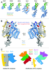
Figure 1. (A) Structure of PCI proteins currently available. PCI-proteins contain a bipartite fold consisting of a N-terminal helical domain (blue) and a C-terminal winged-helix domain (green). The C-terminal winged-helix domain often leads to a C-terminal tail (orange) that interacts with the surface of the helical domain. (B) The Sac3-Thp1-Sem1 structure. The two PCI-proteins Thp1 (gray) and Sac3 (blue) share an extensive interaction interface marked by the insertion of the Thp1 WH “recognition helix” (red) into a cleft formed by the Sac3 WH and HD. The small Sem1 protein (yellow) wraps around the surface of Thp1 and makes little contact with Sac3. Blue spheres indicate the position of side-chains of conserved positive residues that when mutated to a negative charge, resulted in a loss of nucleic acid binding and mRNA export defects in yeast. (C) The role of Sem1 in modulating PCI complexes. Sem1 acts to stabilize the mini-PCI complex formed between Sac3-Thp1-Sem1 by binding to two sites on the Thp1 surface (Sites A and B). In doing so it may be acting as a capping protein shielding the surface of Thp1 from interacting with other PCI-proteins. Within the larger PCI-complex of the 26S proteasome lid Sem1 may act as a modulator of complex assembly, or subunit composition, by binding to and capping specific PCI-protein subunits. Within the schematic representations, the recognition helix (RH) of each WH domains is shown as a protruding tab.

References

    1. Hofmann K, Bucher P. The PCI domain: a common theme in three multiprotein complexes. Trends Biochem Sci. 1998;23:204–5. doi: 10.1016/S0968-0004(98)01217-1. - DOI - PubMed
    1. Pick E, Hofmann K, Glickman MH. PCI complexes: Beyond the proteasome, CSN, and eIF3 Troika. Mol Cell. 2009;35:260–4. doi: 10.1016/j.molcel.2009.07.009. - DOI - PubMed
    1. García-Oliver E, García-Molinero V, Rodríguez-Navarro S. mRNA export and gene expression: The SAGA-TREX-2 connection. Biochim Biophys Acta. 20121819:555–65. - PubMed
    1. Rodríguez-Navarro S, Hurt E. Linking gene regulation to mRNA production and export. Curr Opin Cell Biol. 2011;23:302–9. doi: 10.1016/j.ceb.2010.12.002. - DOI - PubMed
    1. Valkov E., Dean JC, Jani D, Kuhlmann SI, Stewart M. Structural basis for the assembly and disassembly of mRNA nuclear export complexes. Biochim Biophys Acta. 20121819:578–92. - PubMed

Publication types

MeSH terms

Substances

LinkOut - more resources