. 2012 Sep-Oct;1(5):731-41.
doi: 10.1002/wdev.40.

Ink4-Arf locus in cancer and aging

Affiliations

Ink4-Arf locus in cancer and aging

Charles J Sherr. Wiley Interdiscip Rev Dev Biol. 2012 Sep-Oct.

Abstract

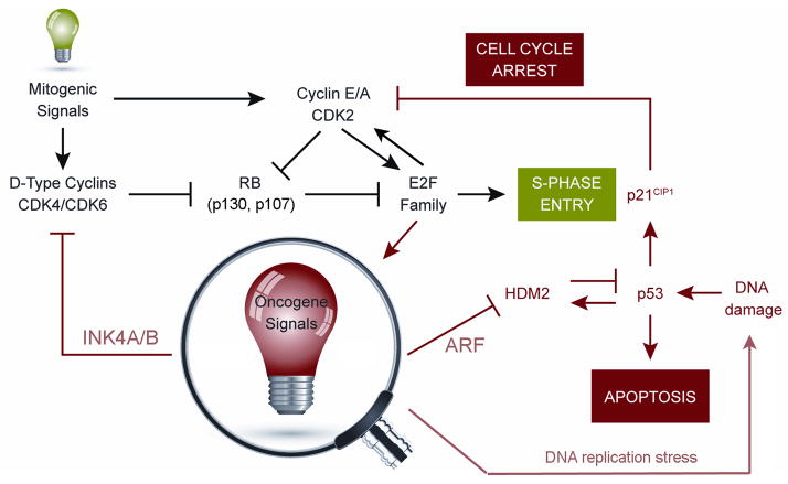
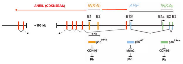
Three tumor suppressor genes at the small (<50 kb) INK4-ARF (CDKN2A/B) locus on human chromosome 9p21 coordinate a signaling network that depends on the activities of the retinoblastoma (RB) protein and the p53 transcription factor. Disruption of this circuitry, frequently by codeletion of INK4-ARF, is a hallmark of cancer, begging the question of why the intimate genetic linkage of these tumor suppressor genes has been maintained in mammals despite the risk of their coinactivation. The INK4-ARF locus is not highly expressed under normal physiologic conditions in young mammals, but its induction becomes more pronounced as animals age. Notably, INK4-ARF is actively silenced en bloc in embryonic, fetal, and adult stem cells but becomes poised to respond to oncogenic stress signals as stem cells lose their self-renewal capacity and differentiate, thereby providing a potent barrier to tumor formation. Epigenetic remodeling of the locus as a whole provides a mechanism for coordinating the activities of RB and p53. A hypothesis is that the INK4-ARF locus may have evolved to physiologically restrict the self-renewal capacities and numbers of stem and progenitor cells with the attendant consequence of limiting tissue regenerative capacity, particularly as animals age. Deletion of INK4-ARF contributes to the aberrant self-renewal capacity of tumor cells and occurs frequently in many forms of human cancer.


Figures

FIGURE 1
FIGURE 1
The INK4-ARF signalling network. Physiologic mitogenic signals (green light, top left) stimulate the transcription of genes encoding D-type cyclins and facilitate their assembly into stable complexes with cyclin-dependent kinases (CDK4 and CDK6). These kinases promote the initial phosphorylation of RB and other RB family members (p130 and p107), cancelling their negative regulation of E2F transcription factors and triggering an E2F-dependent program that stimulates entry into the DNA synthetic (S) phase of the cell division cycle. E2F-responsive genes include those encoding cyclins E and A, which assemble with CDK2 to enforce RB-family protein phosphorylation and drive S-phase entry. Aberrant thresholds of hyperproliferative signals emanating from constitutively active oncogenes (magnified red light, bottom) activate INK4-ARF gene expression to inhibit the activities of cyclin-dependent kinases and HDM2. ARF-mediated inhibition of HDM2 E3 ubiquitin ligase activates the p53 transcriptional program, leading either to apoptosis or cell cycle arrest. Apart from the INK4 proteins, another key mediator of cell cycle arrest is the p53-responsive CDK2 inhibitor, p21Cip1. Multiple types of DNA damage activate p53, including DNA replication errors triggered by oncogenes (bottom right). Many feedback loops regulate the network. Inactivation of p53 leads to increased ARF expression; loss of RB leads to increased p16INK4A levels (not shown). At least one of the transcription factors activating the ARF gene is E2F.
FIGURE 2
FIGURE 2
Expanded view of the INK4-ARF locus. The two INK4 genes and ARF are schematically drawn to scale. Rectangles indicate coding exons of the three genes separated by intronic sequences (black horizontal line). Exons 2 (E2) and three (E3) of the INK4A gene (far right) are translated in alternative reading frames to generate the p16Ink4a protein (green exons and bar) and p19Arf protein (blue exons andbar, p14ARF in human cells). Promoters 5′ to INK4b exon 1 (E1) and to the alternative ARF and INK4a 5′ exons (E1β and E1α, respectively) are noted by arrows. The INK4 proteins are shown to inhibit CDK4/6 to maintain Rb in its growth-suppressive mode. By inhibiting Mdm2, p19Arf activates p53. A long intragenic noncoding RNA (designated ANRIL or CDKN2BAS) is transcribed from the ARF promoter (or from an unidentified promoter element close to exon E1β) in an antisense direction with respect to the primary INK4 and ARF transcripts. ANRIL transcripts are spliced, and putative exons are indicated by red rectangles. A ~60 kb segment within the 100 kb gap region illustrated in the schematic includes single nucleotide polymorphisms on human chromosome 9p21 in proximityto CDKN2A/B that are associated with susceptibility to coronary artery disease, aortic aneurysm, stroke, and type II diabetes.

References

    1. Sherr CJ. The INK4a/ARF network in tumour suppression. Nat Rev Mol Cell Biol. 2001;2:731–737. doi: 10.1038/35096061. - DOI - PubMed
    1. Burkhart DL, Sage J. Cellular mechanisms of tumour suppression by the retinoblastoma gene. Nat Rev Cancer. 2008;8:671–682. doi: 10.1038/nrc2399. - DOI - PMC - PubMed
    1. Vousden KH, Prives C. Blinded by the light: the growing complexity of p53. Cell. 2009;137:413–431. doi: 10.1016/j.cell.2009.04.037. - DOI - PubMed
    1. Juntilla MR, Evan GI. P53 – a jack of all trades but master of none. Nat Rev Cancer. 2009;9:821–829. doi: 10.1038/nrc2778. - DOI - PubMed
    1. Howley PM, Livingston DM. Small DNA tumor viruses: large contributors to biomedical sciences. Virology. 2009;384:256–259. doi: 10.1016/j.virol.2008.12.006. - DOI - PMC - PubMed

Publication types

Substances