. 2012 Sep;24(9):3742-54.
doi: 10.1105/tpc.112.103945. Epub 2012 Sep 7.

Capping protein modulates the dynamic behavior of actin filaments in response to phosphatidic acid in Arabidopsis

Affiliations

Capping protein modulates the dynamic behavior of actin filaments in response to phosphatidic acid in Arabidopsis

Jiejie Li et al. Plant Cell. 2012 Sep.

Abstract

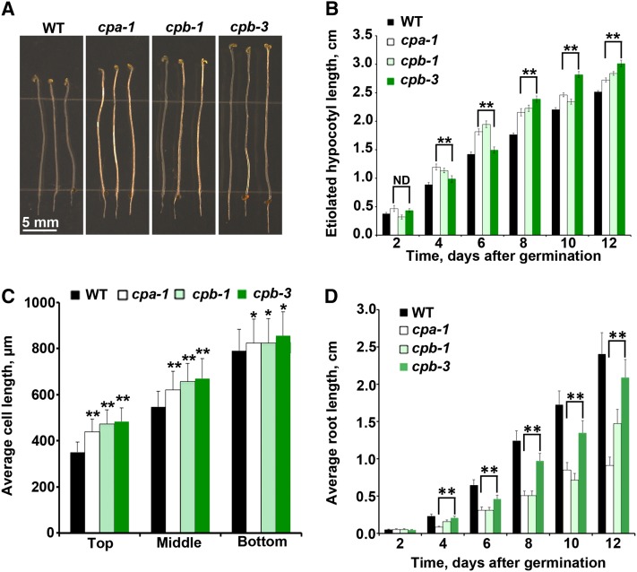
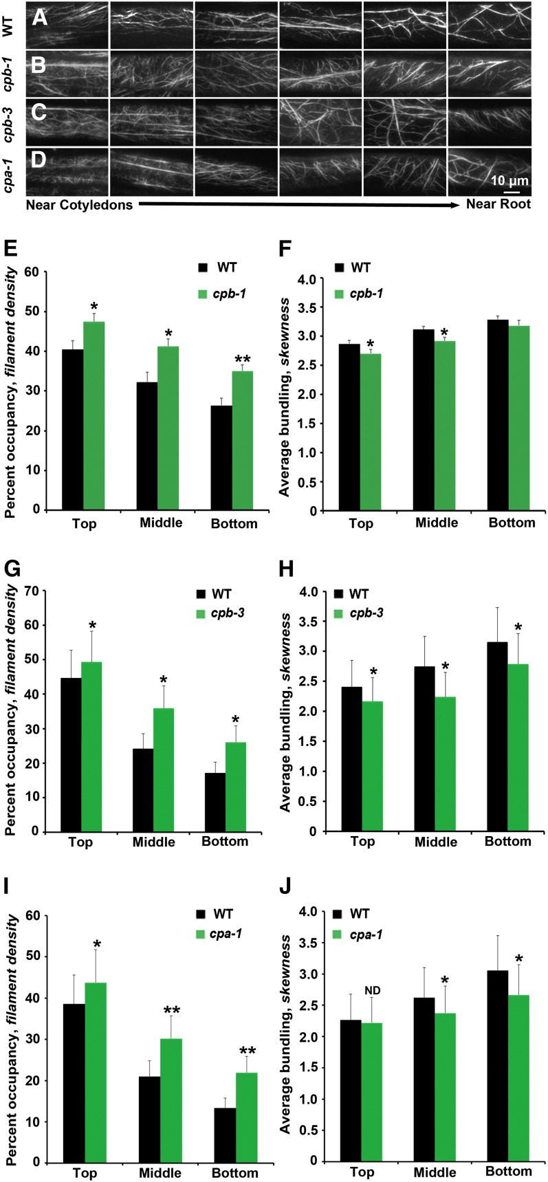
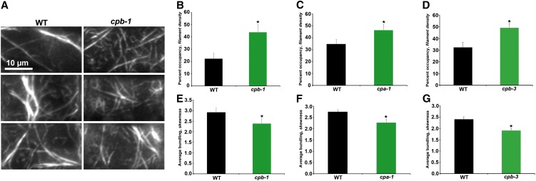
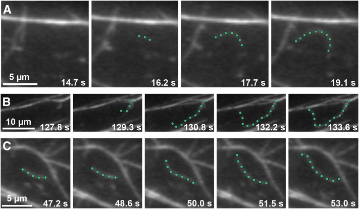
Remodeling of actin filament arrays in response to biotic and abiotic stimuli is thought to require precise control over the generation and availability of filament ends. Heterodimeric capping protein (CP) is an abundant filament capper, and its activity is inhibited by membrane signaling phospholipids in vitro. How exactly CP modulates the properties of filament ends in cells and whether its activity is coordinated by phospholipids in vivo is not well understood. By observing directly the dynamic behavior of individual filament ends in the cortical array of living Arabidopsis thaliana epidermal cells, we dissected the contribution of CP to actin organization and dynamics in response to the signaling phospholipid, phosphatidic acid (PA). Here, we examined three cp knockdown mutants and found that reduced CP levels resulted in more dynamic activity at filament ends, and this significantly enhanced filament-filament annealing and filament elongation from free ends. The cp mutants also exhibited more dense actin filament arrays. Treatment of wild-type cells with exogenous PA phenocopied the actin-based defects in cp mutants, with an increase in the density of filament arrays and enhanced annealing frequency. These cytoskeletal responses to exogenous PA were completely abrogated in cp mutants. Our data provide compelling genetic evidence that the end-capping activity of CP is inhibited by membrane signaling lipids in eukaryotic cells. Specifically, CP acts as a PA biosensor and key transducer of fluxes in membrane signaling phospholipids into changes in actin cytoskeleton dynamics.


Figures

Figure 1.
Figure 1.
Loss of CP Alters Organ and Epidermal Cell Length. (A) Representative images of 11-d-old cp (cpa-1, cpb-1, and cpb-3) homozygous mutants and wild-type (WT) seedlings grown in continuous darkness. Bar = 5 mm. (B) Etiolated hypocotyls from 4- to 12-d-old cpa-1, cpb-1, and cpb-3 mutant seedlings are significantly longer than the wild type (**P < 0.005; ND, no significant difference; t test). Measurements were taken from at least 50 seedlings per genotype on every other day. Values represent means ± se. (C) Epidermal cell lengths were measured on 5-d-old dark-grown hypocotyls. Cell lengths from cp mutants are significantly longer compared with the wild type in the top (**P < 0.001, t test), middle (**P < 0.001, t test), and bottom (*P < 0.05, t test) thirds of hypocotyls. Values are means ± se (n > 50 cells from at least 10 hypocotyls per genotype). (D) Light-grown roots from cpa-1, cpb-1, and cpb-3 mutant seedlings are significantly shorter than wild-type roots (**P < 0.005, t test). Values are means ± se from at least 50 seedlings per genotype taken on alternating days between 2 and 12 d postgermination.
Figure 2.
Figure 2.
The Architecture of Cortical Actin Arrays in Epidermal Cells of cp Mutants Is Altered. (A) to (D) Montages of VAEM micrographs of epidermal cells taken from representative hypocotyls of wild-type (WT) (A), cpb-1 (B), cpb-3 (C), and cpa-1 (D) seedlings expressing GFP-fABD2. Seedlings were grown in continuous darkness for 5 d and images collected to display cells near the cotyledon at left and cells near the root at right. Bar = 10 µm. (E), (G), and (I) Average filament density or percentage of occupancy was measured along the axial gradient of cell expansion in hypocotyls and binned into three regions. The hypocotyls of 5-d-old dark-grown seedlings for all three mutants, cpb-1 (E), cpb-3 (G), and cpa-1 (I), have significantly increased filament density in the top, middle, and bottom thirds of 5-d-old dark-grown seedlings when compared with their respective wild-type siblings. Values given are means ± se (n ≥ 300 images from ≥ 150 cells per region, 30 hypocotyls were measured; t test: *P < 0.05 and **P < 0.01). (F), (H), and (J) The extent of filament bundling (skewness) was measured and binned for the same regions as in density measurements. Epidermal cells from cpb-1 (F), cpb-3 (H), and cpa-1 (J) mutant seedlings have less filament bundling in different regions of the 5-d-old dark-grown seedlings when compared with their respective wild-type siblings. The same images used for (E), (G), and (I) were analyzed for skewness (*P < 0.05; ND, no significant difference; t test). [See online article for color version of this figure.]
Figure 3.
Figure 3.
Actin Architecture in Epidermal Cells from the Root Elongation Zone of cp Mutants Is Altered. (A) Montages of VAEM micrographs from representative epidermal cells taken from the elongation zone of roots from light-grown wild-type (WT) and cpb-1 seedlings. Bar = 10 µm. (B) to (D) Average filament density or percentage of occupancy was measured for epidermal cells within the root elongation zone. cpb-1 (B), cpa-1 (C), and cpb-3 (D) seedlings had significantly increased filament density in 7-d-old light-grown seedlings when compared with wild-type sibling seedlings. (E) to (G) Filament bundling (skewness) was measured on the same images as for density measurements. Filaments in cpb-1 (E), cpa-1 (F), and cpb-3 (G) seedlings had less bundling when compared with wild-type sibling seedlings. Values given are means ± se (n > 60 images per seedling from 25 seedlings per genotype; *P < 0.05, t test). [See online article for color version of this figure.]
Figure 4.
Figure 4.
Growing Actin Filaments Originate from Three Locations in Wild-Type Epidermal Cells. Time-lapse VAEM series show examples of actin filaments originating and growing rapidly from three different cellular locations: de novo (A), the side of a bundle (B), and the end of a preexisting fragment (C) (see Supplemental Movies 1 to 3 online). Representative actin filaments are highlighted with green dots. Images were recorded from epidermal cells in the actively growing region (top third) of 5-d-old dark-grown wild-type hypocotyls. Bars = 5 µm in (A) and (C) and 10 µm in (B).
Figure 5.
Figure 5.
The Dynamic Behavior of Actin Filament Ends in Epidermal Cells Can Be Tracked. (A) Time-lapse VAEM series shows an example of an actin filament elongating from a newly created barbed end. The highlighted filament (green dots) stops growing and is severed (red arrows) into several fragments. One filament end (white arrowheads) resumes growth within 5 s after severing (see Supplemental Movie 4 online). (B) New filaments can be constructed by filament-filament annealing of severed fragments. The highlighted growing filament gets severed (red arrows). Newly created ends join together within ∼1.5 s to form a new filament (yellow arrowheads). Three annealing events occur in the sequential time-lapse frames (see Supplemental Movie 5 online). Images were taken by time-lapse VAEM from cpb-1 epidermal cells. Bars = 5 µm.
Figure 6.
Figure 6.
Density of the Actin Array in cpb-1 Hypocotyl Epidermal Cells Is Insensitive to Exogenous PA Treatment. Percentage of occupancy or density was measured and binned into three regions along the axial gradient of cell expansion in 5-d-old etiolated wild-type (WT) (A) or cpb-1 (B) hypocotyls after incubation with 0, 10, 50, or 100 µM PA or with 100 µM PS for 30 min. Wild-type seedlings treated with PA have significantly increased filament density in the middle and bottom thirds of hypocotyls in a dose-dependent manner (A). By contrast, PA treatments of cpb-1 seedlings did not increase the density, but rather led to a modest decline in density at all concentrations tested (B). Treatments with another acidic phospholipid, PS, had no measurable effect on actin filament levels in either wild-type or cpb-1 seedlings. Values given are means ± se (n = 300 images from 150 cells per region; 10 hypocotyls were measured for each treatment per genotype; *P < 0.05 and **P < 0.001, analysis of variance).
Figure 7.
Figure 7.
Annealing Frequency and Filament Origin Are Insensitive to Exogenous PA Treatment in cpb-1 Epidermal Cells. Annealing frequency and filament origins were measured in cpb-1 and wild-type (WT) epidermal cells from the bottom third of hypocotyls following treatments with PA and PS. (A) Annealing frequency from wild-type (WT) cells treated with PA was approximately threefold higher than untreated cells (blue; *P < 0.05, t test); cpb-1 cells after PA treatment had no obvious increase in annealing frequency compared with control cells (purple). Treatments with PS had no significant effect on annealing frequency in either cpb-1 or wild-type cells. Values given are means ± se (n > 50 filaments from > 10 epidermal cells and at least 10 hypocotyls for each treatment per genotype). (B) The percentage of filaments that originate from ends was increased in wild-type cells after PA treatment. (C) Filament origins in cpb-1 cells treated with phospholipids were not significantly different from untreated cells. Measurements were taken from n = 30 cells from at least 10 hypocotyls for each treatment per genotype; **P < 0.0001, analysis of variance.
Figure 8.
Figure 8.
Density of the Actin Array in cpb-1 Hypocotyl Epidermal Cells Is Insensitive to 1-Butanol Treatment. Percentage of occupancy or density was measured and binned into three regions along the axial gradient of cell expansion in 5-d-old etiolated wild-type (WT) (A) or cpb-1 (B) hypocotyls after incubation with 0, 0.1, 0.5, or 1% 1-butanol or with 1% 2-butanol for 15 min. Wild-type seedlings treated with 1-butanol have significantly decreased filament density in the top, middle, and bottom regions of hypocotyls, and this reduction is dose dependent (A). By contrast, 1-butanol treatments of cpb-1 seedlings did not decrease the density (B). Treatments with 2-butanol had no measurable effect on actin filament levels in either wild-type or cpb-1 seedlings. Values given are means ± se (n = 300 images from 150 cells per region; 10 hypocotyls were measured for each treatment per genotype; *P < 0.05 and **P < 0.001, analysis of variance).

Similar articles

Cited by

References

    1. Abraham V.C., Krishnamurthi V., Taylor D.L., Lanni F. (1999). The actin-based nanomachine at the leading edge of migrating cells. Biophys. J. 77: 1721–1732 - PMC - PubMed
    1. Amatruda J.F., Gattermeir D.J., Karpova T.S., Cooper J.A. (1992). Effects of null mutations and overexpression of capping protein on morphogenesis, actin distribution and polarized secretion in yeast. J. Cell Biol. 119: 1151–1162 - PMC - PubMed
    1. Andrianantoandro E., Blanchoin L., Sept D., McCammon J.A., Pollard T.D. (2001). Kinetic mechanism of end-to-end annealing of actin filaments. J. Mol. Biol. 312: 721–730 - PubMed
    1. Augustine R.C., Pattavina K.A., Tüzel E., Vidali L., Bezanilla M. (2011). Actin interacting protein1 and actin depolymerizing factor drive rapid actin dynamics in Physcomitrella patens. Plant Cell 23: 3696–3710 - PMC - PubMed
    1. Bao C., Wang J., Zhang R., Zhang B., Zhang H., Zhou Y., Huang S. (May 5, 2012). Arabidopsis VILLIN2 and VILLIN3 act redundantly in sclerenchyma development via bundling of actin filaments. Plant J. http://dx.doi.org/.10.1111/j.1365-1313X.2012.05044.x - PubMed

Publication types

MeSH terms