. 2012 Sep;24(9):3630-48.
doi: 10.1105/tpc.112.101063. Epub 2012 Sep 7.

Overexpression of Arabidopsis plasmodesmata germin-like proteins disrupts root growth and development

Affiliations

Overexpression of Arabidopsis plasmodesmata germin-like proteins disrupts root growth and development

Byung-Kook Ham et al. Plant Cell. 2012 Sep.

Abstract

In plants, a population of non-cell-autonomous proteins (NCAPs), including numerous transcription factors, move cell to cell through plasmodesmata (PD). In many cases, the intercellular trafficking of these NCAPs is regulated by their interaction with specific PD components. To gain further insight into the functions of this NCAP pathway, coimmunoprecipitation experiments were performed on a tobacco (Nicotiana tabacum) plasmodesmal-enriched cell wall protein preparation using as bait the NCAP, pumpkin (Cucurbita maxima) PHLOEM PROTEIN16 (Cm-PP16). A Cm-PP16 interaction partner, Nt-PLASMODESMAL GERMIN-LIKE PROTEIN1 (Nt-PDGLP1) was identified and shown to be a PD-located component. Arabidopsis thaliana putative orthologs, PDGLP1 and PDGLP2, were identified; expression studies indicated that, postgermination, these proteins were preferentially expressed in the root system. The PDGLP1 signal peptide was shown to function in localization to the PD by a novel mechanism involving the endoplasmic reticulum-Golgi secretory pathway. Overexpression of various tagged versions altered root meristem function, leading to reduced primary root but enhanced lateral root growth. This effect on root growth was corrected with an inability of these chimeric proteins to form stable PD-localized complexes. PDGLP1 and PDGLP2 appear to be involved in regulating primary root growth by controlling phloem-mediated allocation of resources between the primary and lateral root meristems.


Figures

Figure 1.
Figure 1.
Arabidopsis PDGLP1 and PDGLP2 Are Plasmodesmal Targeted Proteins. (A) Phylogenetic analysis of the relationship between the Arabidopsis GLP family and the tobacco PDGLP1 identified in our tobacco PECP co-IP studies using the pumpkin phloem Cm-PP16 as bait. The phylogenetic tree was constructed in MEGA 3.1; numbers at each branch point represent the bootstrap values for percentage of 1000 replicate trees. (B) PDGLP1 (At1G09560) and PDGLP2 (At1G02335) are colocalized with the PD marker CMV MP-GFP. The C-terminal mCherry (RFP) tagged At1G09560, At1G02335, and At1G18970 (GLP4) were agroinfiltrated into leaves of CMV MP-GFP transgenic N. benthamiana plants. Yellow signal in merged images represents colocalization of At1G09560 and At1G02335 with CMV MP-GFP. Images were taken by CLSM. Bars = 10 µm. (C) PDGLP1-GFP is located within PD. Leaf and root tissues of PDGLP1-GFP transgenic plants were processed for transmission electron microscopy–based double immunogold labeling with anticallose Ab (15-nm gold particles) and anti-GFP Ab (10-nm gold particles). Red arrowheads indicate detection of PDGLP1-GFP within the PD. Bar = 200 nm. [See online article for color version of this figure.]
Figure 2.
Figure 2.
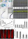
PDGLP1 Expression Profile and Phenotypic Analysis of Mutant and Transgenic PDGLP1-GFP Plant Lines. (A) Histochemical staining of PPDGLP1:GUS transgenic plants reveals PDGLP1 expression pattern in cotyledons and the root tip during early seedling development. HAG, hours after germination. Bar = 1 mm. (B) and (C) GUS staining performed with 7-d-old transgenic PPDGLP1:GUS plants indicates the presence of strong signal in the root system (B), whereas in the first true leaves, GUS staining was restricted to basal trichome cells (C) (area shown is enlarged from the dashed square indicted in [B]). (D) Strong GUS staining in the primary root (area shown is enlarged from the dashed square indicted in [B]). (E) and (F) Phenotype of wild-type (WT), empty vector–transformed transgenic plants (Vec), pdglp1, pdglp2, pdglp1 pdglp2 double mutant, and PPDGLP1:PDGLP1-GFP (PG1-GFP) seedlings at 5 (E) and 10 (F) DAG. Note the short root phenotype for the PPDGLP1:PG1-GFP seedlings. Bars = 10 mm. (G) Average root length of wild-type (open diamonds), Vec (open squares), pdglp1 (closed diamonds), pdglp2 (triangles), pdglp1 pdglp2 (squares), PPDGLP1:PG1-GFP (open circles), and PPDGLP2:PG2-GFP (closed circles) seedlings measured at the indicated DAG. Values represent mean ± sd; n = 40 seedlings for each plant line tested. (H) Transgenic PG1-GFP and PG2-GFP plants displayed enhanced lateral root length compared with wild-type, vec, pdglp1, pdglp2, and pdglp1/pdglp2 seedlings. Asterisk indicates significant differences, P < 0.002, based on Student’s t test. (I) Five-day-old primary roots from wild-type, pdglp1, pdglp2, pdglp1/2, PG1-GFP, and PG2-GFP seedlings. Arrowheads indicate the boundary between the meristem and elongation zones. Images collected by confocal microscopy of propidium iodine–stained roots. Bar = 50 µm. (J) Analysis of meristem size in the indicated plant lines. Confocal images were analyzed to determine the cortical cell number in the meristem zone of each plant line. Values represent mean ± sd; n = 50 roots. Asterisk indicates significant differences, P < 0.002, based on Student’s t test. [See online article for color version of this figure.]
Figure 3.
Figure 3.
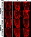
Both PDGLP1-GFP and PDGLP2-GFP Are Localized to the Endodermis and Quiescent Center in Both Primary and Lateral Root Meristems. Confocal microscopy was used to map the cellular domains occupied by PDGLP1-GFP and PDGLP2-GFP in primary and lateral root tips at the indicated DAG. Darts indicate GFP signal in endodermal (white) and quiescent center (yellow) cells. Asterisks indicate propidium iodide–stained dead/dying cells. Bar = 25 µm. [See online article for color version of this figure.]
Figure 4.
Figure 4.
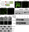
The PDGLP1 SP Is Necessary and Sufficient for Protein Delivery to PD via the ER-Golgi Secretory Pathway. (A) Arabidopsis PDGLP1 and GLP4 constructs used to test the role of the SP in protein targeting to PD. Putative N-terminal SPs shown as a red (PDGLP1) and blue (GLP4) box. GFP was fused to the C terminus of PDGLP1, a SP-deleted PDGLP1 mutant (PDGLP1ΔSP-GFP), and alone with the PDGLP1 SP (PDGLP1SP-GFP) or the GLP4 SP (GLP4SP-GFP). (B) The control constructs tested remained either in the cytoplasm and nucleus (GFP alone) or in the cell wall (GLP4SP-GFP). (C) The PDGLP1ΔSP-GFP mutant is no longer targeted to PD, whereas the PDGLP1SP-GFP construct accumulated in punctate foci along the cell wall, likely representing location to PD. In (B) and (C), the tested constructs were introduced into N. benthamiana leaves by particle bombardment and observed using confocal microscopy after a 24-h incubation period. Bars = 10 µm. (D) PDGLP1 SP is not cleaved during protein targeting to PD. PDGLP1-MHT and PDGLP1SP-GFP-MHT were first agroinfiltrated in N. benthamiana leaves and then 4 d later leaf tissues were extracted for biochemical experiments. IM1 and IM2, internal markers for PDGLP1 lacking (single asterisk) or containing (double asterisk) the SP; P, PECP fraction; S, soluble protein fraction; T, total protein fraction. (E) PDGLP1 uses the secretory pathway for its targeting to PD. A PDGLP1-GFP construct was agroinfiltrated into N. benthamiana leaves and, 24 h later, they were imaged by confocal microscopy prior to infiltration with BFA. Following a 12-h BFA treatment, recovery experiments were performed by infiltration of a 0.5% (v/v) DMSO solution. A parallel set of experiments was performed with a PDGLP1-MHT construct, and these leaves were employed to test for the effect of BFA treatment on PDGLP1-GFP subcellular localization. Here, Nt-NCAPP1 and Rubisco antibodies were used to confirm the purity of the PECP fraction. P, PECP fraction; T, total protein fraction. Bar = 10 µm. (F) PDGLP1 is located on the outside of the Golgi-derived vesicles. PDGLP1-MHT and GLP4-MHT constructs were agroinfiltrated into N. benthamiana leaves and Golgi-derived vesicles were fractionated by velocity sedimentation. Vesicles were either treated with Proteinase K (20 µg/mL) or first pretreated with 0.1% Triton X-100 followed by Proteinase K. Note: PDGLP1-MHT was not detected in Proteinase K–treated vesicles, whereas a combination of Triton X-100 and Proteinase K was necessary to eliminate the GLP4-MHT signal. [See online article for color version of this figure.]
Figure 5.
Figure 5.
PDGLP1 Interacts Directly with the NCAP Cm-PP16. (A) Recombinant CmPP16-GST, NtNCAPP1ΔN1-22-MHT, GFP-MHT, GST, PDGLP1-MHT, and PDGLP1ΔSP-MHT were expressed in and purified from plants using a ZYMV viral vector system. Proteins were separated on a 12% SDS-PAGE gel and visualized by staining with Gelcode Blue (GCB). (B) and (C) An in vitro GST pull-down assay demonstrates that PDGLP1 directly interacts with Cm-PP16. Pull-downed proteins were separated on a 12% SDS-PAGE gel and then subjected to immunoblot analysis using a GST antibody (B) or a His antibody (C). GFP and GST were used as negative controls, and NtNCAPP1ΔN1-22-MHT served as a positive control. [See online article for color version of this figure.]
Figure 6.
Figure 6.
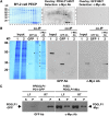
PDGLP1 Interacts with a Subset of Proteins in the PECP Fraction to Form a PDGLP1-Specific Complex. (A) Protein overlay assays establish that PDGLP1-MHT interacts with a range of proteins contained in the BY-2 cell PECP preparation. Proteins in the PECP were separated by fast protein liquid chromatography, and fractions were then separated by SDS-PAGE, followed by transfer to nitrocellulose membrane. Membrane was overlaid with purified PDGLP1-MHT and then subjected to immunoblot analysis, using an anti-c-Myc monoclonal antibody, to identify interacting proteins. GFP-MHT was used as a negative control. (B) PDGLP1-interacting proteins identified by co-IP experiments. Purified recombinant GFP, PDGLP1-GFP (lane 1), or PDGLP1-Myc (lane 2) was incubated with an Arabidopsis PECP fraction (Input) followed by co-IP using anti-GFP or anti-c-Myc monoclonal antibodies (mAb). White, black, and red asterisks indicate immunoprecipitated GFP, PDGLP1-GFP, and PDGLP1-Myc, respectively. GFP served as the negative control. Immunoprecipitated bait proteins were confirmed by immunoblot analysis using anti-GFP (middle panel) or anti-c-Myc mAb (right panel). (C) Both PDGLP1-GFP and PDGLP1-Myc are highly enriched in the Arabidopsis PECP fraction. Transgenic PPDGLP1:PDGLP1-GFP or P35S:PDGLP1-Myc plants were used to prepare soluble and PECP fractions, which were then separated by SDS-PAGE and tested by protein gel blot analysis using GFP or anti-c-Myc monoclonal antibodies. PECP fractions were prepared from leaf (LF) and root (RT) tissues. S, soluble fraction; P, PECP fraction. [See online article for color version of this figure.]
Figure 7.
Figure 7.
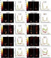
Symplasmic Permeability Is Enhanced in the Root Tips of Transgenic PDGLP1-Myc and PDGLP2-Myc Plants. A brief (10-s) application of CFDA (60 µg/mL) to the primary ([A] to [F]) or lateral ([G] to [J]) root tips was used to load CF into the cytoplasm of Arabidopsis root cap and epidermal cells. CF distribution within the root tip was analyzed by confocal microscopy at 2 and 5 min after CFDA application. In wild-type (WT) (A), pdglp1 (B), pdglp2 (C), and pdglp1 pdglp2 (D) primary root tips, CF was restricted to the outer layer of cells. For the PDGLP1-Myc (E) and PDGLP2-Myc (F) seedlings, CF was detected in the central region of the primary root tip within 5 min of CFDA application. For wild-type (G) and pdglp1 pdglp2 (H) lateral root tips, CF was restricted to the outer layer of cells. By contrast, in the PDGLP1-Myc (I) and PDGLP2-Myc (J) seedlings, CF was detected in the central region of the lateral root tip within 5 min of CFDA application. Right panels present quantification of the fluorescence intensity measurements made at the locations indicated by the white bars on the individual roots. Red and green traces represent fluorescence intensity profiles collected at 2 and 5 min after CFDA application, respectively. Color bars on the left side of the confocal image indicate relative fluorescence intensity. Bars = 60 µm. [See online article for color version of this figure.]
Figure 8.
Figure 8.
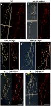
Expressing Myc/GFP-Tagged Forms of PDGLP1 or PDGLP2 in Transgenic Arabidopsis Seedlings Alters the Pattern of Phloem Delivery into the Root System. Phloem translocation into the Arabidopsis root system was examined by applying CFDA (60 µg/mL) onto a single cotyledon of 8-d-old seedlings. Confocal analysis of phloem delivery of CF into the roots was performed 90 min after CFDA application. (A) and (B) In wild-type (WT) (A) and pdglp1 pdglp2 (B) seedlings, CF was unloaded in the root tip regions of both primary and lateral roots. (C) to (F) In PDGLP1-Myc (C), PDGLP2-Myc (D), PDGLP1-GFP (E), and PDGLP2-GFP (F) transgenic seedlings, CF unloading was detected only in the lateral roots. White dotted lines indicate the position of the primary root into which phloem delivery of CF did not take place. Bars = 5 mm. [See online article for color version of this figure.]
Figure 9.
Figure 9.
Primary Roots of Tagged PDGLP1 and PDGLP2 Transgenic Plants Display a Normal Gravitropic Response. (A) Control (Vec), pdglp1 pdglp2 double mutant, PPDGLP1:PG1-GFP, PPDGLP2:PG2-GFP, P35S:PDGLP1-Myc, and P35S:PDGLP2-Myc plants were grown vertically on Murashige and Skoog plates for 8 d and then plates were rotated 90° to test the capacity of these lines to undergo differential growth in response to the altered gravitropic signal. Note that the primary roots of all tagged plant lines displayed a normal gravitropic response. (B) Extent of primary root growth following plate rotation. Values represent mean ± sd; n = 50 roots. Asterisk indicates significant differences, P < 0.002, based on Student’s t test. [See online article for color version of this figure.]

Similar articles

Cited by

References

    1. Aoki K., Kragler F., Xoconostle-Cazares B., Lucas W.J. (2002). A subclass of plant heat shock cognate 70 chaperones carries a motif that facilitates trafficking through plasmodesmata. Proc. Natl. Acad. Sci. USA 99: 16342–16347 - PMC - PubMed
    1. Banerjee A.K., Chatterjee M., Yu Y.Y., Suh S.G., Miller W.A., Hannapel D.J. (2006). Dynamics of a mobile RNA of potato involved in a long-distance signaling pathway. Plant Cell 18: 3443–3457 - PMC - PubMed
    1. Bernier F., Berna A. (2001). Germins and germin-like proteins: Plant do-all proteins. But what do they do exactly? Plant Physiol. Biochem. 39: 545–554
    1. Blackman L.M., Boevink P., Cruz S.S., Palukaitis P., Oparka K.J. (1998). The movement protein of cucumber mosaic virus traffics into sieve elements in minor veins of nicotiana clevelandii. Plant Cell 10: 525–538 - PMC - PubMed
    1. Carlsbecker A., et al. (2010). Cell signalling by microRNA165/6 directs gene dose-dependent root cell fate. Nature 465: 316–321 - PMC - PubMed

Publication types

MeSH terms