. 2012 Nov;61(11):2851-61.
doi: 10.2337/db12-0134. Epub 2012 Sep 6.

Vascular endothelial growth factor-mediated islet hypervascularization and inflammation contribute to progressive reduction of β-cell mass

Affiliations

Vascular endothelial growth factor-mediated islet hypervascularization and inflammation contribute to progressive reduction of β-cell mass

Judith Agudo et al. Diabetes. 2012 Nov.

Abstract

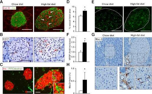
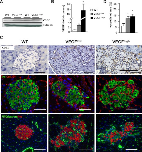
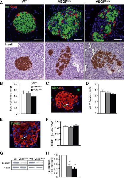
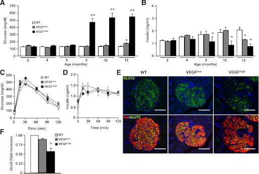
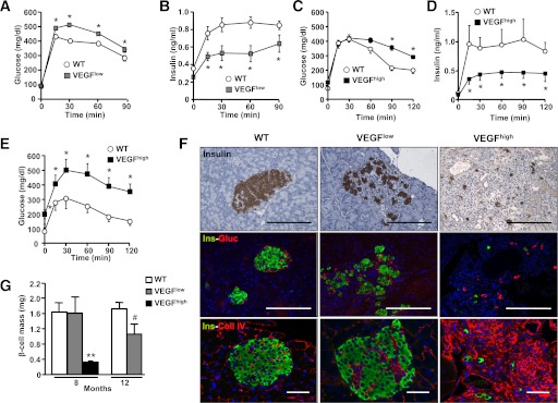
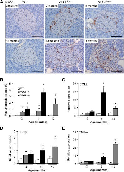
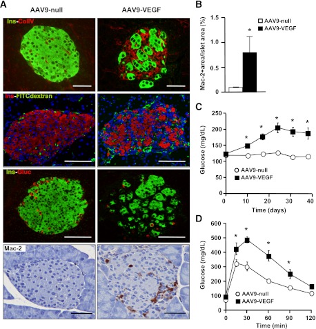
Type 2 diabetes (T2D) results from insulin resistance and inadequate insulin secretion. Insulin resistance initially causes compensatory islet hyperplasia that progresses to islet disorganization and altered vascularization, inflammation, and, finally, decreased functional β-cell mass and hyperglycemia. The precise mechanism(s) underlying β-cell failure remain to be elucidated. In this study, we show that in insulin-resistant high-fat diet-fed mice, the enhanced islet vascularization and inflammation was parallel to an increased expression of vascular endothelial growth factor A (VEGF). To elucidate the role of VEGF in these processes, we have genetically engineered β-cells to overexpress VEGF (in transgenic mice or after adeno-associated viral vector-mediated gene transfer). We found that sustained increases in β-cell VEGF levels led to disorganized, hypervascularized, and fibrotic islets, progressive macrophage infiltration, and proinflammatory cytokine production, including tumor necrosis factor-α and interleukin-1β. This resulted in impaired insulin secretion, decreased β-cell mass, and hyperglycemia with age. These results indicate that sustained VEGF upregulation may participate in the initiation of a process leading to β-cell failure and further suggest that compensatory islet hyperplasia and hypervascularization may contribute to progressive inflammation and β-cell mass loss during T2D.


Figures

FIG. 1.
FIG. 1.
Hypervascularization and macrophage infiltration in pancreatic islets from insulin-resistant HFD-fed mice. A: Islet vessels were revealed by dual collagen IV (red) and insulin (green) immunostaining. Arrows point to thickened basement membrane. B: Endothelial cells were immunostained by CD31 antibody. HFD-fed mice showed more endothelial cells per islet (arrows). C: Pancreatic sections from HFD- and chow-fed FITC-dextran–infused mice showing vessels (green) and insulin (red). Capillaries with a large lumen are shown in the inset. D: HFD animals (black bar) showed increased FITC-dextran area/islet area compared with chow-fed mice (white bar) (n = 3/group). *P < 0.05 HFD vs. chow diet. E: VEGF immunostaining of pancreatic sections from HFD- and chow-fed mice. F: VEGF content in protein extracts of isolated islets from individual animals chow-fed mice (white bar) and HFD mice (black bar) was determined by ELISA (n = 4/group). HFD mice displayed a marked increase in Mac-2–positive macrophage infiltration in islets as shown after immunohistochemical analysis (G) and morphometrical analysis (H) (chow-fed mice [white bar] and HFD mice [black bar]). Infiltrating macrophages at the periphery and inside the islet are shown in the inset (arrows) (n = 3/group). Scale bars, 100 µm. *P < 0.05 HFD vs. chow. (A high-quality digital representation of this figure is available in the online issue.)
FIG. 2.
FIG. 2.
Increased VEGF expression in VEGFlow and VEGFhigh transgenic β-cells leads to islet hypervascularization. A: Representative Western blot showing higher expression of VEGF in both VEGFhigh and VEGFlow. B: A 2.7- and 17-fold increase in VEGF protein levels were found in islets from 2-month-old VEGFlow and VEGFhigh mice measured by ELISA, wild-type (WT) mice (white bar), transgenic VEGFlow mice (gray bar), and transgenic VEGFhigh mice (black bar). *P < 0.05 transgenic vs. WT (n = 8–15/group). C: Top panel: detection of endothelial cells by CD31 immunostaining. Middle panel: collagen IV (red) and insulin (green) immunohistochemical analysis of VEGFlow and VEGFhigh islets. Bottom panel: FITC-dextran (green) together with insulin (red) immunostaining was used to label functional islet vessels. D: Morphometric analysis revealed an increase in the vessel area in both VEGFlow and VEGFhigh mice (n = 3/group): WT mice (white bar), transgenic VEGFlow mice (gray bar), and transgenic VEGFhigh mice (black bar). Scale bars, 100 µm. *P < 0.05 transgenic vs. WT. (A high-quality digital representation of this figure is available in the online issue.)
FIG. 3.
FIG. 3.
Two-month-old VEGFlow and VEGFhigh mice showed disorganization of islet architecture, but normal β-cell mass. A: Top panel: immunohistochemical analysis of insulin (green) and glucagon (red). In transgenic mice, islets appeared disorganized with α-cells in the core. Bottom panel: insulin immunostaining used to visualize islet architecture. B: β-Cell mass was measured in 2-month-old mice (n = 4–7/group): wild-type (WT) mice (white bar), transgenic VEGFlow mice (gray bar), and transgenic VEGFhigh mice (black bar). C: Ki67 (green) and insulin (red) immunostaining were used to label proliferating β-cells (arrow). D: Percentage of Ki67-positive (replicative) β-cells. E: TUNEL (green) and insulin (red) immunostaining showed apoptotic nuclei (arrow). F: Quantification of TUNEL-positive (apoptotic) β-cells. G and H: Western blot analysis of E-cadherin using islet homogenates from 2-month-old mice. G: A representative immunoblot is shown. H: Densitometric analysis of three different immunoblots: WT mice (white bar), transgenic VEGFlow mice (gray bar), and transgenic VEGFhigh mice (black bar). Scale bars, 100 µm. *P < 0.05 transgenic vs. WT. (A high-quality digital representation of this figure is available in the online issue.)
FIG. 4.
FIG. 4.
Glucose homeostasis in VEGFlow and VEGFhigh mice. Blood glucose (A) and insulin (B) levels were monitored in fed mice from 2–12 months: wild-type (WT) mice (white bar), transgenic VEGFlow mice (gray bar), and transgenic VEGFhigh mice (black bar) (n = 15 animals/group). **P < 0.01 *P < 0.05 transgenic vs. WT. Glucose tolerance (C) and in vivo insulin secretion (D) were determined after an intraperitoneal glucose injection (2 g/kg body weight) into 2-month-old mice WT (white circle), VEGFlow mice (gray square), and VEGFhigh mice (black square) (n = 10 animals/group). E: Immunohistochemical analysis of GLUT-2 (green) and insulin (red) in islets from 2-month-old mice is shown. F: GLUT-2 mRNA expression from isolated islets from 2-month-old VEGFlow and VEGFhigh mice was determined by qPCR (n = 3 pools of islets from three mice per pool): WT mice (white bar), transgenic VEGFlow mice (gray bar), and transgenic VEGFhigh mice (black bar). Scale bars, 100 µm. *P < 0.05 vs. WT mice. (A high-quality digital representation of this figure is available in the online issue.)
FIG. 5.
FIG. 5.
Glucose homeostasis in VEGFlow and VEGFhigh mice was impaired with age. Glucose tolerance (A) and insulin in vivo secretion (B) were measured in 12-month-old wild-type (WT) (white circle) and VEGFlow mice (gray square) (n = 12 animals/group). *P < 0.05 VEGFlow vs. WT mice. Glucose tolerance (C) was determined in parallel to in vivo insulin secretion (D) after a glucose load (2 g/kg body weight [b.w.]) in 5-month-old WT mice (white circle) and transgenic VEGFhigh mice (black square). At this age, VEGFhigh mice were normoglycemic, but showed impaired insulin release and glucose intolerance after the glucose load (n = 12 animals/group). *P < 0.05 VEGFhigh vs. WT mice. E: Eight-month-old VEGFhigh mice (black square) were glucose-intolerant compared with age-matched WT (white square) mice as shown by a glucose tolerance test (1 g/kg b.w.) (n = 10 animals/group). *P < 0.05 VEGFhigh vs. WT mice. F: Top panel: representative images of insulin immunostaining of islets. Middle panel: immunohistochemical analysis of insulin (green) and glucagon (red) expression. Bottom panel: immunohistochemical analysis of collagen IV (red) and insulin (green). Islets from 12-month-old WT and VEGFlow and 8-month-old VEGFhigh mice are shown. Scale bars, 100 µm. G: β-Cell mass was determined by insulin immunostaining in 8- and 12-month-old VEGFlow and 8-month-old VEGFhigh mice and compared with age-matched WT mice, as indicated in Research Design and Methods (WT mice [white bar], transgenic VEGFlow mice [gray bar], and transgenic VEGFhigh mice [black bar]) (n = 3 sections/mouse and 4 animals/group). **P < 0.001 VEGFhigh vs. WT mice, #P < 0.1 VEGFlow vs. WT. (A high-quality digital representation of this figure is available in the online issue.)
FIG. 6.
FIG. 6.
VEGF-overexpressing islets showed progressive inflammation. A: Macrophage infiltration was determined by Mac-2 immunostaining in islets from 2-month-old wild-type (WT), VEGFlow, and VEGFhigh mice (top panel) and 12-month-old WT and VEGFlow and 8-month-old VEGFhigh mice (bottom panel). B: Quantification of Mac-2–positive area in pancreas sections from 2-, 8-, and 12-month-old WT, in 2-, 8-, and 12-month-old VEGFlow, and in 2- and 8-month-old VEGFhigh mice (n = 4 animals/group): WT mice (white bar), transgenic VEGFlow mice (gray bar), and transgenic VEGFhigh mice (black bar). CE: Islets from VEGFlow and VEGFhigh mice displayed increased expression of key inflammatory cytokines. CCL-2 (C), IL-1β (D), and TNF-α (E) were measured by qPCR from isolated islets from 2- and 12-month-old VEGFlow and 2- and 5-month-old VEGFhigh mice (n = 4 pools of islets from three mice per pool): WT mice (white bar), transgenic VEGFlow mice (gray bar), and transgenic VEGFhigh mice (black bar). Scale bars, 100 µm. *P < 0.05 vs. age-matched WT mice. (A high-quality digital representation of this figure is available in the online issue.)
FIG. 7.
FIG. 7.
AAV-mediated VEGF overexpression in β-cells increased islet vascularization and inflammation. Two-month-old wild-type (WT) mice were injected with VEGF-expressing (AAV9-VEGF) or nonexpressing (null) AAV9 vectors (1012 vector genomes/mouse). A: Ten days after AAV injection vasculature structure was revealed by immunostaining for collagen IV (red) and insulin (green) (top panel). VEGF-treated islets showed increased basement membrane compared with AAV-null. FITC-dextran (green) together with insulin (red) immunostaining was used to label functional blood vessels (top middle panel). Insulin (green) and glucagon (red) expression showed islet disorganization (bottom middle panel). Macrophage infiltration in AAV-VEGF–treated animals was determined by Mac-2 immunostaining 10 days after AAV injection (bottom panel). Scale bars, 100 µm. B: AAV-VEGF–injected animals showed increased Mac-2–positive area/islet area when compared with AAV-null–treated mice as early as 10 days after injection: AAV-null–treated mice (white bar) and AAV-VEGF–treated mice (black bar) (n = 4 mice/group). C: Fed blood glucose levels were determined before AAV injection (day 0) and at several time points thereafter: AAV-null-treated mice (white circle) and AAV-VEGF–treated mice (black square) (n = 10 mice/group). D: Glucose tolerance was measured 20 days after AAV administration (2 g/kg body weight) (n = 10 animals/group). *P < 0.05 VEGF vs. null. (A high-quality digital representation of this figure is available in the online issue.)

References

    1. Mathis D, Vence L, Benoist C. beta-Cell death during progression to diabetes. Nature 2001;414:792–798 - PubMed
    1. Rhodes CJ. Type 2 diabetes-a matter of beta-cell life and death? Science 2005;307:380–384 - PubMed
    1. Kahn SE, Hull RL, Utzschneider KM. Mechanisms linking obesity to insulin resistance and type 2 diabetes. Nature 2006;444:840–846 - PubMed
    1. Sakuraba H, Mizukami H, Yagihashi N, Wada R, Hanyu C, Yagihashi S. Reduced beta-cell mass and expression of oxidative stress-related DNA damage in the islet of Japanese Type II diabetic patients. Diabetologia 2002;45:85–96 - PubMed
    1. Butler AE, Janson J, Bonner-Weir S, Ritzel R, Rizza RA, Butler PC. Beta-cell deficit and increased beta-cell apoptosis in humans with type 2 diabetes. Diabetes 2003;52:102–110 - PubMed

Publication types

MeSH terms

Substances