. 2012 Oct;18(10):1525-30.
doi: 10.1038/nm.2896. Epub 2012 Sep 9.

Early infection with respiratory syncytial virus impairs regulatory T cell function and increases susceptibility to allergic asthma

Affiliations

Early infection with respiratory syncytial virus impairs regulatory T cell function and increases susceptibility to allergic asthma

Nandini Krishnamoorthy et al. Nat Med. 2012 Oct.

Abstract

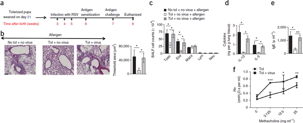
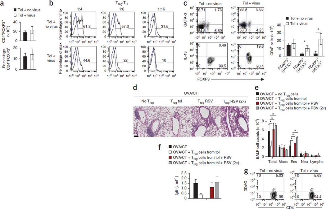
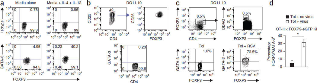
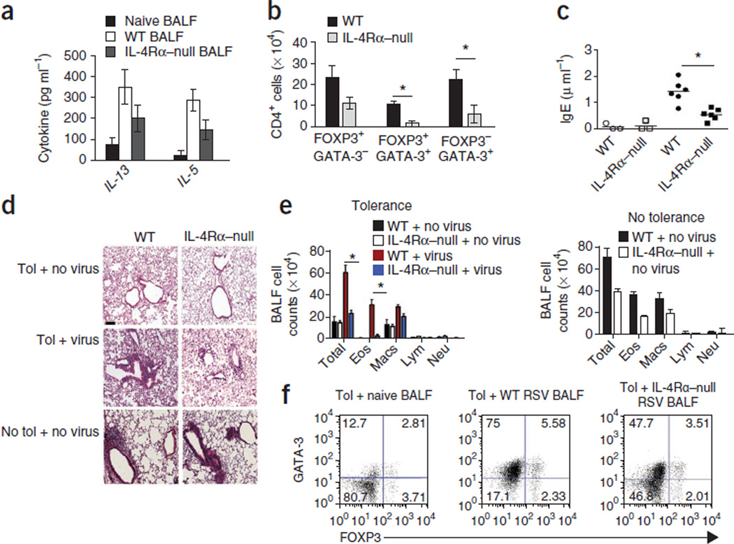
Immune tolerance is instituted early in life, during which time regulatory T (T(reg)) cells have an important role. Recurrent infections with respiratory syncytial virus (RSV) in early life increase the risk for asthma in adult life. Repeated infection of infant mice tolerized to ovalbumin (OVA) through their mother's milk with RSV induced allergic airway disease in response to OVA sensitization and challenge, including airway inflammation, hyper-reactivity and higher OVA-specific IgE, as compared to uninfected tolerized control mice. Virus infection induced GATA-3 expression and T helper type 2 (T(H)2) cytokine production in forkhead box P3 (FOXP3)(+) T(reg) cells and compromised the suppressive function of pulmonary T(reg) cells in a manner that was dependent on interleukin-4 receptor α (IL-4Rα) expression in the host. Thus, by promoting a T(H)2-type inflammatory response in the lung, RSV induced a T(H)2-like effector phenotype in T(reg) cells and attenuated tolerance to an unrelated antigen (allergen). Our findings highlight a mechanism by which viral infection targets a host-protective mechanism in early life and increases susceptibility to allergic disease.


Figures

Figure 1
Figure 1. Recurrent RSV infection compromises maternally transferred tolerance to inhaled allergen
(a) Experimental scheme for measuring the effects of tolerization and RSV infection in mice. (b) Histology, as evaluated by PAS staining. Scale bar, 100 µm. The threshold values shown indicate the quantification of the number of inflammatory cells and PAS staining in the different groups using MetaMorph. Tol, tolerization. (c) Quantification of total cell count, eosinophils (Eos), macrophages (Macs), lymphocytes (Lym) and neutrophils (Neu) in BALF cytospins. (d) Concentration of cytokines in the lung homogenates by ELISA. (e) Concentration of OVA-specific IgE in the serum. For be, the results shown are from an experiment performed five times. All data in be are means ± s.d. n = 4–5 mice per group. *P < 0.05, **P < 0.01 by two-way analysis of variance (ANOVA) with Bonferroni’s post-hoc test. (f) Central airway resistance (Newtonian resistance, Rn) in response to increasing doses of methacholine in uninfected and infected mice tolerized mice. Data are means ± s.d. *P < 0.05, **P < 0.01, ***P < 0.001 by Student’s unpaired two-tailed t test.
Figure 2
Figure 2. RSV infection triggers inflammation in lung-draining lymph nodes
(a) The total number of cells in the pooled lymph nodes of uninfected and infected mice tolerized as shown in Figure 1a, as determined by light microscopy after discounting dead cells on the basis of trypan blue exclusion. (b) Cytokine concentrations in the supernatant from lymph node cell cultures after stimulation with OVA (100 µg/ml) for 3 d as determined by ELISA. (c) Expression of GATA-3, FOXP3 and IL-13 on lymph node cells isolated from tolerized mice that were uninfected or infected with RSV after stimulation with phorbol 12-myristate 13-acetate (PMA) and ionomycin for 6 h. Cells were gated on CD4. The numbers in the corners of the FACS dot plots are the percentage of each cell population within that quadrant as a fraction of the total CD4 cells. All data are means ± s.d. and are representative of two independent experiments. n = 4–5 mice per group. *P < 0.05 by Student’s unpaired two-tailed t test.
Figure 3
Figure 3. RSV infection alters the phenotype and function of Treg cells in the lung
(a) The numbers (top) and the frequencies (bottom) of Treg (CD4+FOXP3+) cells in the lungs of tolerized FOXP3-eGFP knock-in pups in the presence or absence of virus infection. (b) Proliferation of naive responder T cells (TH) cocultured with different ratios of Treg (FOXP3+) cells isolated from uninfected and infected DO11.10 pups in the presence of OVA (100 µg/ml), as determined by the dilution of DDAO-labeled cells. (c) The expression of GATA-3, IL-13 and FOXP3 in CD4+ T cells isolated from the lungs of tolerized mice with and without RSV infection after stimulation with PMA and ionomycin. Cells shown in the upper images were gated on CD4 expression, and cells shown in the lower images were gated on FOXP3 expression. The numbers in the corners of the FACS dot plots are the percentage of each cell population within that quadrant. (d) PAS staining of lung sections of mice that received adoptively transferred eGFP+CD4+ T cells from tolerized mice that were uninfected or infected by RSV and then were sensitized to OVA/CT and challenged by OVA aerosols. (e) Number of immune cells in BALF recovered from different groups of mice. RSV (2×) indicates mice that received double the number of eGFP+CD4+ cells as compared to the RSV group (third group). (f) Serum IgE concentrations in different groups measured by ELISA. All data in a,c,e and f are means ± s.d. n = 4–5 mice per group (af). *P < 0.01 by Student’s unpaired two-tailed t test (c) or two-way ANOVA with Bonferroni’s post-hoc test (e). (g) Frequency of DDAO-labeled CD4+FOXP3+ (eGFP+) cells isolated from uninfected and RSV-infected mice in the lungs of OVA/CT-immunized recipient mice challenged with OVA. Data shown are representative of two independent experiments. The numbers in the corners of the FACS dot plots are the percentage of each cell population within that quadrant.
Figure 4
Figure 4. Treg cells acquire a TH2-like effector phenotype in tolerized RSV-infected mice
(a) Expression of FOXP3 and GATA-3 in CD4+FOXP3+ T cells isolated from tolerized mice and cultured with OVA and antibodies to CD28 with or without IL-4 and IL-13 for 48 h. (b) FOXP3 expression (top) and the absence of GATA-3 expression (bottom) in in vitro–generated Treg cells sorted on high CD25 expression. (c) Gating strategy (top) and GATA-3 expression in FOXP3+ cells isolated from tolerized uninfected and infected mice adoptively transferred with in vitro–generated DDAO-labeled Treg cells. (d) Percentage of FOXP3+GATA-3+ Treg cells isolated from the lungs of tolerized C57BL/6 mice with or without RSV infection. Treg cells were generated in vitro from splenocytes of OT-II × FOXP3-eGFP knock-in (KI) mice and labeled with DDAO dye prior to transfer. Total DDAO+ cells within the CD4 gate is shown. *P < 0.05 by Student’s two-tailed t test. Data are the means ± s.d. The experiments were performed twice with similar results and used a minimum of three mice in each group.
Figure 5
Figure 5. Host IL-4Rα signaling is important to breach tolerance
(a) Cytokine concentrations determined by ELISA 8 d after RSV infection in the BALF from naive, RSV-infected WT or IL-4Rα–null mice. Data are means ± s.d. n = 4–5 mice per group. Data are representative of two independent experiments. (b) Number of CD4+ cells expressing FOXP3 and/or GATA-3 in the lungs of WT and IL-4Rα–null mice (both on (BALB/c background) tolerized to OVA and infected with RSV. Data are the mean of values pooled from individual mice of the number of CD4+ T cells within each population ± s.d. n = 4–5 mice per group. *P < 0.05 by Student’s unpaired two-tailed t test. (c) Serum concentration of OVA-specific IgE in WT and IL-4Rα–null mice after transfer of 3 × 10⁶ CD4+ T cells sorted from DO11.10 × Rag2−/− mice and exposed to OVA aerosol. *P < 0.05. Open symbols denote the transfer of TH2 cells into tolerized uninfected mice, and closed symbols indicate transfer into tolerized infected mice. (d,e) H&E-stained lung sections (d) and cell counts in the BALF (e) of the recipient mice described in (c). *P < 0.05. Scale bar, 100 µm. (f) FOXP3 and GATA-3 expression in CD4+ T cells from tolerized mice cocultured with dendritic cells, OVA and cell-free BALF from naive mice or from WT or IL-4Rα–null mice infected by RSV. The experiment was performed twice with similar results, and the results of one representative experiment are shown. The numbers in the corners of the FACS dot plots are the percentage of each cell population within that quadrant.

References

    1. Levy O. Innate immunity of the newborn: basic mechanisms and clinical correlates. Nat. Rev. Immunol. 2007;7:379–390. - PubMed
    1. Noble V, et al. Respiratory status and allergy nine to 10 years after acute bronchiolitis. Arch. Dis. Child. 1997;76:315–319. - PMC - PubMed
    1. Sigurs N, Bjarnason R, Sigurbergsson F, Kjellman B. Respiratory syncytial virus bronchiolitis in infancy is an important risk factor for asthma and allergy at age 7. Am. J. Respir. Crit. Care Med. 2000;161:1501–1507. - PubMed
    1. Sigurs N, et al. Severe respiratory syncytial virus bronchiolitis in infancy and asthma and allergy at age 13. Am. J. Respir. Crit. Care Med. 2005;171:137–141. - PubMed
    1. Wu P, et al. Evidence of a causal role of winter virus infection during infancy in early childhood asthma. Am. J. Respir. Crit. Care Med. 2008;178:1123–1129. - PMC - PubMed

Publication types

MeSH terms