. 2012 Sep 25;109(39):15740-5.
doi: 10.1073/pnas.1204547109. Epub 2012 Sep 7.

Revisiting the contribution of negative charges on the chaperonin cage wall to the acceleration of protein folding

Affiliations

Revisiting the contribution of negative charges on the chaperonin cage wall to the acceleration of protein folding

Fumihiro Motojima et al. Proc Natl Acad Sci U S A. .

Abstract

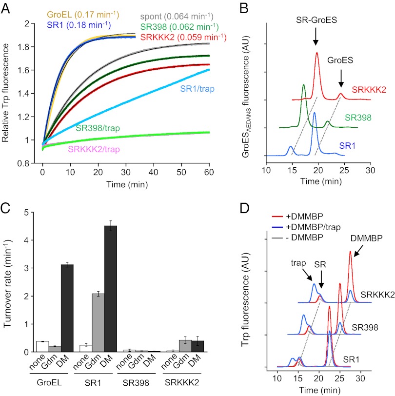
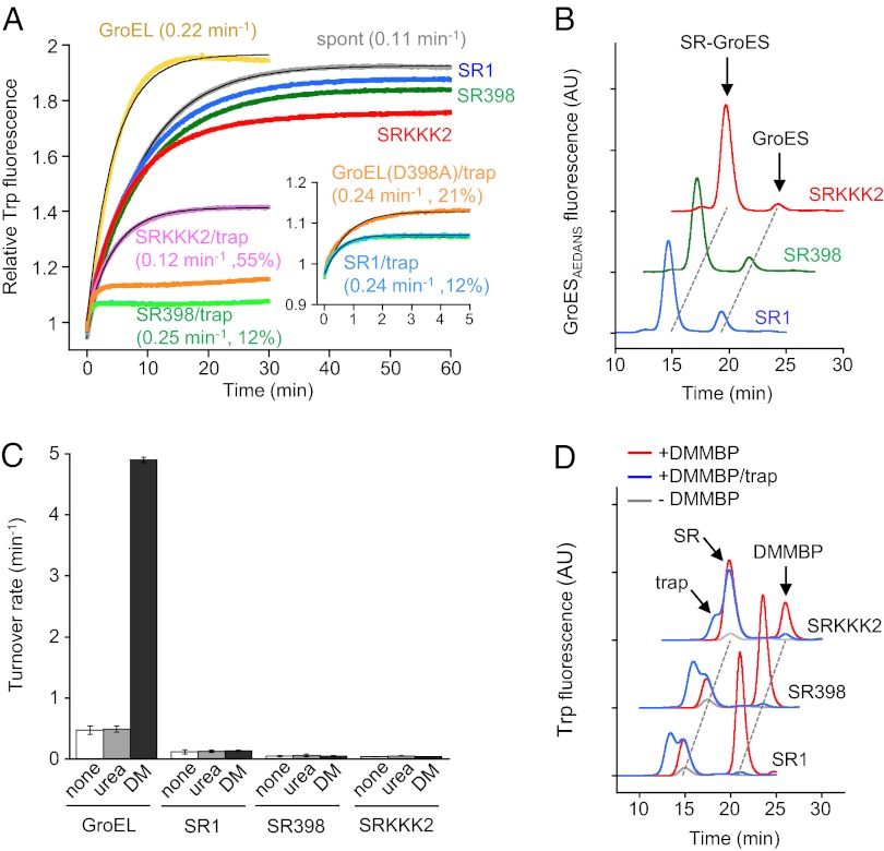
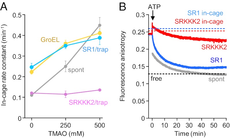
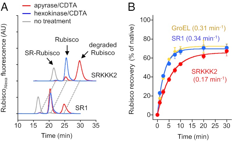
Chaperonin GroEL mediates the folding of protein encapsulated in a GroES-sealed cavity (cage). Recently, a critical role of negative charge clusters on the cage wall in folding acceleration was proposed based on experiments using GroEL single-ring (SR) mutants SR1 and SRKKK2 [Tang YC, et al. (2006) Cell 125:903-914; Chakraborty K, et al. (2010) Cell 142:112-122]. Here, we revisited these experiments and discovered several inconsistencies. (i) SR1 was assumed to bind to GroES stably and to mediate single-round folding in the cage. However, we show that SR1 repeats multiple turnovers of GroES release/binding coupled with ATP hydrolysis. (ii) Although the slow folding observed for a double-mutant of maltose binding protein (DMMBP) by SRKKK2 was attributed to mutations that neutralize negative charges on the cage wall, we found that the majority of DMMBP escape from SRKKK2 and undergo spontaneous folding in the bulk medium. (iii) An osmolyte, trimethylamine N-oxide, was reported to accelerate SRKKK2-mediated folding of DMMBP by mimicking the effect of cage-wall negative charges of WT GroEL and ordering the water structure to promote protein compaction. However, we demonstrate that in-cage folding by SRKKK2 is unaffected by trimethylamine N-oxide. (iv) Although it was reported that SRKKK2 lost the ability to assist the folding of ribulose-1,5-bisphosphate carboxylase/oxygenase, we found that SRKKK2 retains this ability. Our results argue against the role of the negative charges on the cage wall of GroEL in protein folding. Thus, in chaperonin studies, folding kinetics need to be determined from the fraction of the real in-cage folding.


Conflict of interest statement

The authors declare no conflict of interest.

Figures

Fig. 1.
Fig. 1.
Folding of GdmCl-denatured DMMBP. (A) Folding of GdmCl-denatured DMMBP diluted into buffer B containing 0.5 μM GroES (spont, gray) or buffer B containing 0.5 μM GroES and 0.2 μM SRs (or 0.1 μM GroEL) (GroEL, yellow; SR1, blue; SR398, green; SRKKK2, red) at 25 °C. ATP was added at time 0. Trap(D87K) was added before ATP when indicated (trap, final = 0.1 μM). Folding was monitored by the increase in Trp fluorescence of DMMBP. SDs of rate constants from three independent experiments are shown in Table S1. The folding rate constant is shown in parentheses. The baseline fluorescence of chaperonin was subtracted from the data. (B) Gel-filtration analysis of fluorescently labeled GroESAEDANS associated with SRs in buffer B containing diluted GdmCl-denatured DMMBP (final GdmCl = 60 mM). Excess nonlabeled GroES (1 μM) was added 10 s after the start of the reaction. The folding solutions were analyzed 1 h after the start of the folding reaction. (C) ATP hydrolysis activity of GroEL and SRs in buffer B containing none, 60 mM GdmCl (Gdm), or GdmCl-denatured DMMBP (DM) (final GdmCl = 60 mM). The averaged turnover rates for a ring (7 subunits of GroEL) and SDs from three independent experiments are shown. (D) Gel-filtration analysis of the folding solution of SR-mediated folding of GdmCl-denatured DMMBP. Elution was monitored by Trp fluorescence of DMMBP. The folding solutions in A were analyzed 1 h after the start of the folding reaction. Red, folded DMMBP; blue, folded DMMBP in the presence of trap(D87K); gray baseline without DMMBP. AU, arbitrary units.
Fig. 2.
Fig. 2.
Folding of urea-denatured DMMBP. Experimental procedures were the same as in Fig. 1, except that urea-denatured DMMBP and HKM buffer were used. Colors of data and labels in figures are the same as in Fig. 1. (A) Folding of urea-denatured DMMBP in HKM buffer. (Inset) First 5 min of folding by SR1, SR398, and GroEL(D398A) in the presence of trap(D87K). The folding rate constant and in-cage yield are shown in parentheses. SDs of rate constants from three independent experiments are shown in Table S1. The residuals of curve fitting are shown in Fig. S1E. (B) Gel-filtration analysis of GroESAEDANS associated with SRs in HKM buffer containing diluted urea-denatured DMMBP. Excess nonlabeled GroES (1 μM) was added at 10 s, and the folding solutions were analyzed 1 h after the start of the folding reaction. AU, arbitrary units. (C) ATP hydrolysis activity of GroEL and SRs in HKM buffer containing none, 80 mM urea (urea), or urea-denatured DMMBP (DM). (D) Gel-filtration analysis of the folding solution of SR-mediated folding of urea-denatured DMMBP. The folding solutions in A were analyzed 1 h after the start of the folding reaction.
Fig. 3.
Fig. 3.
Effect of TMAO and anisotropy change of urea-denatured DMMBP. (A) In-cage folding rate constant of urea-denatured DMMBP in the presence of trap(D87K) at various concentrations of TMAO. The concentration of DMMBP is 0.05 μM. Other folding conditions were the same as described in Fig. 2A. (B) Fluorescence anisotropy change during folding of urea-denatured DMMBP(A52C)Alexa in HKM buffer (final = 0.02 μM). Spontaneous folding (spont, gray), SR1-mediated folding (blue), and SRKKK2-mediated folding (red) are shown. Dashed lines represent anisotropy values of folded DMMBP(A52C)Alexa in the medium (black) and in the cages of SR1 (blue) and SRKKK2 (red).
Fig. 4.
Fig. 4.
Folding of Rubisco by SRKKK2. (A) Gel-filtration analysis of the SR1- and SRKKK2-mediated folding solutions of urea/acid-denatured fluorescently labeled RubiscoAlexa before (gray) or after GroES-detaching treatments. The folding solutions 30 min after the start of the reaction were treated with apylase/CDTA for 30 min at 25 °C (red) or with hexokinase/CDTA, followed by freezing with liquid nitrogen (blue). The positions of the SR/Rubisco complex, Rubisco dimer, and degraded Rubisco are shown by arrows. AU, arbitrary units. (B) Recovery time course of Rubisco activity at 25 °C by GroEL (yellow), SR1 (blue), and SRKKK2 (red) measured after hexokinase/CDTA treatment.

Similar articles

Cited by

References

    1. Hartl FU, Hayer-Hartl M. Molecular chaperones in the cytosol: From nascent chain to folded protein. Science. 2002;295:1852–1858. - PubMed
    1. Horwich AL, Fenton WA. Chaperonin-mediated protein folding: Using a central cavity to kinetically assist polypeptide chain folding. Q Rev Biophys. 2009;42(2):83–116. - PubMed
    1. Weissman JS, et al. Mechanism of GroEL action: Productive release of polypeptide from a sequestered position under GroES. Cell. 1995;83:577–587. - PubMed
    1. Apetri AC, Horwich AL. Chaperonin chamber accelerates protein folding through passive action of preventing aggregation. Proc Natl Acad Sci USA. 2008;105:17351–17355. - PMC - PubMed
    1. Brinker A, et al. Dual function of protein confinement in chaperonin-assisted protein folding. Cell. 2001;107:223–233. - PubMed

Publication types

MeSH terms