. 2012 Oct;19(10):1011-7.
doi: 10.1038/nsmb.2381. Epub 2012 Sep 9.

Dxo1 is a new type of eukaryotic enzyme with both decapping and 5'-3' exoribonuclease activity

Affiliations

Dxo1 is a new type of eukaryotic enzyme with both decapping and 5'-3' exoribonuclease activity

Jeong Ho Chang et al. Nat Struct Mol Biol. 2012 Oct.

Abstract

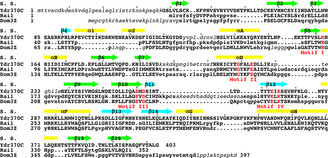
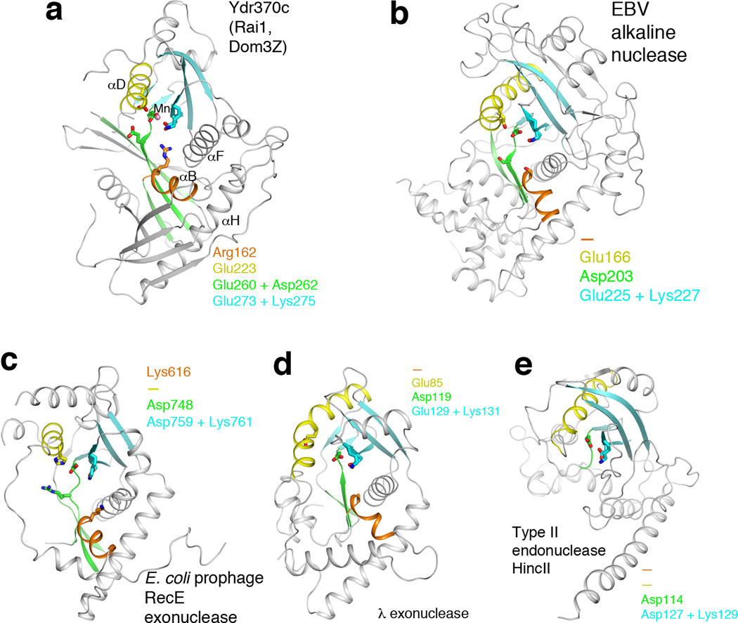
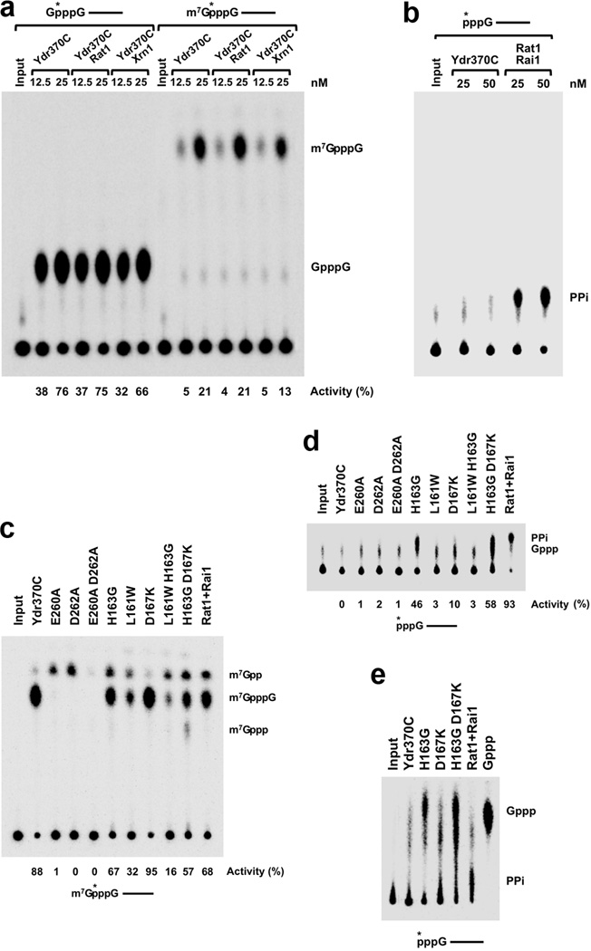
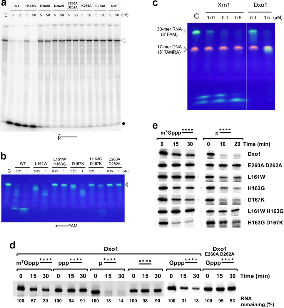
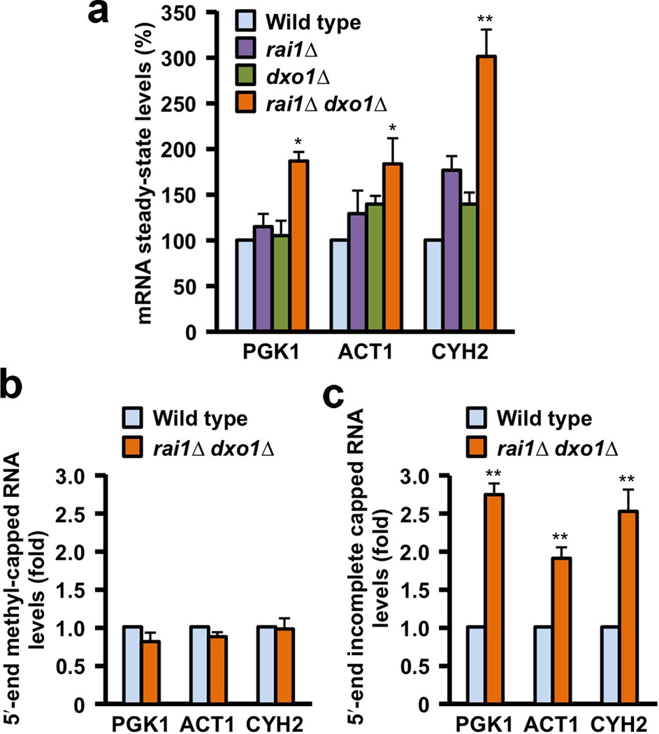
Recent studies showed that Rai1 is a crucial component of the mRNA 5'-end-capping quality-control mechanism in yeast. The yeast genome encodes a weak homolog of Rai1, Ydr370C, but little is known about this protein. Here we report the crystal structures of Ydr370C from Kluyveromyces lactis and the first biochemical and functional studies on this protein. The overall structure of Ydr370C is similar to Rai1. Ydr370C has robust decapping activity on RNAs with unmethylated caps, but it has no detectable pyrophosphohydrolase activity. Unexpectedly, Ydr370C also possesses distributive, 5'-3' exoRNase activity, and we propose the name Dxo1 for this new eukaryotic enzyme with both decapping and exonuclease activities. Studies of yeast in which both Dxo1 and Rai1 are disrupted reveal that mRNAs with incomplete caps are produced even under normal growth conditions, in sharp contrast to current understanding of the capping process.


Figures

Figure 1
Figure 1
Sequence conservation among Ydr370C/Dxo1, Rai1, and Dom3Z. Structure-based sequence alignment of K. lactis Ydr370C, S. pombe Rai1, and mouse Dom3Z. The secondary structure elements in the Ydr370C structure are shown (S. S.), and the four conserved sequence motifs are shown in red and labeled. Residues in Rai1 and Dom3Z that are located with 3 Å of the equivalent residue in Ydr370C are shown in uppercase. Residues that are disordered in the structures are shown in italic in lowercase.
Figure 2
Figure 2
Crystal structures of Ydr370C/Dxo1. (a). Schematic drawing of the crystal structure of the K. lactis Ydr370C free enzyme. Strands in the two β-sheets are colored in cyan and green, respectively. The conserved motifs in the active site are shown as stick models, with carbon atoms in green. Red dashed lines indicate the bidentate ion-pair interactions between Arg162 and Glu273. (b). Structure of the active site region of K. lactis Ydr370C in complex with Mn2+. Side chains from the four conserved motifs are colored green, and other side chains in yellow. Mn2+ is shown as a pink sphere, and two waters associated with it as red spheres. (c). Conformational differences in the active site region of Ydr370C between the free enzyme (in color) and the Mn2+ complex (in gray). A citrate molecule is bound in the free enzyme (cyan), and the position of GDP in the complex with Dom3Z is also shown. (d). Molecular surface of Ydr370C in the active site region. The Mn2+ ion is shown as a pink sphere and labeled. The α2-αA and β7-αD connections in the structure of S. pombe Rai1 are shown for reference (in violet). A single-stranded nucleic acid (four nucleotides, labeled) was positioned in the active site pocket (in cyan) based on the structure of the HincII endonuclease in complex with substrate . All the structure figures were produced with PyMOL (www.pymol.org).
Figure 3
Figure 3
Remote structural similarity to D-(D/E)XK nucleases. (a). Schematic drawing of the structure of Ydr370C. Motif I and helix αB are shown in orange, motif II and helix αD in yellow, motif III and its associated β-strands in green, motif IV and its associated β-strands in cyan. The Mn2+ ion is shown as a sphere in pink. (b). Structure of herpesvirus alkaline nuclease ,, in the same view as that for Ydr370C (panel a). (c). Structure of E. coli prophage RecE 5′-3′ exonuclease . (d). Structure of λ phage 5′-3′ exonuclease . (e). Structure of type II restriction endonuclease HincII . Residues in the four conserved motifs of Ydr370C, and their equivalents in the other enzymes, are indicated in each of the panels. A dash means that a functionally equivalent residue is absent.
Figure 4
Figure 4
Ydr370C/Dxo1 has strong decapping activity but no PPH activity. (a). Decapping assay monitored by thin-layer chromatography (TLC). Decapping activity of Ydr370C toward capped but unmethylated and mature, methylated RNA. The percentage of substrate turnover is indicated at the bottom. Rat1 and Xrn1 have no effect on the decapping activity. A schematic of the RNA used is indicated at the top with the asterisk denoting the position of the 32P-labeling. (b). PPH assay monitored by TLC. Ydr370C had essentially no PPH activity under the assay condition tested. Rai1 was used as a positive control. (c). The effects of mutations in the active site region on the decapping activity of Ydr370C. Most of the mutants were also able to release small amounts of a new product, m7Gpp. (d). PPH assay monitored by TLC. The H163G, H163G D167K, and D167K mutations in the active site region confer activity toward an RNA with 5′-end triphosphate. (e). The product released by the H163G, H163G D167K, and D167K mutants from an RNA with 5′-end triphosphate is GTP (Gppp), rather than pyrophosphate (PPi). A different running buffer was used to more clearly separate PPi from Gppp. Gppp in the sample lanes migrates slightly differently compared to the marker, as the samples are in a buffer and also contain protein while the marker is in water.
Figure 5
Figure 5
Ydr370C/Dxo1 has distributive, 5′-3′ exoribonuclease activity. (a). Activity of wild-type Ydr370C and various mutants toward a 240-nt RNA substrate labeled at the 5′-end. The position of unreacted, full-length substrate is marked with the arrowhead, and the dot indicates the migration of single nucleotides at the front of the gel. C: no enzyme control. (b). Activity of Ydr370C toward a 30-nt RNA substrate with 5′-end monophosphate and labeled at the 3′-end with the fluorophore FAM, confirming the distributive, 5′-3′ exonuclease activity. (c). Activity of Ydr370C toward an RNA-DNA heteroduplex substrate . K. lactis Xrn1 (residues 1–1245) was included as a comparison. (d). Activity of Ydr370C/Dxo1 on a panel of RNAs with different 5′-end modifications. The E260A D262A mutant was used as a negative control. (e). Activity of wild-type Ydr370C/Dxo1 and mutants toward body-labeled RNA with 5′-end methylated cap or monophosphate.
Figure 6
Figure 6
Dxo1 is involved in mRNA 5′-end capping quality control. (a). RT-PCR quantitation of PGK1, ACT1 and CYH2 mRNAs. Levels of PGK1, ACT1 and CYH2 mRNAs in the various yeast cells are shown, under normal growth conditions. Data are presented relative to the 18S rRNA with the level of mRNA in wild-type cells set to 100. A significant increase in steady-state mRNA levels was observed in the rai1Δ dxo1Δ doubly disrupted strain for all three mRNAs. Error bars indicate standard deviation, from three independent experiments. (b). Levels of methylated capped RNAs, immunoprecipitated using monoclonal anti-trimethylguanosine antibody column from the indicated yeast strains, are shown, normalized relative to the amount of total input RNA and levels in the respective wild-type cells were set to 1. (c). Levels of mRNA lacking a 5′-end methylated cap. Levels were normalized relative to 18S rRNA and mRNA levels in wild-type cells were set to 1. Error bars indicate standard deviation, from six independent experiments. * represents P < 0.01; ** represents P < 0.001. The P values were determined with the Student’s t-test.

References

    1. Shatkin AJ, Manley JL. The ends of the affair: capping and polyadenylation. Nature Struct. Biol. 2000;7:838–842. - PubMed
    1. Shuman S. The mRNA capping apparatus as drug target and guide to eukaryotic phylogeny. Cold Spring Harbor Symp. Quant. Biol. LXVI. 2001:301–312. - PubMed
    1. Hocine S, Singer RH, Grunwald D. RNA processing and export. Cold Spring Harbor Perspect. Biol. 2010;2:a000752. - PMC - PubMed
    1. Ghosh A, Lima CD. Enzymology of RNA cap synthesis. Wiley Interdiscip. Rev. RNA. 2010;1:152–172. - PMC - PubMed
    1. Coller J, Parker R. Eukaryotic mRNA decapping. Ann. Rev. Biochem. 2004;73:861–890. - PubMed

Publication types

MeSH terms