

Hypoperfusion predicts lesion progression in cerebral X-linked adrenoleukodystrophy
- PMID: 22961546
- PMCID: PMC3437030
- DOI: 10.1093/brain/aws206
Hypoperfusion predicts lesion progression in cerebral X-linked adrenoleukodystrophy
Abstract
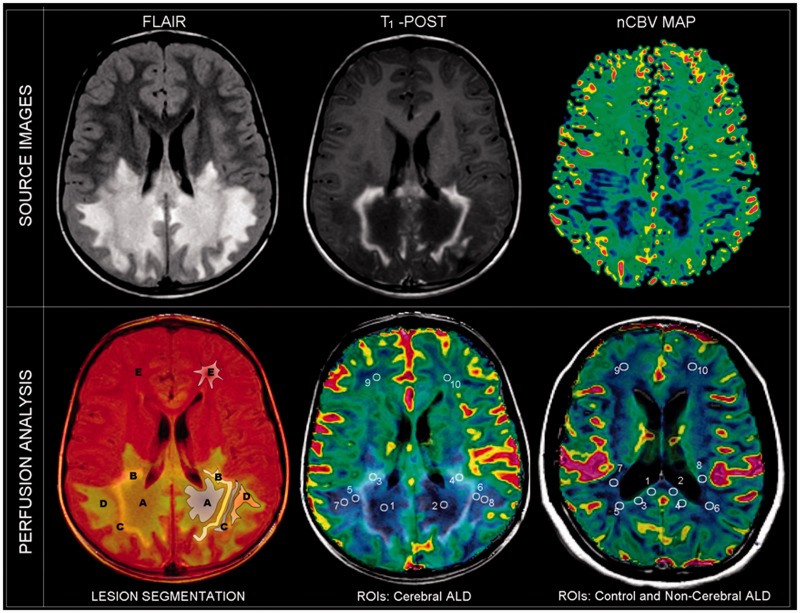
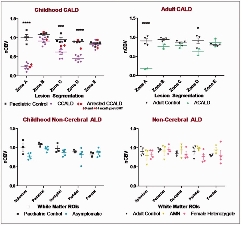
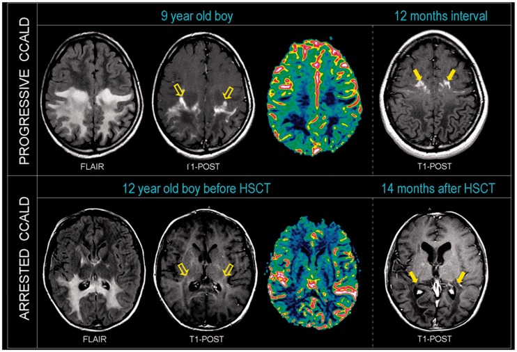
Magnetic resonance imaging sequences such as diffusion and spectroscopy have been well studied in X-linked adrenoleukodystrophy, but no data exist on magnetic resonance perfusion imaging. Since inflammation is known to modulate the microcirculation, we investigated the hypothesis that changes in the local perfusion might be one of the earliest signs of lesion development. Twenty patients with different phenotypes of adrenoleukodystrophy and seven age-matched controls were evaluated between 2006 and 2011. Fluid attenuated inversion recovery, post-contrast T(1)-weighted and normalized dynamic susceptibility contrast magnetic resonance perfusion cerebral blood volume maps were co-registered, segmented when cerebral lesion was present, and normalized cerebral blood volume values were analysed using a Food and Drug Association approved magnetic resonance perfusion software (NordicICE). Clinical and imaging data were reviewed to determine phenotype and status of progression. All eight patients with cerebral adrenoleukodystrophy had an average 80% decrease in normalized cerebral blood volume at the core of the lesion (P < 0.0001). Beyond the leading edge of contrast enhancement cerebral perfusion varied, patients with progressive lesions showed an average 60% decrease in normalized cerebral blood volume (adults P < 0.05; children P < 0.001), while one child with arrested progression normalized cerebral blood volume in this region. In six of seven patients with cerebral adrenoleukodystrophy lesions and follow-up imaging (2-24 month interval period), we found progression of contrast enhancement into the formerly hypoperfused perilesional zone. Asymptomatic, adrenomyeloneuropathy and female heterozygote patients had no significant changes in cerebral perfusion. Our data indicate that decreased brain magnetic resonance perfusion precedes leakage of the blood-brain barrier as demonstrated by contrast enhancement in cerebral adrenoleukodystrophy and is an early sign of lesion progression.
Figures
References
-
- Arnold AC, Pepose JS, Hepler RS, Foos RY. Retinal periphlebitis and retinitis in multiple sclerosis. I. Pathologic characteristics. Ophthalmology. 1984;91:255–62. - PubMed
-
- Eichler FS, Barker PB, Cox C, Edwin D, Ulug AM, Moser HW, et al. Proton MR spectroscopic imaging predicts lesion progression on MRI in X-linked adrenoleukodystrophy. Neurology. 2002;58:901–7. - PubMed
-
- Eichler FS, Ren JQ, Cossoy M, Rietsch AM, Nagpal S, Moser AB, et al. Is microglial apoptosis an early pathogenic change in cerebral X-linked adrenoleukodystrophy? Ann Neurol. 2008;63:729–42. - PubMed
-
- Eichler F, Van Haren K. Immune response in leukodystrophies. Pediatr Neurol. 2007;37:235–44. - PubMed
