. 2012 Sep;135(Pt 9):2684-98.
doi: 10.1093/brain/aws205.

Early-onset Lafora body disease

Affiliations

Early-onset Lafora body disease

Julie Turnbull et al. Brain. 2012 Sep.

Abstract

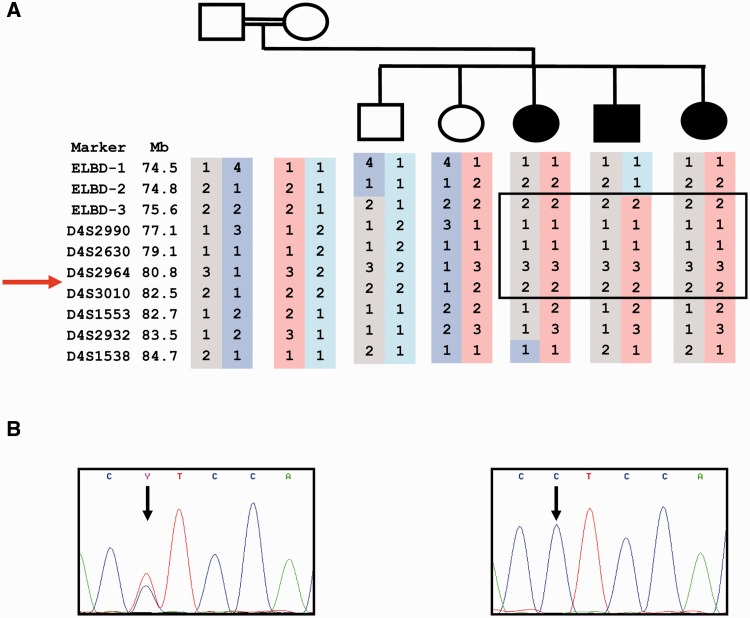
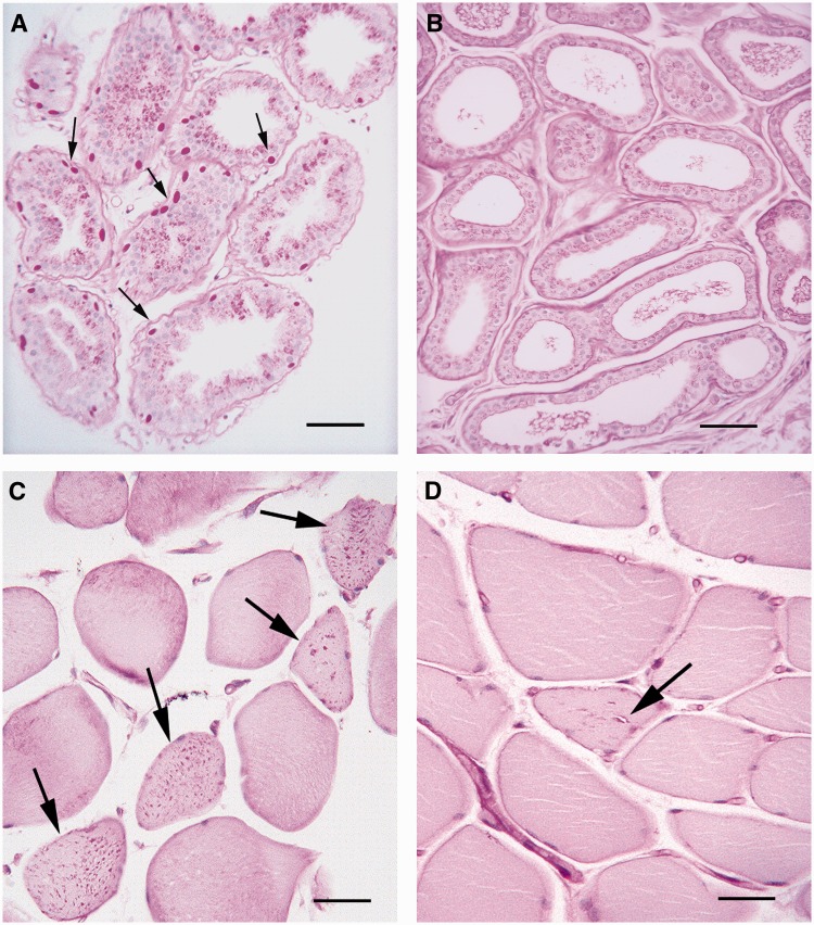
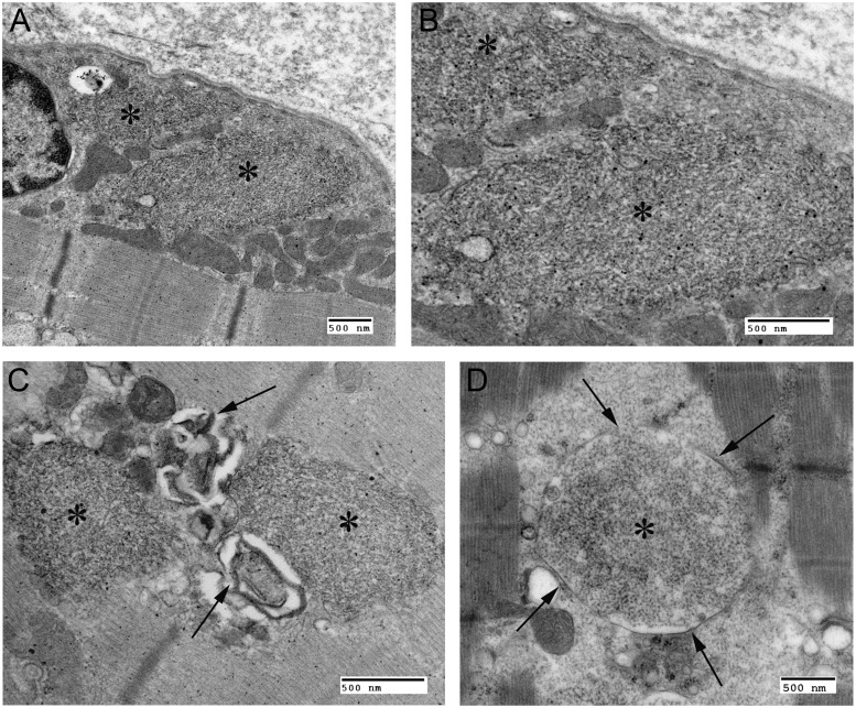
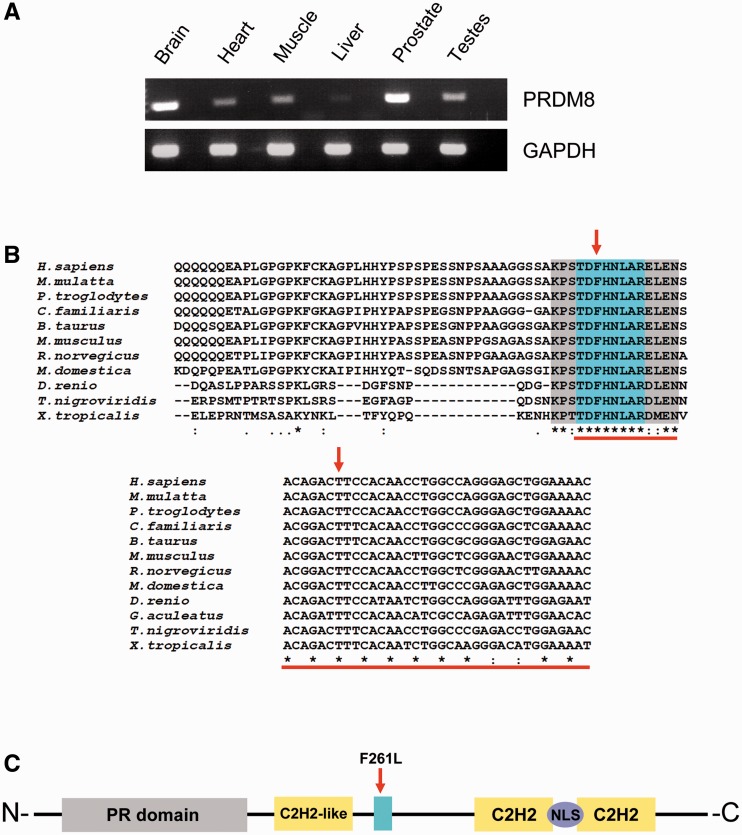
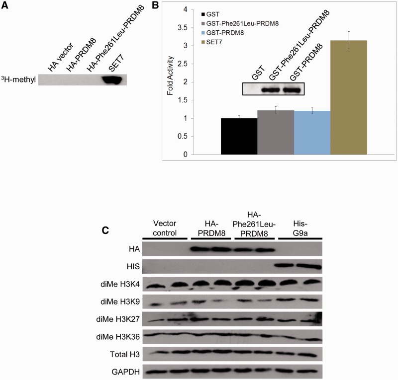
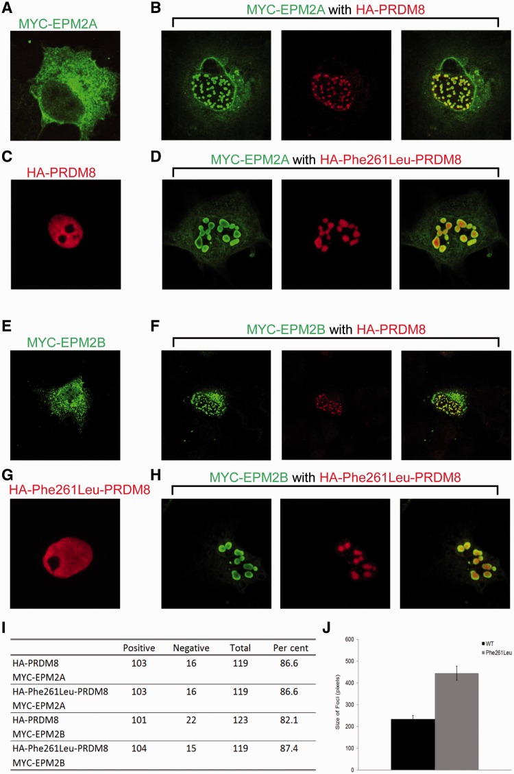
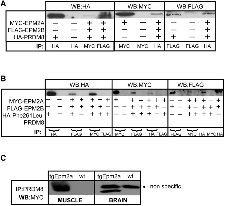
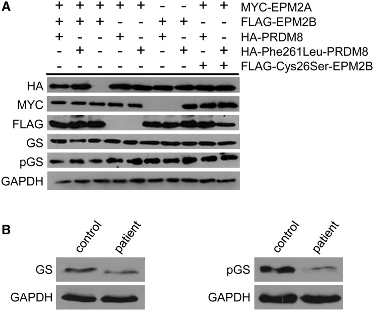
The most common progressive myoclonus epilepsies are the late infantile and late infantile-variant neuronal ceroid lipofuscinoses (onset before the age of 6 years), Unverricht-Lundborg disease (onset after the age of 6 years) and Lafora disease. Lafora disease is a distinct disorder with uniform course: onset in teenage years, followed by progressively worsening myoclonus, seizures, visual hallucinations and cognitive decline, leading to a vegetative state in status myoclonicus and death within 10 years. Biopsy reveals Lafora bodies, which are pathognomonic and not seen with any other progressive myoclonus epilepsies. Lafora bodies are aggregates of polyglucosans, poorly constructed glycogen molecules with inordinately long strands that render them insoluble. Lafora disease is caused by mutations in the EPM2A or EPM2B genes, encoding the laforin phosphatase and the malin ubiquitin ligase, respectively, two cytoplasmically active enzymes that regulate glycogen construction, ensuring symmetric expansion into a spherical shape, essential to its solubility. In this work, we report a new progressive myoclonus epilepsy associated with Lafora bodies, early-onset Lafora body disease, map its locus to chromosome 4q21.21, identify its gene and mutation and characterize the relationship of its gene product with laforin and malin. Early-onset Lafora body disease presents early, at 5 years, with dysarthria, myoclonus and ataxia. The combination of early-onset and early dysarthria strongly suggests late infantile-variant neuronal ceroid lipofuscinosis, not Lafora disease. Pathology reveals no ceroid lipofuscinosis, but Lafora bodies. The subsequent course is a typical progressive myoclonus epilepsy, though much more protracted than any infantile neuronal ceroid lipofuscinosis, or Lafora disease, patients living into the fourth decade. The mutation, c.781T>C (Phe261Leu), is in a gene of unknown function, PRDM8. We show that the PRDM8 protein interacts with laforin and malin and causes translocation of the two proteins to the nucleus. We find that Phe261Leu-PRDM8 results in excessive sequestration of laforin and malin in the nucleus and that it therefore likely represents a gain-of-function mutation that leads to an effective deficiency of cytoplasmic laforin and malin. We have identified a new progressive myoclonus epilepsy with Lafora bodies, early-onset Lafora body disease, 101 years after Lafora disease was first described. The results to date suggest that PRDM8, the early-onset Lafora body disease protein, regulates the cytoplasmic quantities of the Lafora disease enzymes.


Figures

Figure 1
Figure 1
Gene locus and mutation. (A) Pedigree and locus in chromosome 4q21.21; microsatellite haplotype shown; ELBD-1, 2, 3, un-named microsatellites identified in-house; arrow indicates position of the PRDM8 gene. (B) Non-synonymous single nucleotide replacement is present in homozygous state in all three affected (proband’s electropherogram shown, right panel) and in none of the unaffected individuals (heterozygous change in the father shown, left panel). ELBD = early-onset Lafora body disease.
Figure 2
Figure 2
Absence of Lafora bodies in skin and the presence in skeletal muscle. (A) Apocrine glands in skin from a patient with typical Lafora disease; note numerous Lafora bodies (arrows) in the myoepithelia surrounding the glands. Scale bar = 50 µm. (B) Comparable apocrine glands from the proband of the present family, no Lafora bodies are seen. Scale bar = 50 µm. (C) Skeletal muscle from the proband; numerous Lafora bodies are present throughout the sarcoplasm of several muscle fibres (arrows). Scale bar = 100 µm. (D) Vacuolation in skeletal muscle from the proband. Arrow indicates a sarcoplasmic vacuole surrounded by Lafora bodies. All slides stained with periodic acid-Schiff following diastase treatment. Scale bar = 100 µm.
Figure 3
Figure 3
Electron microscopy of skeletal muscle Lafora bodies. (A) Transmission electron micrograph from the proband: asterisks indicate Lafora bodies in perinuclear sarcoplasm. (B) Higher magnification of A: note filamentous nature of Lafora bodies and absence of any surrounding membrane. (C) Vacuolation (arrows) in association with Lafora bodies in the proband. (D) Transmission electron microscope image of skeletal muscle from a patient with Lafora disease; note membrane-bound Lafora bodies (arrows and asterisk). All scale bars = 500 nm.
Figure 4
Figure 4
PRDM8 expression, domain conservation and structure. (A) Semiquantitative reverse transcriptase-PCR using human tissue complementary DNAs and primers amplifying across the first PRDM8 exon–exon boundary: the gene is expressed in brain and skeletal muscle, among other human tissues; lanes between bands are reverse transcriptase-PCRs of preceding tissues with reverse transcriptase omitted. (B) ClustalW alignments of PRDM8 protein and gene sequences from different vertebrate species show strict evolutionary conservation of the substituted amino acid, the protein domain containing this amino acid and the mutated nucleotide. (C) Five conserved domains with predicted functions are present in PRDM8, the PR domain, three zinc finger domains (C2H2 and C2H2-like) and the nuclear localization signal (NLS); blue highlight indicates position of the conserved domain containing the replaced amino acid.
Figure 5
Figure 5
PRDM8 histone methyltransferase assays. (A and B) Wild-type and mutant PRDM8 have no above-baseline methyltransferase activities against mixed core histones. Haemagglutinin (HA)-tagged proteins were expressed in mammalian cells and activities detected using autoradiography; glutathione S-transferase (GST)-tagged proteins were bacterially expressed/purified and activities measured by scintillation counting, expressed as fold increase normalized to glutathione S-transferase alone. SET7 = positive control. Inset in (B) shows western blot of purified PRDM8 proteins used in the assay. (C) Levels of endogenous histone H3 dimethylated at lysines 4, 9, 27 and 36 are not increased following transfection with wild-type (or mutant) haemagglutinin-tagged PRDM8 (each lane represents a separate transfection); first two rows, levels of transfected proteins; ‘Total H3’ row, levels of total histone H3; bottom row, GAPDH loading control; pcDNA-His-G9a is a specific methyltransferase positive control for diMe H3K9.
Figure 6
Figure 6
PRDM8 modifies the subcellular distributions of laforin (EPM2A) and malin (EPM2B). (A) Representative image of subcellular distribution of overexpressed laforin; note predominant cytoplasmic expression in reticular pattern and minor nuclear presence. (B) PRDM8 and laforin co-expression: green signal, laforin; red, PRDM8. (C) Nuclear distribution of overexpressed PRDM8. (D) Mutant Phe261Leu-PRDM8 and laforin co-expression: green, laforin; red, PRDM8. (E) Representative image of subcellular distribution of overexpressed malin: cytoplasmic expression in reticular pattern and diffuse nuclear expression. (F) PRDM8 and malin co-expression: green, malin; red, PRDM8. (G) Nuclear distribution of overexpressed Phe261Leu-PRDM8. (H) Mutant Phe261Leu-PRDM8 and malin co-expression: green, malin; red, PRDM8. (I) Percentage of transfected cells with nuclear sequestration foci of laforin and malin by wild-type versus Phe261Leu-PRDM8. (J) Size of nuclear foci with laforin, malin and either wild-type PRDM8 or Phe261Leu-PRDM8. Size is shown as means ± SEM and significance calculated using an unpaired student’s t-test (P < 2.54 × 10⁻⁹).
Figure 7
Figure 7
Laforin and malin co-precipitate with both PRDM8 and mutant Phe261Leu-PRDM8. (A and B) The three proteins co-expressed, immunoprecipitating one co-precipitates the other two. MYC-EPM2A = myc-tagged laforin; FLAG-EPM2B = FLAG-tagged malin; HA-PRDM8 = haemagglutinin-tagged PRDM8; HA-Phe261Leu-PRDM8 = haemagglutinin-tagged mutant Phe261Leu PRDM8; IP = antibody used for imunoprecipitation; WB = antibody used for western blot; (+) indicates that the particular plasmid is transfected and (–) that it is not. (C) PRDM8 immunoprecipitates myc-tagged laforin from skeletal muscle and brain of a transgenic mouse (tgEpm2a) overexpressing myc laforin; wt = wild-type mouse tissues.
Figure 8
Figure 8
Total and phosphorylated glycogen synthase levels with Phe261Leu-PRDM8 overexpression in early-onset Lafora body disease. (A) Western blots of extracts of cell cultures overexpressing indicated (+) proteins. GS = glycogen synthase; pGS = phosphorylated glycogen synthase; FLAG-Cys26Ser-EPM2B = a ubiquitin ligase-inactive malin mutant, used as a control; GAPDH = loading control; note that neither glycogen synthase nor phosphorylated glycogen synthase are altered with Phe261Leu-PRDM8 compared with wild-type PRDM8. (B) Western blots of extracts from skeletal muscle from a patient with early-onset Lafora body disease and a normal control with antibodies against total (left) and phosphorylated (right) glycogen synthase.

References

    1. Abecasis GR, Cherny SS, Cookson WO, Cardon LR. Merlin—rapid analysis of dense genetic maps using sparse gene flow trees. Nat Genet. 2002;30:97–101. - PubMed
    1. Andrade DM, Ackerley CA, Minett TS, Teive HA, Bohlega S, Scherer SW, et al. Skin biopsy in Lafora disease: genotype-phenotype correlations and diagnostic pitfalls. Neurology. 2003;61:1611–4. - PubMed
    1. Andrade DM, del Campo JM, Moro E, Minassian BA, Wennberg RA. Nonepileptic visual hallucinations in Lafora disease. Neurology. 2005;64:1311–2. - PubMed
    1. Badhwar A, Berkovic SF, Dowling JP, Gonzales M, Narayanan S, Brodtmann A, et al. Action myoclonus-renal failure syndrome: characterization of a unique cerebro-renal disorder. Brain. 2004;127:2173–82. - PubMed
    1. Bassuk AG, Wallace RH, Buhr A, Buller AR, Afawi Z, Shimojo M, et al. A homozygous mutation in human PRICKLE1 causes an autosomal-recessive progressive myoclonus epilepsy-ataxia syndrome. Am J Hum Genet. 2008;83:572–81. - PMC - PubMed

Publication types

MeSH terms