. 2012 Sep;135(Pt 9):2736-49.
doi: 10.1093/brain/aws196.

PTEN deletion enhances survival, neurite outgrowth and function of dopamine neuron grafts to MitoPark mice

Affiliations

PTEN deletion enhances survival, neurite outgrowth and function of dopamine neuron grafts to MitoPark mice

YaJun Zhang et al. Brain. 2012 Sep.

Abstract

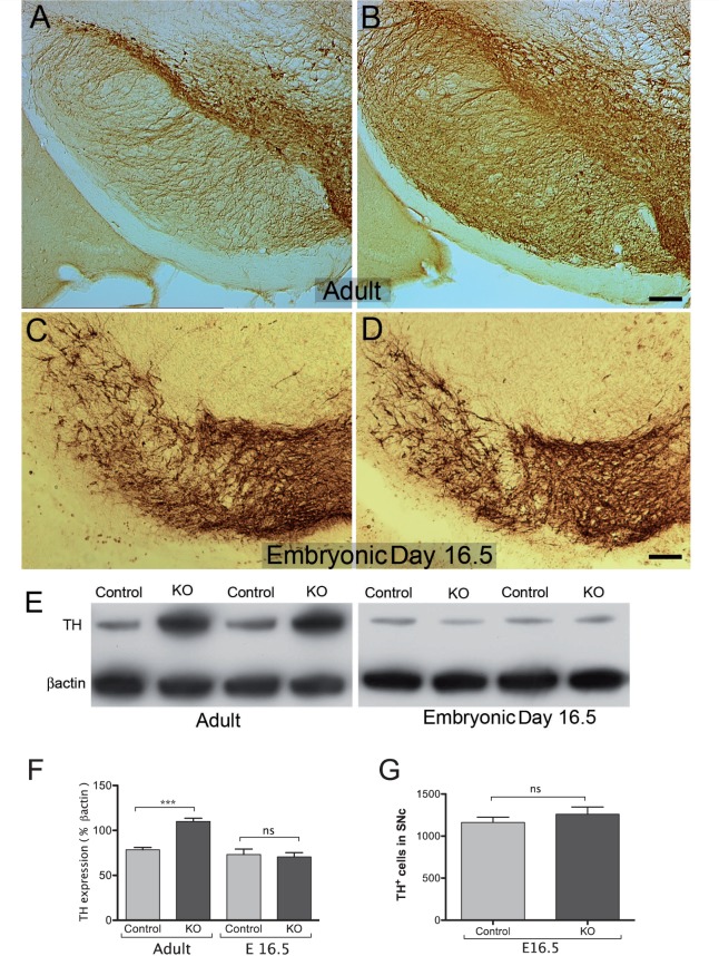
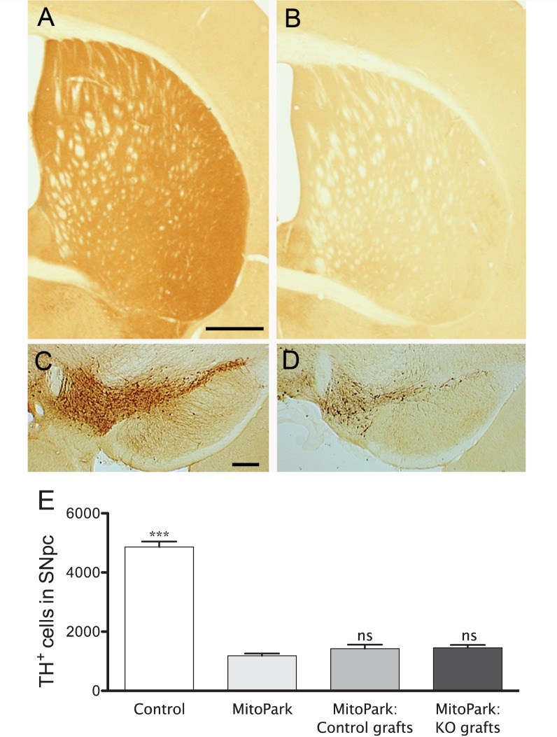
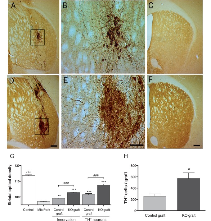
Clinical trials in Parkinson's disease have shown that transplants of embryonic mesencephalic dopamine neurons form new functional connections within the host striatum, but the therapeutic benefits have been highly variable. One obstacle has been poor survival and integration of grafted dopamine neurons. Activation of Akt, a serine/threonine kinase that promotes cell survival and growth, increases the ability of neurons to survive after injury and to regenerate lost neuronal connections. Because the lipid phosphatase, phosphatase and tensin homolog (PTEN) inhibits Akt, we generated a mouse with conditional knock-out of PTEN in dopamine neurons, leading to constitutive expression of Akt in these neurons. Ventral mesencephalic tissue from dopamine phosphatase and tensin homologue knock-out or control animals was then transplanted bilaterally into the dopamine depleted striata of MitoPark mice that express a parkinsonian phenotype because of severe respiratory chain dysfunction in dopamine neurons. After transplantation into MitoPark mice, PTEN-deficient dopamine neurons were less susceptible to cell death, and exhibited a more extensive pattern of fibre outgrowth compared to control grafts. Voltammetric measurements demonstrated that dopamine release and reuptake were significantly increased in the striata of animals receiving dopamine PTEN knock-out transplants. These animals also displayed enhanced spontaneous and drug-induced locomotor activity, relative to control transplanted MitoPark mice. Our results suggest that disinhibition of the Akt-signalling pathway may provide a valuable strategy to enhance survival, function and integration of grafted dopamine neurons within the host striatum and, more generally, to improve survival and integration of different forms of neural grafts.


Figures

Figure 1
Figure 1
Overview of the present experiment. Behavioural measurements were obtained for all animal groups included in the study starting at 16 weeks of age. After recording baseline behavioural activity at 20 weeks, MitoPark mice were grafted with control or DA-PTEN-KO ventral mesencephalon tissue. This was followed by a sequence of behavioural tests during the subsequent 16 weeks post-grafting.
Figure 2
Figure 2
Tyrosine hydroxylase immunocytochemistry in the substantia nigra of adult control (A) and DA-PTEN-KO animals (B). Adult DA-PTEN-KO animals contain a significantly greater number of tyrosine hydroxylase-positive neurons and fibres in the ventral midbrain region. Significant differences in tyrosine hydroxylase-positive immunocytochemistry were not observed in embryonic Day 16.5 DA-PTEN-KO embryos (D) compared with controls (C). These findings were verified by western blot analyses of tyrosine hydroxylase (TH) protein in ventral mesencephalon tissue homogenates from control and DA-PTEN-KO adult and embryonic Day 16.5 (E16.5) mice (E and F, one-way ANOVA, ***P < 0.001), and (G) by quantification of tyrosine hydroxylase-positive (TH+) neurons in the substantia nigra pars compacta (SNc) from Day 16.5 control and DA-PTEN-KO embryos (two-tailed t-test). Data presented as mean ± SEM. Scale bars: A and B = 150 µm, C and D = 100 µm. ns = non-significant.
Figure 3
Figure 3
Spontaneous and drug-induced locomotor activity following transplants in MitoPark mice. (A and B) Spontaneous locomotor behavior was examined every 4 weeks, starting at ∼16 weeks of age. Mitopark animals received control and DA-PTEN-KO intrastriatal grafts at 20 weeks of age, after baseline behavioural recordings, and behavioural evaluations continued for an additional 16 weeks. MitoPark mice showed reduced spontaneous locomotor activity at 16 weeks of age. Total distance (A) and vertical activity (B) of MitoPark mice continued to decline, and animals were severely impaired at 36 weeks of age (two-way ANOVA; Bonferroni post hoc analysis *P < 0.05, **P < 0.01). MitoPark animals grafted with DA-PTEN-KO tissue showed a significant improvement in spontaneous ambulatory activity at 36 weeks, relative to control grafted and naïve MitoPark mice. Vertical activity did not change significantly in MitoPark animals grafted with either DA-PTEN-KO or control tissue, although there was a trend to increased vertical activity in the former group. (C) At 36 weeks of age (or 16 weeks after grafting), animals were evaluated for nomifensine (15 mg/kg, intraperitoneal)-induced locomotor behaviour. Thirty-six-week old MitoPark mice did not respond to nomifensine treatment, while locomotor activity of DA-PTEN-KO grafted MitoPark mice was similar to that from age-matched controls, and significantly higher when compared with both control-grafted MitoPark animals and naïve MitoPark mice (one-way ANOVA; Dunn’s post hoc analysis **P < 0.01). (D) At 35 weeks of age, locomotor activity was measured every hour for 24 h, starting at 11:00 am, during the light phase. All groups showed a normal circadian rhythm by moving less during the light phase, after habituation to the new environment and more during the dark phase. MitoPark animals grafted with DA-PTEN-KO tissue showed a significant increase in the total distance in the nocturnal cycle as compared with all other groups. Significant differences shown compared to MitoPark control graft group (two-way ANOVA; Bonferroni post hoc analyses for all groups: *P < 0.05, **P < 0.01, ***P < 0.001). Data presented as mean ± SEM; ns = non-significant.
Figure 4
Figure 4
Tyrosine hydroxylase immunoreactivity in striatum of a 36-week-old control (A) and naïve MitoPark (B) mouse illustrates the extensive degeneration following TFAM deletion in dopamine neurons. At the level of the midbrain, there was a near complete loss of tyrosine hydroxylase-positive dopamine neurons in the substantia nigra pars compacta of MitoPark mice (D) when compared with controls (C). Tyrosine hydroxylase-positive neurons in the ventral tegmental area were partially spared in MitoPark mice (C and D). (E) Quantification of tyrosine hydroxylase-positive neurons in the substantia nigra pars compacta of control and MitoPark mice confirmed a significant and comparable loss of tyrosine hydroxylase-positive neurons in both naïve and transplanted MitoPark mice (one-way ANOVA; Bonferroni post hoc analyses for all groups: ***P < 0.001). Significant differences are shown compared to the MitoPark group. Scale bars: A = 250 µm, B = 200 µm. ns = non-significant. KO = knock-out; SNpc = substantia nigra pars compacta; TH = tyrosine hydroxylase; ns = non-significant.
Figure 5
Figure 5
Photomicrographs illustrating a control (A, B and C) and DA-PTEN-KO (D, E and F) graft in the striatum of 36-week-old MitoPark mice. For each group, one section through the core of the graft and one section from the same animal, rostral to the graft placement site, are shown. Boxed areas in A and D are enlarged and illustrate the appearance of tyrosine hydroxylase-positive neurons in control (B) and DA-PTEN-KO transplants (E). Tyrosine hydroxylase-positive neurons in DA-PTEN-KO transplants are more densely packed and are larger in size. Neurites extending from DA-PTEN-KO grafts are increased and extend into more distal areas of the MitoPark striatum, when compared with control grafts (C and F). (F) To determine differences in innervation among the transplanted animals, optical density values were obtained separately from striatal sections containing tyrosine hydroxylase-positive cell bodies (see A and D: TH+ neurons), and sections containing only fibres and no cell bodies (see C and F: innervation). (G) While MitoPark mice transplanted with both control and DA-PTEN-KO embryonic tissue showed a significant increase in striatal tyrosine hydroxylase density when compared with naïve MitoPark mice (two-way ANOVA; Bonferroni post hoc analysis, *P < 0.05, ***P < 0.001), MitoPark mice transplanted with DA-PTEN-KO grafts showed significantly higher tyrosine hydroxylase density values when compared with control grafts (two-way ANOVA; Bonferroni post hoc analysis, ###P < 0.001). Data presented as mean ± SEM. (H) Quantification of tyrosine hydroxylase-positive neurons in the striatum of grafted MitoPark mice confirmed a significant increase in the number of tyrosine hydroxylase-positive neurons present in DA-PTEN-KO grafts as compared with control grafts. Two-tailed t-test, *P < 0.05. Data presented as mean ± SEM. Scale bars: D and F = 200 µm, E = 100 µm, KO = knock-out.
Figure 6
Figure 6
Voltammetric assessment of graft function in 36-week-old MitoPark mice. (A) Fast scan cyclic voltammetry pseudocolour plots of signals obtained in a striatal slice obtained from a 36-week-old control mouse and a MitoPark mouse. Release was elicited by a single, 1 ms pulse (100 µA). Time and applied potential are indicated on the x- and y-axes, respectively; current is represented in pseudocolour. Dashed white line indicates the potential at which voltammograms below each colour plot were obtained. Average current versus time plots (mean ± SEM, indicated by dashed line) are shown for all slices tested (control, nine slices; MitoPark, nine slices). (B) Representative signals taken from the striatum of a control-grafted (top) and a DA-PTEN-KO grafted (bottom) MitoPark mouse. Following recording, sections were processed for tyrosine hydroxylase immunohistochemistry. Numbers indicate the approximate location of the recording site within each slice. Note the clear difference in innervation density (left), corresponding to the differences in the signals (right). (C) Summary of input–output curves in 36-week-old MitoPark mice grafted with control (pooled data from 60 recording sites in 15 slices) or DA-PTEN-KO tissue (65 recording sites in 14 slices). Signals are averaged (SEM indicated by dashed lines) responses to 100 µA, single pulse stimulation (arrow). Significantly greater release was observed in slices from DA-PTEN-KO grafted MitoPark mice [two-way repeated measures ANOVA, genotype effect; F(1,27) = 19.46, P < 0.001]. (D) Summary of decay time constants for voltammetric signals. Upper panel shows the averaged signals elicited by 100 µA stimulation, in DA-PTEN-KO grafted striatum (red trace, n = 14) and control-grafted striatum (blue trace, n = 15) normalized to the amplitude of control, non-MitoPark mice (control, green, n = 9). Decay time constants from DA-PTEN-KO grafted striatum did not significantly differ from control striatum (one-way ANOVA, Tukey post hoc analysis P > 0.05). Note that decay time constants could not be calculated for age-matched, non-grafted MitoPark mice owing to the minimal nature of the signal (A). DA = dopamine.

Similar articles

Cited by

References

    1. Abe N, Cavalli V. Nerve injury signaling. Curr Opin Neurobiol. 2008;18:276–83. - PMC - PubMed
    1. Ahn YH, Bensadoun JC, Aebischer P, Zurn AD, Seiger A, Bjorklund A, et al. Increased fiber outgrowth from xeno-transplanted human embryonic dopaminergic neurons with co-implants of polymer-encapsulated genetically modified cells releasing glial cell line-derived neurotrophic factor. Brain Res Bull. 2005;66:135–42. - PubMed
    1. Andereggen L, Meyer M, Guzman R, Ducray AD, Widmer HR. Effects of GDNF pretreatment on function and survival of transplanted fetal ventral mesencephalic cells in the 6-OHDA rat model of Parkinson's disease. Brain Res. 2009;1276:39–49. - PubMed
    1. Antonini A, Schwarz J, Oertel WH, Pogarell O, Leenders KL. Long-term changes of striatal dopamine D2 receptors in patients with Parkinson's disease: a study with positron emission tomography and [11C]raclopride. Mov Disord. 1997;12:33–8. - PubMed
    1. Backman CM, Malik N, Zhang Y, Shan L, Grinberg A, Hoffer BJ, et al. Characterization of a mouse strain expressing Cre recombinase from the 3' untranslated region of the dopamine transporter locus. Genesis. 2006;44:383–90. - PubMed

Publication types

MeSH terms

Substances