. 2013 Jan;57(1):339-50.
doi: 10.1002/hep.26053.

A novel murine model to deplete hepatic stellate cells uncovers their role in amplifying liver damage in mice

Affiliations

A novel murine model to deplete hepatic stellate cells uncovers their role in amplifying liver damage in mice

Juan E Puche et al. Hepatology. 2013 Jan.

Abstract

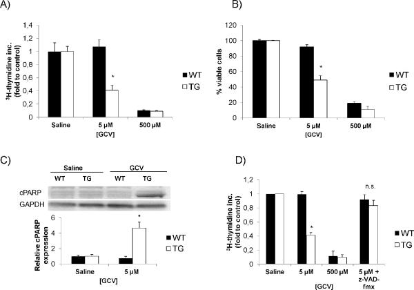
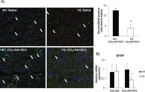
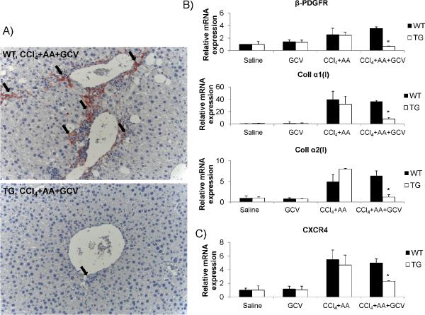
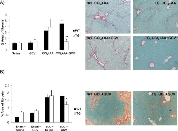
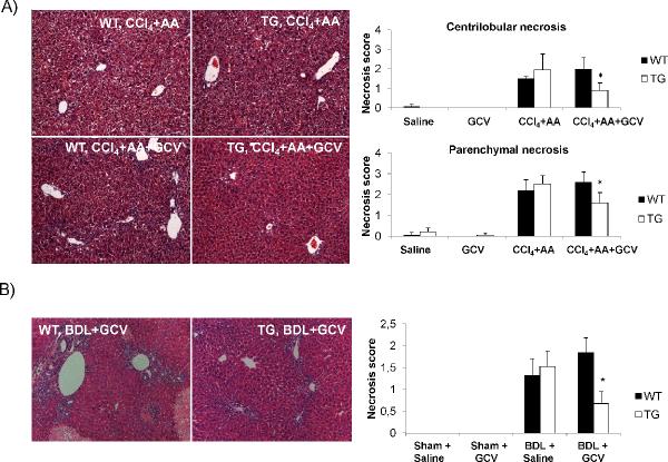
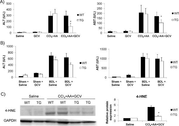
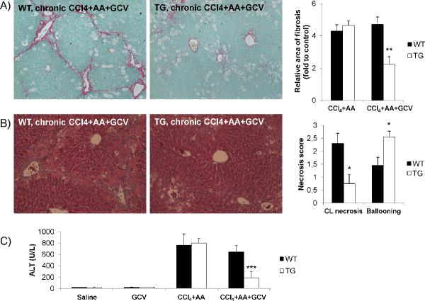
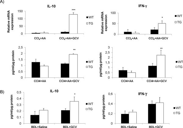
We have developed a novel model for depleting mouse hepatic stellate cells (HSCs) that has allowed us to clarify their contributions to hepatic injury and fibrosis. Transgenic (Tg) mice expressing the herpes simplex virus thymidine kinase gene (HSV-Tk) driven by the mouse GFAP promoter were used to render proliferating HSCs susceptible to killing in response to ganciclovir (GCV). Effects of GCV were explored in primary HSCs and in vivo. Panlobular damage was provoked to maximize HSC depletion by combining CCl(4) (centrilobular injury) with allyl alcohol (AA) (periportal injury), as well as in a bile duct ligation (BDL) model. Cell depletion in situ was quantified using dual immunofluorescence (IF) for desmin and GFAP. In primary HSCs isolated from both untreated wild-type (WT) and Tg mice, GCV induced cell death in ≈ 50% of HSCs from Tg, but not WT, mice. In TG mice treated with CCl(4) +AA+GCV, there was a significant decrease in GFAP and desmin-positive cells, compared to WT mice (≈ 65% reduction; P < 0.01), which was accompanied by a decrease in the expression of HSC-activation markers (alpha smooth muscle actin, beta platelet-derived growth factor receptor, and collagen I). Similar results were observed after BDL. Associated with HSC depletion in both fibrosis models, there was marked attenuation of fibrosis and liver injury, as indicated by Sirius Red/Fast Green, hematoxylin and eosin quantification, and serum alanine/aspartate aminotransferase. Hepatic expression of interleukin-10 and interferon-gamma was increased after HSC depletion. No toxicity of GCV in either WT or Tg mice accounted for the differences in injury.

Conclusion: Activated HSCs significantly amplify the response to liver injury, further expanding this cell type's repertoire in orchestrating hepatic injury and repair.


Figures

Figure 1
Figure 1. In vitro optimization of the GCV dose to provoke HSC depletion
(A) Characterization of HSC proliferation mediated by GCV in isolated HSC from TG mice as determined by 3H-thymidine incorporation. A concentration of 5 μM GCV could specifically decrease TG HSC proliferation (B) Doses higher than 10 μM were toxic towards primary HSCs and hepatocytes from WT and TG mice as determined by trypan blue staining. (C) Immunoblotting for cleaved PARP expression in primary HSCs from both WT and TG mice. TG HSCs treated with GCV expressed more cleaved PARP consistent with increased apoptosis as mechanism for cell death. (D) Incubation with the pan-caspase inhibitor z-VAD-fmx abrogated the decrease in 3H-thymidine incorporation of GCV on TG HSC. Data are mean values from three independent experiments. *p<0.05.
Figure 2
Figure 2. Validation of HSC depletion in vivo in TG mice treated with CCl4+AA+GCV
(A) Reduction of desmin (green) and GFAP (red) positive cells, and merged images (yellow) by 65% (p<0.01) in TG mice treated with CCl4+AA+GCV, compared to WT mice as assessed by dual-immunofluorescence. (B) Relative GFAP mRNA expression (by qPCR) demonstrates a significant reduction in TG mice undergoing HSC depletion. Data are mean values from three independent experiments normalized to saline-treated mice. GCV treatment (without CCl4+AA) did not differ from saline treated group. Original magnification ×100. *p<0.05.
Figure 3
Figure 3. Reduced markers of HSC activation following HSC depletion
(A) Reduced α-SMA positive cells in TG mice undergoing HSC depletion as assessed by immunohistochemistry. (B) Relative mRNA expression genes associated with HSC activation (β-PDGFR and collagen I). There is a significant reduction of these markers in TG mice with HSC depletion. (C) Relative mRNA expression of CXCR4 is reduced in TG mice undergoing HSC depletion. Original magnification ×200. *p<0.05.
Figure 4
Figure 4. Attenuation of liver fibrosis in mice with HSC depletion
(A) Fibrosis content was quantified by Sirius Red/Fast Green staining and Bioquant computerized morphometry in (A) CCl4+AA+GCV treated mice as well as in (B) BDL+GCV treated mice. Original magnification ×100. *p<0.05; **p<0.01.
Figure 5
Figure 5. Reduced hepatic necrosis in mice with HSC depletion
(A) H&E staining and its histological scoring demonstrates a significant reduction in both centrilobular and parenchymal necrosis in TG mice undergoing HSC depletion by CCl4+AA+GCV depletion treatment. (B) GCV treatment following BDL led to a consistent decrease in the extent of hepatic necrosis in TG mice undergoing HSC depletion. Original magnification ×100. *p<0.05.
Figure 6
Figure 6. Reduced serum AST/ALT levels in mice with HSC and hepatic 4-HNE expression
ALT and AST levels are lower in TG mice treated with (A) CCl4+AA+GCV and (B) in BDL+GCV treated animals compared to WT mice. (C) Protein from whole liver tissue was analyzed for 4-HNE expression by Western blot. Less oxidative damage was observed in TG mice undergoing HSC depletion than in WT mice. *p<0.05.
Figure 7
Figure 7. Effect of HSC depletion on chronic liver damage with CCl4
(A) Fibrosis deposition is decreased in TG mice with HSC depletion undergoing chronic liver damage, compared to WT animals. Values are normalized to saline treated WT and TG mice. (B) H&E staining and histological scoring for hepatocellular necrosis and ballooning. TG mice displayed decreased necrosis compared to WT animals but increased ballooning. (C) TG mice undergoing HSC depletion had lower serum ALT levels compared to WT animals following chronic liver damage. CL, centrilobular. Original magnification ×100. *p<0.05, **p<0.01 and ***p<0.001.
Figure 8
Figure 8. Increased IL-10 and IFN-γ in whole liver following HSC depletion
(A) Relative mRNA expression and protein concentration of IL-10 and IFN-γ were increased in TG mice undergoing HSC depletion by CCl4+AA+GCV. (B) Protein concentrations for IL-10 and and IFN-γ in whole liver lysates of BDL+GCV treated mice. qPCR data are mean values from three independent experiments, normalized to saline-treated mice. GCV treatment (without CCl4+AA) did not differ from saline treated group. *p<0.05, **p<0.01 and ***p<0.001.

References

    1. Friedman SL. Hepatic Stellate Cells – Protean, Multifunctional, and Enigmatic Cells of the Liver. Physiological Reviews. 2008;88:125–172. - PMC - PubMed
    1. Orr JG, Leel V, Cameron GA, Marek CJ, Haughton EL, Elrick LJ, Trim JE, et al. Mechanism of action of the antifibrogenic compound gliotoxin in rat liver cells. Hepatology. 2004;40:232–242. - PubMed
    1. Ebrahimkhani MR, Oakley F, Murphy LB, Mann J, Moles A, Perugorria MJ, Ellis E, et al. Stimulating healthy tissue regeneration by targeting the 5-HT2B receptor in chronic liver disease. Nat Med. 2011;17:1668–1673. - PMC - PubMed
    1. Douglass A, Wallace K, Parr R, Park J, Durward E, Broadbent I, Barelle C, et al. Antibody-targeted myofibroblast apoptosis reduces fibrosis during sustained liver injury. J Hepatol. 2008;49:88–98. - PubMed
    1. Hagens WI, Olinga P, Meijer DK, Groothuis GM, Beljaars L, Poelstra K. Gliotoxin non-selectively induces apoptosis in fibrotic and normal livers. Liver Int. 2006;26:232–239. - PubMed

Publication types

MeSH terms