. 2013 Feb;57(2):716-26.
doi: 10.1002/hep.26055. Epub 2013 Jan 8.

Intrahepatic cholestasis of pregnancy levels of sulfated progesterone metabolites inhibit farnesoid X receptor resulting in a cholestatic phenotype

Affiliations
Free PMC article

Intrahepatic cholestasis of pregnancy levels of sulfated progesterone metabolites inhibit farnesoid X receptor resulting in a cholestatic phenotype

Shadi Abu-Hayyeh et al. Hepatology. 2013 Feb.
Free PMC article

Abstract

Intrahepatic cholestasis of pregnancy (ICP) is the most prevalent pregnancy-specific liver disease and is associated with an increased risk of adverse fetal outcomes, including preterm labor and intrauterine death. The endocrine signals that cause cholestasis are not known but 3α-sulfated progesterone metabolites have been shown to be elevated in ICP, leading us to study the impact of sulfated progesterone metabolites on farnesoid X receptor (FXR)-mediated bile acid homeostasis pathways. Here we report that the 3β-sulfated progesterone metabolite epiallopregnanolone sulfate is supraphysiologically raised in the serum of ICP patients. Mice challenged with cholic acid developed hypercholanemia and a hepatic gene expression profile indicative of FXR activation. However, coadministration of epiallopregnanolone sulfate with cholic acid exacerbated the hypercholanemia and resulted in aberrant gene expression profiles for hepatic bile acid-responsive genes consistent with cholestasis. We demonstrate that levels of epiallopregnanolone sulfate found in ICP can function as a partial agonist for FXR, resulting in the aberrant expression of bile acid homeostasis genes in hepatoma cell lines and primary human hepatocytes. Furthermore, epiallopregnanolone sulfate inhibition of FXR results in reduced FXR-mediated bile acid efflux and secreted FGF19. Using cofactor recruitment assays, we show that epiallopregnanolone sulfate competitively inhibits bile acid-mediated recruitment of cofactor motifs to the FXR-ligand binding domain.

Conclusion: Our results reveal a novel molecular interaction between ICP-associated levels of the 3β-sulfated progesterone metabolite epiallopregnanolone sulfate and FXR that couples the endocrine component of pregnancy in ICP to abnormal bile acid homeostasis.


Figures

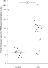
Fig. 1
Fig. 1
Levels of epiallopregnanolone-sulfate (PM5S) are supraphysiologically raised in ICP. Serum concentrations of PM5S in women with ICP and uncomplicated pregnancies at 33-38 weeks of gestation. Black line represents mean serum concentrations of PM5S. Measurements were carried out by UPLC/MSMS on a minimum of n = 12 samples. *P < 0.05 for control pregnant versus ICP serum samples as determined by Student's t test.
Fig. 2
Fig. 2
PM5S exacerbates hypercholanemia in the mouse. (A) UPLC/MSMS-derived serum bile acid concentrations represented in two graphs according to the range of their concentrations and (B) hepatic gene expression levels of Bsep, Mdr2, Ost-β, and Sult2a1 in mice gavaged with vehicle, cholic acid, or cholic acid and PM5S at four timepoints over 2 days. *P < 0.05 for vehicle or cholic acid-gavaged group versus cholic acid and PM5S co-gavaged group. #P < 0.05 for vehicle versus cholic acid-gavaged group as determined by one-way analysis of variance (ANOVA). Values represent mean ± standard error of the mean (SEM) of n = 6. CA, cholic acid; T-CA, taurocholic acid; G-CA, glycocholic acid; CDCA, chenodeoxycholic acid; T-CDCA, taurochenodeoxycholic acid; DCA, deoxycholic acid; UDCA, ursodeoxycholic acid; T-UDCA, tauroursodeoxycholic acid; T-LCA, taurolithocholic acid; T-αMCA, tauro-α muricholic acid; T-βMCA, tauro-β muricholic acid.
Fig. 3
Fig. 3
PM5S inhibits function and expression of FXR target genes. (A) Huh7 cells were treated overnight with vehicle or 0.5 μM GW4064 ± 25-50 μM PM5S, following which cells were washed for 1.5 hours and allowed to effluxHTC for 5 minutes. (B) Representative western blot and graphical representation of normalized BSEP to GAPDH levels in Huh7 cells treated with vehicle or 0.5 μM GW4064 ± 0-100 μM PM5S for 24 hours. (C) Huh7 cells were treated with vehicle or 0.5 μM GW4064 / 50 μM CDCA ±0-100 μM PM5S for 24 hours, after which cells were analyzed with qPCR for relative BSEP gene expression. (D) GW4064-mediated secretion of FGF19 is inhibited by PM5S. Secreted FGF19 was assayed in cell culture media taken from Huh7 cells incubated with 0.5 μM GW4064 ±0-100 μM PM5S for 24 hours. (E) GW4064-mediated FGF19 gene expression is inhibited by PM5S. Experiments were performed as in (C). (F) GW4064-mediated SHP and MDR3 gene expression is inhibited by PM5S. Experiments were performed as in (C). *P < 0.05 for 0.5 μM GW4064/50 μM CDCA versus vehicle control. #P < 0.05 for 0.5 μM GW4064/50 μM CDCA versus cotreatment group as determined by one-way ANOVA. Values represent mean ± SEM of n = 3.
Fig. 4
Fig. 4
PM5S reduces ligand-mediated FXR activity. (A) Huh7 cells were transfected with the human expression constructs RXR, FXRα2 / empty vector, FXR-luciferase reporter and renilla construct for 24 hours. Transfected cells were cotreated with 100 μM CDCA and increasing doses of PM5S. RLU, relative light units. n = 3 ± SEM. (B) PM5S inhibits ligand activated FXR recruitment of SRC-1. GST-tagged FXR-LBD and biotinylated LxxLL SRC-1 peptide incubated in the presence of 9 or 100 μM CDCA and 0-600 μM PM5S and HTRF measured after 1 hour incubation at room temperature with orbital shaking. n = 3 ± standard deviation (SD). (C) PM5S exhibits dose-dependent mild FXR agonistic characteristics. Transfection experiments were performed as in (A). Huh7 cells were treated with 0-100 μM PM5S. RLU, relative light units. n = 3 ± SEM. (D) PM5S is unable to recruit the LxxLL motif to the FXR-LBD. Experiments were performed as in (B). HTRF was measured following a 1-hour incubation of HTRF mixture and increasing CDCA/PM5S doses. n = 3 ± SD.
Fig. 5
Fig. 5
3β-Sulfated progesterone-based compounds modulate FXR activity. (A) Huh7 cells were transfected with the human expression constructs for RXR, FXRα2 / empty vector, FXR-luciferase reporter, and renilla construct for 24 hours. Fifty μM of compound or 1 μM GW4064 was used to treat the transfected cells for 24 hours. *P < 0.05 for treatment group versus vehicle control. RLU, relative light units. n = 3 ± SEM. Dose response curves of 0-100 μM EPAS and EPS, which were identified in the compound screen. RLU, relative light units. n = 3 ± SEM. (B) EPAS and EPS are unable to recruit the LxxLL motif to the FXR-LBD. GST-FXR-LBD and biotinylated LxxLL SRC-1 peptide were incubated in the presence of increasing EPAS, EPS, and CDCA concentrations and HTRF measured after 1-hour incubation at room temperature with orbital shaking. n = 3 ± SD. (C) Huh7 cells were transfected as in (A) and cotreated with 100 μM CDCA and increasing doses of EPAS and EPS. n = 3 ± SEM. (D) EPAS and EPS inhibit ligand-activated FXR recruitment of SRC-1. HTRF experiments were performed as in (B). HTRF reaction mix was incubated in the presence of 9 or 100 μM CDCA and 0-600 μM EPAS/EPS. n = 3 ± SD.
Fig. 6
Fig. 6
Depletion of FXR in primary human hepatocytes blunts the response to PM5S and CDCA. Primary human hepatocytes were transfected with siRNA against FXR (hatched bar) or scrambled siRNA (black bar) for 6 hours, following which cells were allowed to recover overnight and then treated with 50 μM PM5S or 50 μM CDCA for 24 hours. Relative messenger RNA (mRNA) expression levels are shown for the genes (A) FXR, (B) BSEP, (C) SHP, and (D) CYP7A1. *P < 0.05 for treatment group versus vehicle siScrambled vehicle control as determined by one-way ANOVA. Values represent mean ± SEM of n = 3.

References

    1. Geenes V, Williamson C. Intrahepatic cholestasis of pregnancy. World J Gastroenterol. 2009;15:2049–2066. - PMC - PubMed
    1. Fisk NM, Storey GN. Fetal outcome in obstetric cholestasis. Br J Obstet Gynaecol. 1988;95:1137–1143. - PubMed
    1. Glantz A, Marschall HU, Mattsson LA. Intrahepatic cholestasis of pregnancy: relationships between bile acid levels and fetal complication rates. HEPATOLOGY. 2004;40:467–474. - PubMed
    1. Van Mil SW, Milona A, Dixon PH, Mullenbach R, Geenes VL, Chambers J, et al. Functional variants of the central bile acid sensor FXR identified in intrahepatic cholestasis of pregnancy. Gastroenterology. 2007;133:507–516. - PubMed
    1. Pauli-Magnus C, Lang T, Meier Y, Zodan-Marin T, Jung D, Breymann C, et al. Sequence analysis of bile salt export pump (ABCB11) and multidrug resistance p-glycoprotein 3 (ABCB4, MDR3) in patients with intrahepatic cholestasis of pregnancy. Pharmacogenetics. 2004;14:91–102. - PubMed

Publication types

Supplementary concepts