

doi: 10.1002/anie.201205676.
Epub 2012 Sep 7.
Using ligand-mapping simulations to design a ligand selectively targeting a cryptic surface pocket of polo-like kinase 1
Affiliations
- PMID: 22961729
- PMCID: PMC3547296
- DOI: 10.1002/anie.201205676
Item in Clipboard
Using ligand-mapping simulations to design a ligand selectively targeting a cryptic surface pocket of polo-like kinase 1
Angew Chem Int Ed Engl.
.
Free PMC article
No abstract available
Figures
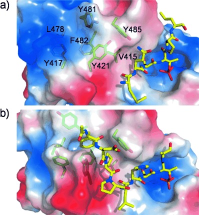
Binding pockets of Plk1 PBD as revealed by phosphopeptide ligands. Residues of the hydrophobic binding site are shown in green sticks and ligands in yellow sticks. Regions of negative, positive and neutral electrostatic potential on the protein surface are red, blue and white, respectively. a) Crystal structure of a phosphopeptide bound to the positively charged phosphopeptide binding pocket (PDB code 1Q4K), with closed hydrophobic pocket. b) Crystal structure of a phosphopeptide bound to both binding pockets (PDB code 3P37), with open hydrophobic pocket.
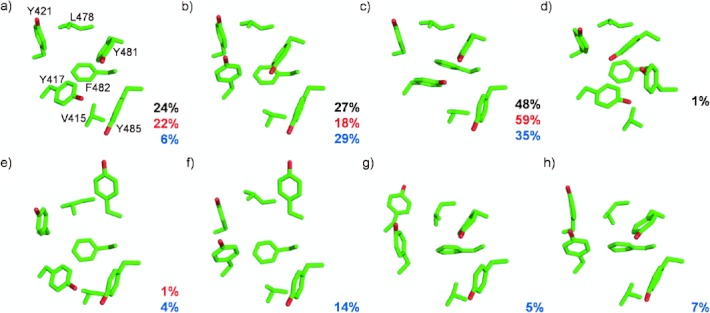
Conformations of the hydrophobic binding pocket in Plk1 PBD. Percentage populations of the conformations observed in the single long, multiple short and ligand-mapping simulations are indicated in black, red and blue, respectively. a–c) Closed conformations observed in crystal structures. d) Closed conformation observed only in the single long MD simulation. e,f) Open conformations observed in crystal structures. g,h) New conformations observed in ligand-mapping MD simulations.
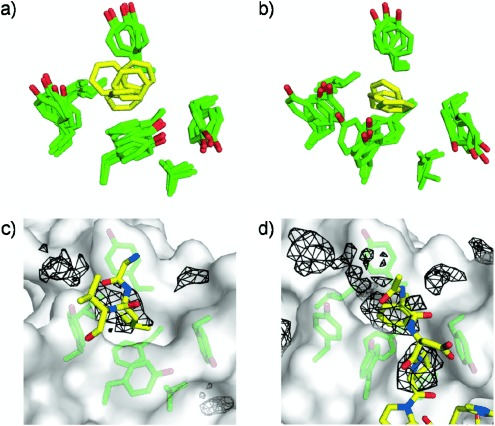
Reproduction of crystallographic binding modes at PBD hydrophobic pocket by ligand-mapping simulations. a,b) Superposition of five snapshots of the partially and fully open binding pocket, respectively, from ligand-mapping simulations. Pocket residues are green and benzene molecules are yellow. c) Benzene probes mimic the crystal-packing interaction of Leu394 from a neighboring protomer with the binding pocket (PDB code 3P35). Interacting ligands and crystal contact residues are shown in yellow sticks while regions sampled by benzene probes for this protein conformation are represented as black mesh (see Supporting Information for details). Only the part of the protomer binding to the pocket is shown. d) Benzene probes mimic the interactions of Phe and Pro from the peptide ligand with the binding pocket in the PBD/FDPPLHSpTA complex (PDB code 3P37).
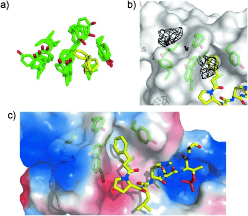
Design of a chimeric peptide ligand based on an alternative binding mode obtained from ligand-mapping simulations. a) Superposition of five snapshots of PBD binding pocket (green) from a ligand-mapping MD trajectory, showing an alternative binding mode of benzene (yellow). b) Binding mode of chimeric peptide (yellow sticks) at PBD pocket (green sticks) overlaid with benzene probability isosurfaces (black mesh) for the protein structure shown. c) Crystal structure of PBD complexed with chimeric peptide (yellow sticks). Regions of positive and negative electrostatic potential on the PBD surface are colored blue and red, respectively.
References
-
- Fuentes G, Dastidar SG, Madhumalar A, Verma CS. Drug Dev. Res. 2011;72:26–35.
-
- Betts MJ, Sternberg MJE. Protein Eng. 1999;12:271–283. - PubMed
-
- Withers IM, Mazanetz MP, Wang H, Fischer PM, Laughton CA. J. Chem. Inf. Model. 2008;48:1448–1454. - PubMed
-
- DeLano WL, Ultsch MH, de Vos AM, Wells JA. Science. 2000;287:1279–1283. - PubMed
Publication types
MeSH terms
Substances
Grants and funding
LinkOut - more resources
Full Text Sources
