Review
. 2012 Nov-Dec;28(6):1409-18.
doi: 10.1002/btpr.1629. Epub 2012 Oct 18.

Expanding the concepts and tools of metabolic engineering to elucidate cancer metabolism

Affiliations
Review

Expanding the concepts and tools of metabolic engineering to elucidate cancer metabolism

Mark A Keibler et al. Biotechnol Prog. 2012 Nov-Dec.

Abstract

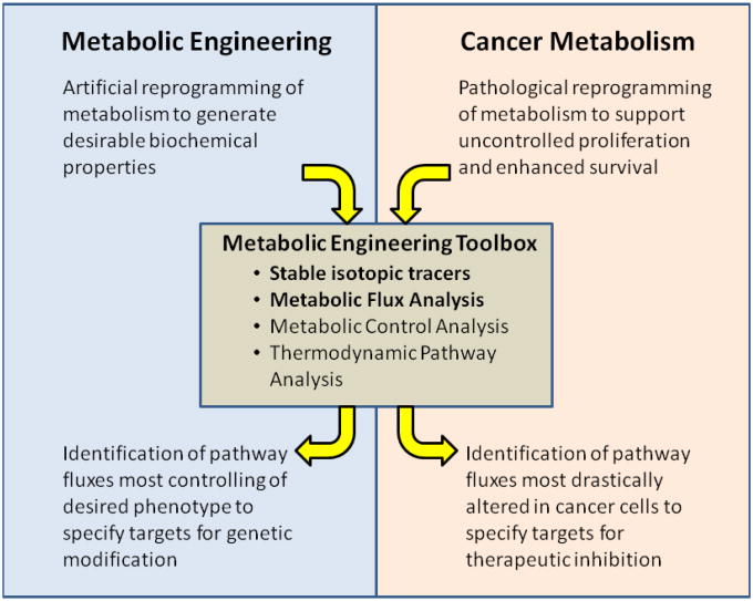
The metabolic engineer's toolbox, comprising stable isotope tracers, flux estimation and analysis, pathway identification, and pathway kinetics and regulation, among other techniques, has long been used to elucidate and quantify pathways primarily in the context of engineering microbes for producing small molecules of interest. Recently, these tools are increasingly finding use in cancer biology due to their unparalleled capacity for quantifying intracellular metabolism of mammalian cells. Here, we review basic concepts that are used to derive useful insights about the metabolism of tumor cells, along with a number of illustrative examples highlighting the fundamental contributions of these methods to elucidating cancer cell metabolism. This area presents unique opportunities for metabolic engineering to expand its portfolio of applications into the realm of cancer biology and help develop new cancer therapies based on a new class of metabolically derived targets.


Figures

Figure 1
Figure 1
A schematic overview of cancer cell metabolism. Oncogenes and tumor suppressor genes (blue) mutated in cancer reprogram metabolism to support elevated proliferation and enhanced survival, increasing flux (wide arrows) through pathways such as aerobic glycolysis, lipogenesis, and nucleotide/NADPH synthesis (products in green). This leads certain enzymes (red) to have particular oncological significance, as they may be distinctly activated, expressed, or mutated in cancer cells. This figure is courtesy of Christian Metallo (University of California, San Diego).
Figure 2
Figure 2
Both metabolic engineering and cancer cell metabolism involve the reprogramming of physiological metabolic pathways for some alternative purpose; for metabolic engineers, this purpose is typically the overproduction of some chemical, while for cancer cells, it is primarily accelerated proliferation. Tools such as stable isotopic tracers, metabolic flux analysis, metabolic control analysis, and thermodynamic pathway analysis can be used to identify key pathways in each; in metabolic engineering, these are reactions that contribute most to the intended product formation (and are hence potential targets of genetic modification), and in cancer metabolism, these are reactions that most drastically contribute to the tumor cell phenotype (and are hence potential targets of therapeutic inhibition).
Figure 3
Figure 3
13C isotopic tracers can help identify the differential contributions of fluxes to metabolic pathways. Cells are cultured in the presence of labeled media; after a prolonged period, their metabolites are harvested, analyzed using GC/MS (or another isotope-sensitive detection instrument), and catalogued into mass isotopomer distributions (MIDs). (a) Contribution of pyruvate carboxylase (PC) flux to the TCA cycle can be measured by culturing cells in [3-13C] glucose, which is eventually metabolized to [1-13C] pyruvate. Because the labeled carbon is retained only in the PC reaction, quantification of M1 (singly-enriched) TCA cycle intermediates from the generated MIDs enables estimation of PC activity. (b) In addition, M4 and M5 citrate levels from [U-13C5] glutamine-cultured cells can be used to determine the total amount of citrate synthesized from glutamine. (c) The fractional contribution of reductive carboxylation to citrate formation is directly given by the M1 citrate isotopomer in [1-13C] glutamine-cultured cells, and likewise, (d) its contribution to lipid synthesis by the palmitate MID in [5-13C] glutamine-cultured cells.
Figure 3
Figure 3
13C isotopic tracers can help identify the differential contributions of fluxes to metabolic pathways. Cells are cultured in the presence of labeled media; after a prolonged period, their metabolites are harvested, analyzed using GC/MS (or another isotope-sensitive detection instrument), and catalogued into mass isotopomer distributions (MIDs). (a) Contribution of pyruvate carboxylase (PC) flux to the TCA cycle can be measured by culturing cells in [3-13C] glucose, which is eventually metabolized to [1-13C] pyruvate. Because the labeled carbon is retained only in the PC reaction, quantification of M1 (singly-enriched) TCA cycle intermediates from the generated MIDs enables estimation of PC activity. (b) In addition, M4 and M5 citrate levels from [U-13C5] glutamine-cultured cells can be used to determine the total amount of citrate synthesized from glutamine. (c) The fractional contribution of reductive carboxylation to citrate formation is directly given by the M1 citrate isotopomer in [1-13C] glutamine-cultured cells, and likewise, (d) its contribution to lipid synthesis by the palmitate MID in [5-13C] glutamine-cultured cells.

Similar articles

Cited by

References

    1. Stephanopoulos G. Metabolic fluxes and metabolic engineering. Metab Eng. 1999;1(1):1–11. - PubMed
    1. Wiechert W. 13C metabolic flux analysis. Metab Eng. 2001;3:195–206. - PubMed
    1. Wiechert W, de Graaf aa. In vivo stationary flux analysis by 13C labeling experiments. Adv Biochem Eng Biotechnol. 1996;54:109–54. - PubMed
    1. Zamboni N, Fendt SM, Rühl M, Sauer U. (13)C-based metabolic flux analysis. Nat Protoc. 2009;4(6):878–92. - PubMed
    1. Henry CS, Broadbelt LJ, Hatzimanikatis V. Thermodynamics-based metabolic flux analysis. Biophys J. 2007;92(5):1792–805. - PMC - PubMed

Publication types