. 2012 Nov 1;303(9):G1004-16.
doi: 10.1152/ajpgi.00238.2012. Epub 2012 Sep 6.

Insights from a novel model of slow-transit constipation generated by partial outlet obstruction in the murine large intestine

Affiliations

Insights from a novel model of slow-transit constipation generated by partial outlet obstruction in the murine large intestine

Dante J Heredia et al. Am J Physiol Gastrointest Liver Physiol. .

Abstract

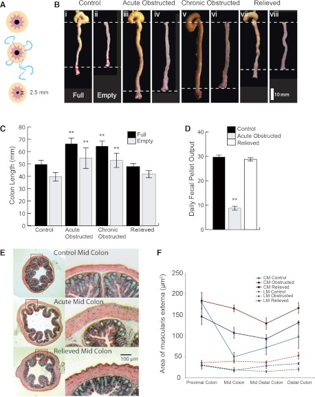
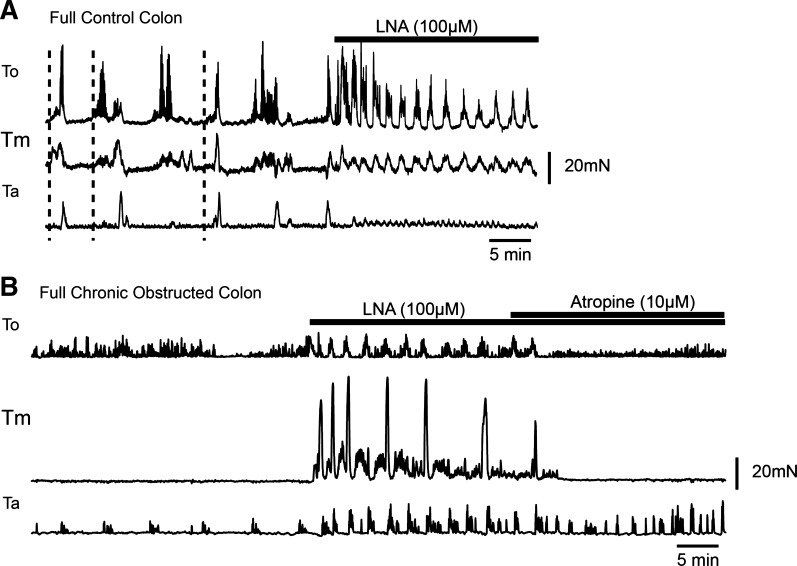
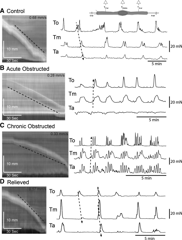
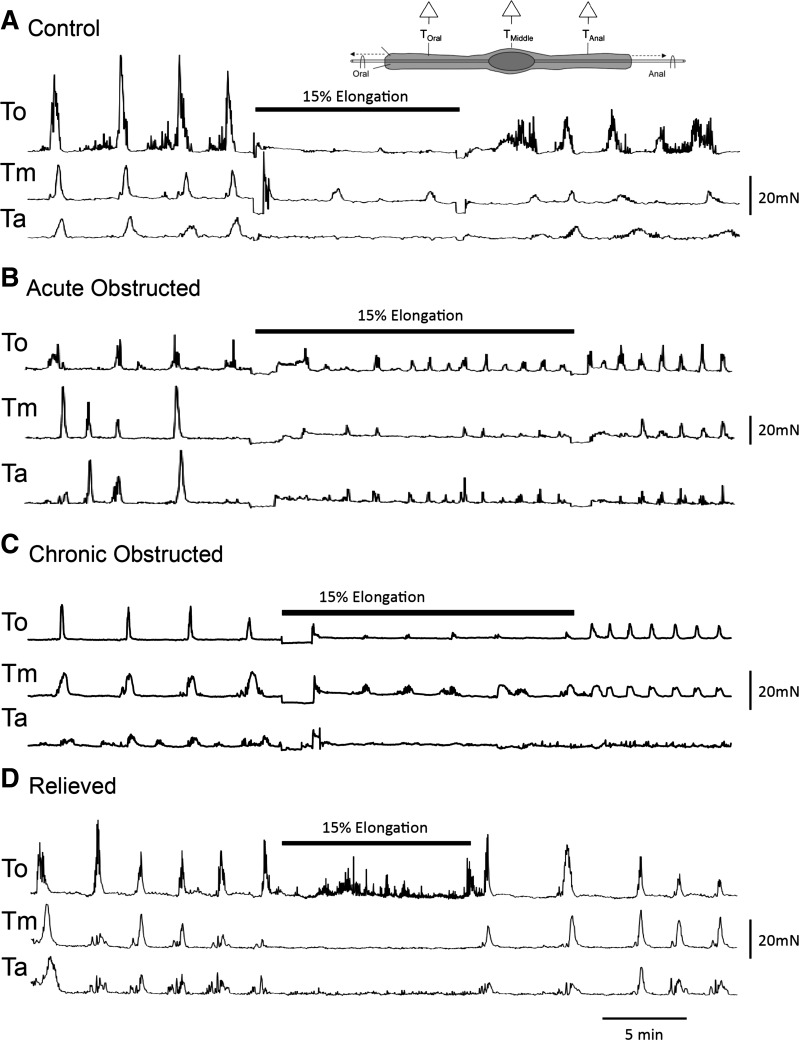
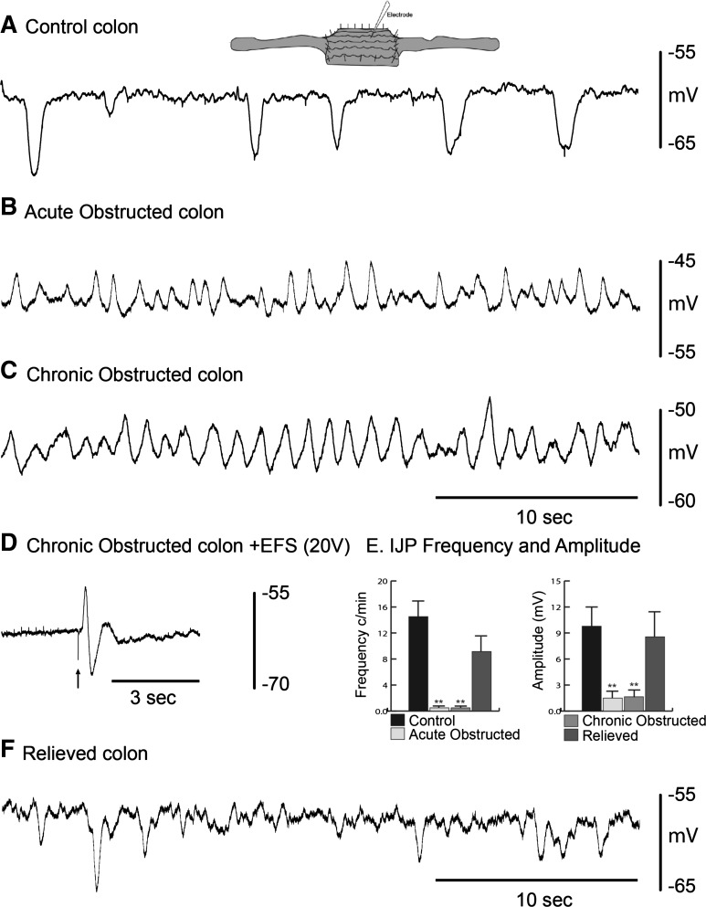
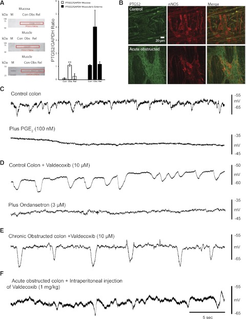
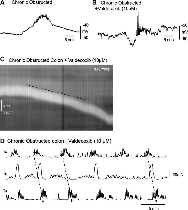
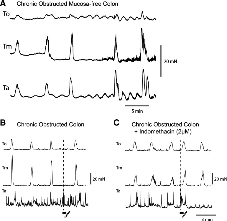
The mechanisms underlying slow-transit constipation (STC) are unclear. In 50% of patients with STC, some form of outlet obstruction has been reported; also an elongated colon has been linked to patients with STC. Our aims were 1) to develop a murine model of STC induced by partial outlet obstruction and 2) to determine whether this leads to colonic elongation and, consequently, activation of the inhibitory "occult reflex," which may contribute to STC in humans. Using a purse-string suture, we physically reduced the maximal anal sphincter opening in C57BL/6 mice. After 4 days, the mice were euthanized (acutely obstructed), the suture was removed (relieved), or the suture was removed and replaced repeatedly (chronically obstructed, over 24-31 days). In partially obstructed mice, we observed increased cyclooxygenase (COX)-2 levels in muscularis and mucosa, an elongated impacted large bowel, slowed transit, nonpropagating colonic migrating motor complexes (CMMCs), a lack of mucosal reflexes, a depolarized circular muscle with slow-wave activity due to a lack of spontaneous inhibitory junction potentials, muscle hypertrophy, and CMMCs in mucosa-free preparations. Elongation of the empty obstructed colon produced a pronounced occult reflex. Removal of the obstruction or addition of a COX-2 antagonist (in vitro and in vivo) restored membrane potential, spontaneous inhibitory junction potentials, CMMC propagation, and mucosal reflexes. We conclude that partial outlet obstruction increases COX-2 leading to a hyperexcitable colon. This hyperexcitability is largely due to suppression of only descending inhibitory nerve pathways by prostaglandins. The upregulation of motility is suppressed by the occult reflex activated by colonic elongation.


Figures

Fig. 1.
Fig. 1.
Anatomic consequences of partial outlet obstruction. A: purse-string suture applied to perianal region restricts sphincter outlet to 2.5 mm. B: length changes in full and empty control (i and ii), acutely obstructed (iii and iv), chronically obstructed (v and vi), and relieved (vii and viii) colons. C: changes in colon length. D: daily fecal pellet output in control and acutely obstructed colons. **P < 0.001. E: hematoxylin-eosin-stained sections of acutely obstructed midproximal region of colon. F: area of longitudinal muscle (LM) and circular muscle (CM) along the length of acutely obstructed colon.
Fig. 2.
Fig. 2.
Contractile activity in obstructed mice. A: tension recordings from a full normal colon along 3 points [oral (To), middle (Tm), and anal (Ta)]. N′-nitro-l-arginine (l-NA) was added to block nitric oxide (NO). B: tension recordings from a full chronically obstructed colon at To, Tm, and Ta. l-NA increased amplitude and frequency of contractile activity, and atropine depressed contractile activity.
Fig. 3.
Fig. 3.
Pellet propulsion and colonic migrating motor complexes (CMMCs) in an empty obstructed colon. Left: spatiotemporal maps of artificial pellet propagation in control, acutely obstructed, chronically obstructed, and relieved colons. Right: tension recordings from a colon that contained a fixed fecal pellet along points To, Tm, and Ta. Oral-to-anal propagation is less likely in B and C than in A and D.
Fig. 4.
Fig. 4.
Occult reflexes in obstructed colon. A–D: tension recordings of occult reflex in response to elongation (15%) in control, acutely obstructed, chronically obstructed, and relieved empty colons.
Fig. 5.
Fig. 5.
Spontaneous inhibitory junction potentials (IJPs) in whole colons. A: intracellular recordings from circular smooth muscle showing spontaneous IJPs in a control colon. B and C: slow waves, but no IJPs, in acutely and chronically obstructed colon. D: electrical field stimulation (EFS) evokes an excitatory junction potential and IJP in a chronically obstructed colon. E: IJP frequency and amplitude in control, acutely/chronically obstructed, and relieved colon. **P < 0.001. F: spontaneous IJPs in a relieved colon.
Fig. 6.
Fig. 6.
Consequences of cyclooxygenase (COX)-2 inhibition. A: Western blots indicating an increase in prostaglandin-endoperoxide synthase II (PTGS2) levels in muscularis externa (including myenteric plexus) and mucosa of obstructed control (Con), obstructed (Obs), and relieved (Rel) colon (left; M, molecular weight marker) and plot illustrating increased PTGS2-to-GAPDH ratio in muscularis externa and mucosa of obstructed colon (right). **P < 0.001. B: immunohistochemical labeling of myenteric ganglia for COX-2 (PTGS2 in green) and neuronal nitric oxide synthase (nNOS, red) in control and acutely obstructed colon. Merge shows COX-2 labeling of nNOS-positive neurons. C: spontaneous IJPs in control colon before and after PGE2. D: spontaneous IJPs in control colon after valdecoxib and ondansetron. E: spontaneous IJPs in chronically obstructed colon following valdecoxib. F: spontaneous IJPs in acutely obstructed colon following intraperitoneal injection of valdecoxib.
Fig. 7.
Fig. 7.
Effect of COX-2 inhibition on CMMCs in obstructed colon. A and B: electrical recording of CMMC in obstructed colon before and after valdecoxib (10 μM), where CMMC is preceded by a hyperpolarization. C and D: valdecoxib (10 μM) increases transit of a fecal pellet (C) and causes propagation of CMMCs in obstructed colon (D).
Fig. 8.
Fig. 8.
Effect of mucosa removal and mucosal reflexes in obstructed colon. A: tension recordings from a mucosa-free chronically obstructed colon (note CMMCs occurred simultaneously along the colon). B and C: mucosal stimulation (3 strokes) of the rectum before (B, no response) and after (C) indomethacin. Note premature CMMC at oral recording sites.
Fig. 9.
Fig. 9.
Hypothetical nerve circuit controlling colonic motility. A and B: electrical and tension recordings of CMMCs. A: ongoing tonic inhibition, consisting of spontaneous inhibitory junction potentials (IJPs, blue downward deflections) in circular muscle and inhibition of pacemaker interstitial cells of Cajal, between CMMCs. Before the CMMC, there can be an increased burst of IJPs/relaxation that determines its oral-to-anal direction of propagation and fecal pellet propulsion. A CMMC consists of activation of excitatory motor neurons (EMNs), which produce a slow depolarization, upon which are superimposed fast oscillations that trigger action potentials and contraction (red). IMN, inhibitory motor neurons; TK, tachydinin; NO, nitric oxide. B: between CMMCs, there is little muscle tone because of ongoing tonic inhibition. When a fecal pellet is present, a small relaxation, due to increased IJP activity (blue), can precede the CMMC. C: spontaneous release of 5-HT from enterochromaffin cells (ECC) activates 5-HT3 receptors on projections of afterhyperpolarizing (AH) neurons in the mucosa (circuit 1); these neurons synapse with ascending interneurons in the ascending excitatory pathway (AEP), which synapse with excitatory motor neurons that are responsible for the CMMC (A and B). When fecal pellets are present, they are more likely to activate different AH sensory neurons (located around the pellet) by circumferential stretch or by mucosal distortion, which releases 5-HT onto the 5-HT3 receptors of AH neurons in the mucosa (circuit 2); these neurons synapse with descending serotonergic interneurons in the descending inhibitory pathway (DIP), which synapse with inhibitory motor neurons (A and B). Elongation activates mechanosensitive descending nNOS-positive interneurons, which release NO to inhibit ascending interneurons and AH sensory neurons, thereby decreasing colonic motility (circuit 3, occult reflex); partial outlet obstruction increases prostaglandins, which depress the DIP (circuit 2), by inhibiting serotonergic neurons or IMNs; in addition, increased prostaglandins may inhibit 5-HT release from ECC in the mucosa and block mucosal reflexes. Nic, nicotinic receptor; ICC-MY, ICCs in the myenteric region; ICC-SM, ICCs along the submucosal surface.

Similar articles

Cited by

References

    1. Bayguinov PO, Hennig GW, Smith TK. Calcium activity in different classes of myenteric neurons underlying the migrating motor complex in the murine colon. J Physiol 588: 399–421, 2010 - PMC - PubMed
    1. Bayguinov PO, Hennig GW, Smith TK. Ca2+ imaging of activity in ICC-MY during local mucosal reflexes and the colonic migrating motor complex in the murine large intestine. J Physiol 588: 453–474, 2010 - PMC - PubMed
    1. Bayguinov PO, Broadhead MJ, Okamoto T, Hennig GW, Smith TK. Activity in varicosities within the myenteric plexus between and during the colonic migrating motor complex in the isolated murine large intestine. Neurogastroenterol Motil 24: e185–e201, 2012 - PMC - PubMed
    1. Bayliss WM, Starling EH. The movements and innervation of the small intestine. J Physiol 24: 99–143, 1899 - PMC - PubMed
    1. Bayliss WM, Starling EH. The movements and innervation of the large intestine. J Physiol 26: 107–118, 1900 - PMC - PubMed

Publication types

MeSH terms

Substances