. 2012 Dec;30(12):2672-82.
doi: 10.1002/stem.1228.

ALKBH1 is a histone H2A dioxygenase involved in neural differentiation

Affiliations

ALKBH1 is a histone H2A dioxygenase involved in neural differentiation

Rune Ougland et al. Stem Cells. 2012 Dec.

Abstract

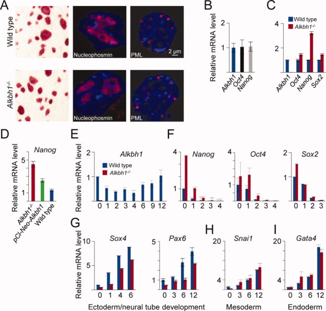
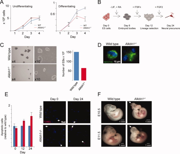
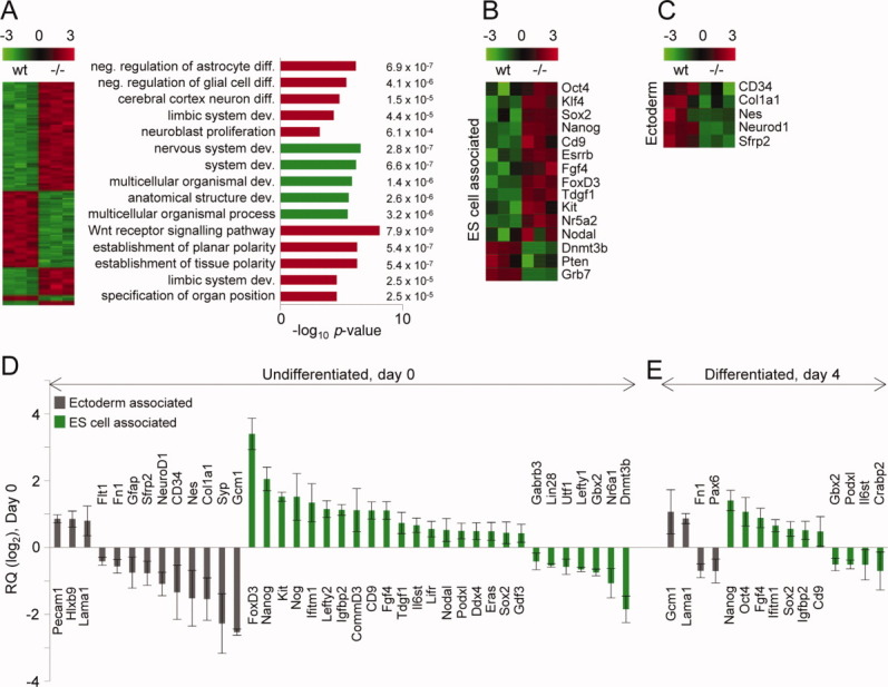
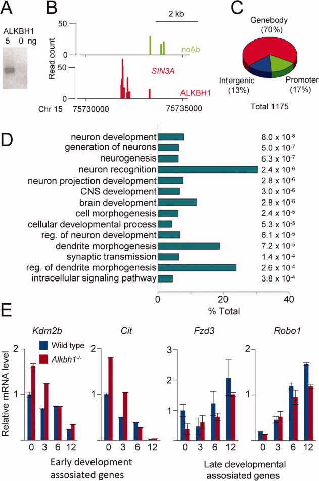
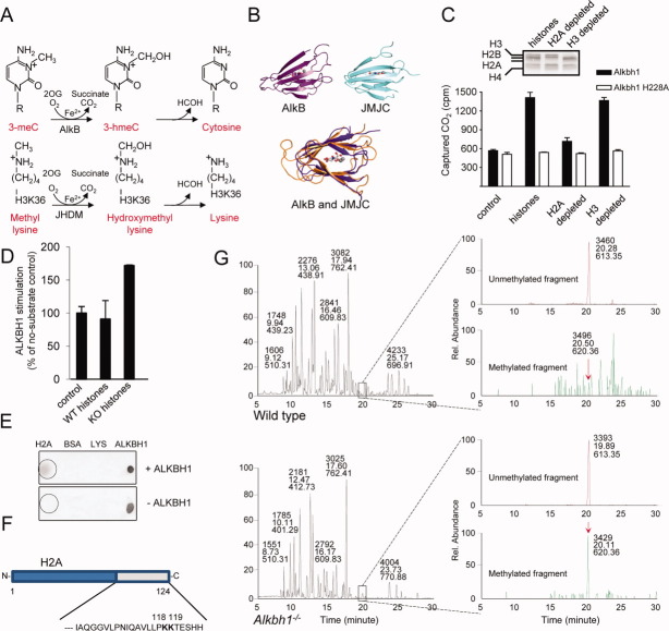
AlkB homolog 1 (ALKBH1) is one of nine members of the family of mammalian AlkB homologs. Most Alkbh1(-/-) mice die during embryonic development, and survivors are characterized by defects in tissues originating from the ectodermal lineage. In this study, we show that deletion of Alkbh1 prolonged the expression of pluripotency markers in embryonic stem cells and delayed the induction of genes involved in early differentiation. In vitro differentiation to neural progenitor cells (NPCs) displayed an increased rate of apoptosis in the Alkbh1(-/-) NPCs when compared with wild-type cells. Whole-genome expression analysis and chromatin immunoprecipitation revealed that ALKBH1 regulates both directly and indirectly, a subset of genes required for neural development. Furthermore, our in vitro enzyme activity assays demonstrate that ALKBH1 is a histone dioxygenase that acts specifically on histone H2A. Mass spectrometric analysis demonstrated that histone H2A from Alkbh1(-/-) mice are improperly methylated. Our results suggest that ALKBH1 is involved in neural development by modifying the methylation status of histone H2A.


Figures

Figure 1
Figure 1
The ALKBH1 protein localizes to the nuclei of human embryonic stem cells (ESCs). Staining of human ESCs (H9) revealed that ALKBH1 is predominantly nuclear. The mitomarker is red (Alexa-555), ALKBH1 is green (Alexa-488), and 4′,6–diamidino–2–phenylindole (DAPI) is blue. Abbreviation: ALKBH1, AlkB homolog 1.
Figure 2
Figure 2
Differentiation is delayed in Alkbh1−/− embryonic stem cells (ESCs). (A): Alkaline phosphatase (AP) staining of wild-type (WT) and Alkbh1−/− mouse ESCs (mESCs). Alkbh1−/− mESCs express markers of pluripotency. (B): The measurement of Alkbh1 expression (blue bar) in undifferentiated mESCs showed expression at the same level as the core pluripotency transcription factors (represented by Oct4 and Nanog). (C): Quantitation of the master regulators of pluripotency revealed upregulation of genes encoding all three transcription factors (Oct4, Nanog, and Sox2) in undifferentiated Alkbh1−/− mESCs. The Nanog gene displayed the greatest (more than fourfold) increase in expression. (D): Quantitative real-time PCR (qRT-PCR) analysis showed that expression of ALKBH1 in Alkbh1−/− mESCs (pCl-Neo-Alkbh1) reduced Nanog expression, albeit not to levels as low as those found in WT mESCs. (E): The qRT-PCR analysis of WT mESCs that were forced to differentiate along an ectodermal path (i.e., by incubation in media without leukemia inhibitory factor and supplemented with 1 μM all-trans retinoic acid) revealed that Alkbh1 was downregulated during the first 4 days of differentiation, before it was upregulated. (F): As expected, levels of expression of the pluripotency markers Oct4, Sox2, and Nanog decreased, albeit at a slower rate in Alkbh1−/− mESCs than in WT mESCs, suggesting delayed differentiation of Alkbh1−/− mESCs. (G): The impeded differentiation of Alkbh1−/− mESCs was confirmed by demonstrating the induction of Sox4 and Pax6 at a later time in the Alkbh1−/− mESCs than in WT mESCs. (H): Expression of the mesodermal marker Snai1 was identical in Alkbh1−/− mESCs and WT mESCs. (I): Expression of the endodermal marker Gata4 was identical in Alkbh1−/− mESCs and WT mESCs. Vertical lines represent the 1 ± SEM. Abbreviations: ALKBH1, AlkB homolog 1; PML, promyelocytic leukemia protein.
Figure 3
Figure 3
Differentiation of Alkbh1−/− mouse ESCs leads to increased apoptosis. (A): Proliferation rate of mouse ESCs cultured under conditions favoring self-renewal showed no difference between WT and Alkbh1−/−. Upon induction of differentiation, the culture of Alkbh1−/− mouse ESCs had a slower growth rate and increased cell death from differentiation day 3. (B): Timeline showing the steps of the neural differentiation protocol of mouse ESCs. (C):Alkbh1−/− mouse ESCs form embryoid bodies that are fewer and smaller than their WT counterpart. (D): After 24 days of differentiation, the mouse ESCs formed NESTIN-positive neural progenitor cells (NPCs). (E): TUNEL staining reveals an increased rate of apoptosis in the NPCs derived from Alkbh1−/− mESCs. (F): Ten percentage of E10.5-E11.5 Alkbh−/− embryos exhibits neural tube defects (white arrowheads) when compared with WT littermates [26]. Abbreviations: ALKBH1, AlkB homolog 1; ESC, embryonic stem cell; FGF2, fibroblast growth factor 2; LIF, leukemia inhibitory factor; RA, retinolic acid; WT, wild type.
Figure 4
Figure 4
Alkbh1−/− mouse ESCs show severe dysregulation of genes involved in differentiation and pluripotency. (A): An Affymetrix GeneChip Mouse Genome 430 2.0 Array revealed that 248 transcripts were differentially expressed between WT and Alkbh1−/− mouse ESCs (fold change >1.5 [log2], false discovery rate (FDR) 5%). Five discrete clusters were identified by visual inspection. Gene ontology classification according to biological process (TopGO, Bioconductor) showed an enrichment of terms related to differentiation, the development of the nervous system, and Wnt signaling. (B): Selected ESC-related genes and genes related to pluripotency. (C): Selected genes related to ectodermal development. (D): Analysis involving the TaqMan Mouse Stem Cell Pluripotency Array v.2 (Applied Biosystems) was performed to compare the expression of lineage marker genes and pluripotency-associated genes in WT and Alkbh1−/− mouse ESCs grown under self-renewal conditions. The Gapdh gene was used as an internal control. Selected genes with Ct <32 were included. Gray, ectoderm-associated genes; green, pluripotency- and ESC-associated genes. (E): As in (D), but with mESCs allowed to differentiate for 4 days. Vertical lines represent the 1 ± SEM. Abbreviations: ESC, embryonic stem cell; WT, wild type.
Figure 5
Figure 5
ChIP-seq analysis reveals that ALKBH1 occupies genes that encode developmental regulators in human embryonic stem cells. (A): The specificity of the antibody used in the ChIP-seq assay was confirmed by Western blotting of recombinant human ALKBH1 protein. (B): Image from the UCSC Genome Browser (http://genome.ucsc.edu/). This view of the paired amphipathic helix protein (SIN3A) illustrates the read distribution over one ALKBH1-binding region designated as a read. The locations of the amplicons detected by ChIP-qPCR are indicated at the bottom. Green bars represent reads from the control sample lacking antibody, whereas red bars represent reads from the ALKBH1 immunopreciptated sample. (C): Analysis of the distribution of ALKBH1 reads over various genomic features revealed that the majority of reads reside in the gene body (70%), whereas 13% and 17% are associated with intergenic and promoter regions, respectively. (D): Gene ontology analysis of genes occupied by ALKBH1 revealed a significant enrichment for regulators of neural development. (E): Analysis by quantitative real-time PCR of a selection of the occupied genes confirmed that ALKBH1 regulates their expression. Vertical lines represent the 1 ± SEM. Abbreviation: ALKBH1, AlkB homolog 1.
Figure 6
Figure 6
Histone H2A from Alkbh1−/− mouse embryonic fibroblast (MEF) cells contains a methylation group not present in wild-type (WT) histones. (A): Analogous hydroxylation mechanisms for demethylation of 3-methyladenine by the AlkB repair enzyme (top panel) and the hydroxylation of monomethyl lysine by a JmjC domain-containing histone demethylase, resulting in a similar loss of one methyl group and the generation of unmodified lysine (bottom panel). (B): The Fe(II)-2OG dioxygenase cores of E. coli AlkB (upper left) and the JmjC domain of human JMJD2A (upper right), shown in the same orientation. This shows the common “jellyroll” structural fold of the AlkB and JmjC-domain superfamilies. (C): Dioxygenase activity was evaluated by the CO2-capture assay using purified ALKBH1 with either purified HeLa core histones or core histones that were immunodepleted of H2A or H3. The ALKBH1 H228A variant is an inactive protein harboring a mutation in the Fe(II)-binding domain. Data are presented as the mean of two replicates. Error bars represent one SD. We used Coomassie-stained SDS-PAGE to analyze HeLa core histones immunodepleted for histones H2A and H3. (D): Histones purified from WT and Alkbh1−/− mESC show that Alkbh1−/− histones stimulate ALKBH1 activity approximately 80% more efficiently than WT histones. (E): Dot blot showing the physical interaction between the ALKBH1 and H2A proteins in vitro. (F): Representation of histone H2A, highlighting the C-terminal sequence harboring the region targeted by ALKBH1. (G): Analysis of histone H2A purified from WT or Alkbh1−/− MEFs using mass spectrometry. The maintenance of modifications after purification is shown in Supporting Information Figure S3A. Chromatograms on the left represent signals obtained from the entire run. Chromatograms on the right represent signals found for the peptide indicated in (E). The signal for the unmethylated peptides (upper chromatograms of the WT or Alkbh1−/−, respectively) is 6.68 × 10⁵ for WT MEFs and 4.30 × 10⁵ for Alkbh1−/− MEFs. The lower chromatograms represent the same peptide with the addition of 28.0314 Da (the mass of a dimethyl group or two monomethyl groups). The methylated peptide was undetectable in the WT sample, whereas there was a signal of 1.87 × 10⁴ for the Alkbh1−/− sample. The x-axis represents the elution time and the y-axis indicates the relative abundances of the peptides. Abbreviation: ALKBH1, AlkB homolog 1.

Similar articles

Cited by

References

    1. Loebel DA, Watson CM, De Young RA, et al. Lineage choice and differentiation in mouse embryos and embryonic stem cells. Dev Biol. 2003;264:1–14. - PubMed
    1. Smith AG. Embryo-derived stem cells: Of mice and men. Annu Rev Cell Dev Biol. 2001;17:435–462. - PubMed
    1. Bernstein BE, Mikkelsen TS, Xie X, et al. A bivalent chromatin structure marks key developmental genes in embryonic stem cells. Cell. 2006;125:315–326. - PubMed
    1. Chambers I, Colby D, Robertson M, et al. Functional expression cloning of Nanog, a pluripotency sustaining factor in embryonic stem cells. Cell. 2003;113:643–655. - PubMed
    1. Masui S, Nakatake Y, Toyooka Y, et al. Pluripotency governed by Sox2 via regulation of Oct3/4 expression in mouse embryonic stem cells. Nat Cell Biol. 2007;9:625–635. - PubMed

Publication types

MeSH terms

Substances