. 2012 Nov;194(22):6131-42.
doi: 10.1128/JB.01419-12. Epub 2012 Sep 7.

SurA is involved in the targeting to the outer membrane of a Tat signal sequence-anchored protein

Affiliations

SurA is involved in the targeting to the outer membrane of a Tat signal sequence-anchored protein

Arnaud Rondelet et al. J Bacteriol. 2012 Nov.

Abstract

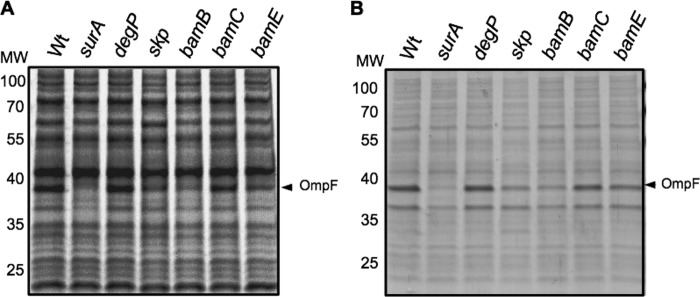
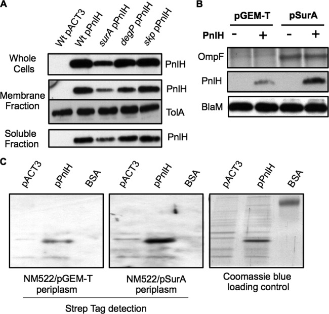
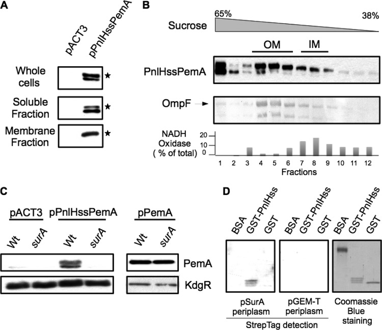
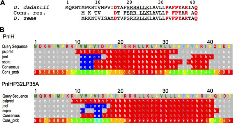
The twin arginine translocation (Tat) pathway exports folded proteins from the cytoplasm to the periplasm of bacteria. The targeting of the exported proteins to the Tat pathway relies on a specific amino-terminal signal sequence, which is cleaved after exportation. In the phytopathogen Dickeya dadantii, the pectin lyase homologue PnlH is exported by the Tat pathway without cleavage of its signal sequence, which anchors PnlH into the outer membrane. In proteobacteria, the vast majority of outer membrane proteins consists of β-barrel proteins and lipoproteins. Thus, PnlH represents a new kind of outer membrane protein. In Escherichia coli, periplasmic chaperones SurA, Skp, and DegP work together with the β-barrel assembly machinery (Bam) to target and insert β-barrel proteins into the outer membrane. In this work, we showed that SurA is required for an efficient targeting of PnlH to the outer membrane. Moreover, we were able to detect an in vitro interaction between SurA and the PnlH signal sequence. Since the PnlH signal sequence contains a highly hydrophobic region, we propose that SurA protects it from the hydrophobic periplasm during targeting of PnlH to the outer membrane. We also studied the nature of the information carried by the PnlH signal sequence responsible for its targeting to the outer membrane after exportation by the Tat system.


Figures

Fig 1
Fig 1
Porin depletion in D. dadantii mutants with mutations in the OMP-targeting pathway. Protein profiles of whole cells of D. dadantii wild-type (Wt) strain A350 and derivative strains (indicated at the top) carrying the chromosomal mutation (A) and protein profiles of membrane fractions of the same strains (B). Equivalent amounts of cell materials from exponential-phase cultures of the different strains were separated by SDS-PAGE, followed by Coomassie blue staining. The arrowhead indicates the D. dadantii major porin OmpF. MW, molecular weight (in thousands).
Fig 2
Fig 2
Role of periplasmic chaperones SurA, Skp, and DegP in the targeting of PnlH-6His to the OM of D. dadantii. (A) Immunodetection of PnlH in whole-cell, membrane, and soluble fractions of D. dadantii wild-type strain and derivative strains carrying a chromosomal mutation (indicated at the top) and either empty pACT3 vector or pPnlH. Equivalent amounts of cell material from exponential-phase cultures of the different strains were loaded in each well. Immunodetection of the inner membrane protein TolA was used as a loading control. (B) Complementation of a surA mutant carrying either empty pACT3 vector or pPnlH with either empty pGEM-T vector or pSurA. Equivalent amounts of whole cells of steady-state cultures were analyzed. Proteins were separated on an SDS-polyacrylamide gel and transferred onto a PVDF membrane for immunodetection of PnlH and BlaM (loading control). OmpF was detected by SDS-PAGE, followed by Coomassie blue staining. (C) The in vitro interaction between PnlH-6His and SurA-Strep was assayed by overlay. BSA and cell lysates of NM522 carrying either empty pACT3 vector or pPnlH were separated by SDS-PAGE and transferred onto a PVDF membrane. The membrane was next incubated with the periplasm of NM552 carrying either empty pGEM-T vector or pSurA. SurA-Strep bound to proteins on the membrane was detected with streptavidin conjugated with HRP. The amounts of protein loaded on the gel were estimated by Coomassie blue staining.
Fig 3
Fig 3
Role of the PnlH signal sequence in the interaction with SurA. (A) Cell fractionation of D. dadantii A350 strains carrying either empty pACT3 vector or pPnlHssPemA. PnlHssPemA was detected with antibodies against PemA. Equivalent amounts of cell materials from exponential-phase cultures of the different strains were loaded in each well. (B) The membrane fraction from strain A350 carrying pPnlHssPemA was separated by flotation sucrose gradient centrifugation and analyzed by immunoblotting with PemA antibodies or stained with Coomassie blue to detect the major porin, OmpF, which reflects the position of the outer membrane. NADH oxidase activity indicates the position of the inner membrane. (C) Immunodetection of PemA in whole cells of D. dadantii wild-type strain A350 and a surA mutant carrying either empty pACT3 vector or pPnlHssPemA. Equivalent amounts of cell material from steady-state cultures were loaded in each well. The cytoplasmic protein KdgR was used as a loading control. (D) The in vitro interaction between the PnlH signal sequence and SurA-Strep was assayed by overlay. Purified BSA, GST, and GST fused to the PnlH signal sequence (GST-PnlHss) were run on an SDS-polyacrylamide gel and transferred onto a PVDF membrane. The membrane was next incubated with the periplasm of NM552 carrying either empty pGEM-T vector or pSurA. SurA-Strep bound to proteins on the membrane was detected with streptavidin conjugated with HRP. The amounts of protein loaded on the gel were estimated by Coomassie blue staining.
Fig 4
Fig 4
In silico analysis of the PnlH signal sequence. (A) Amino acid sequence alignment of PnlH from D. dadantii (upper line, GenBank accession number YP_003883450.1) and D. zeae (lower line, GenBank accession number YP_003005603.1). Conserved residues (Cons. res.) are indicated in the middle line. The Tat consensus is underlined. Residues that were substituted are in red bold. (B) Secondary structure prediction of the first 41 residues of PnlH and PnlHP32LP235A by the Phyre protein fold recognition software (available at www.sbg.bio.ic.ac.uk/∼phyre/). Letters indicate the type of secondary structure predicted at the corresponding amino acid site (h, alpha helix; c, coil; e, β sheet). Numbers indicate the reliability of the prediction on a scale of from 0 (low) to 9 (high).
Fig 5
Fig 5
Study of the PnlH signal sequence by directed mutagenesis. (A) Cell fractionation of D. dadantii A350 strains carrying either empty pACT3 vector, pPnlH, or pPnlHΔ28-41. PnlH and PnlHΔ28-41 were detected with antibodies against PnlH. Equivalent amounts of cell materials from exponential-phase cultures of the different strains were loaded in each well. (B) The membrane fraction from strain A350 carrying pPnlHΔ28-41 was separated by flotation sucrose gradient centrifugation and analyzed by immunoblotting with PemA antibodies or stained with Coomassie blue to detect the major porin OmpF, which reflects the position of the outer membrane. Immunoblotting with TolA antibodies indicates the position of the inner membrane. (C) Immunodetection of PnlH in whole-cell lysates of D. dadantii A350 strains carrying either empty pACT3 vector (−) or pPnlH (PnlH) or pPnlH derivatives carrying one or two substitutions in the PnlH signal sequence. Equivalent amounts of cell material from steady-state cultures were loaded in each well. Immunoblotting with KdgR antibodies was used as a loading control. (D) Membrane fraction of A350 strains carrying either empty pACT3 vector (−), pPnlH (PnlH), pD16A (D16A), pL30SL31T (L30SL31T), or pP32LP35A (P32LP35A). PnlH and derivatives were detected with antibodies against PnlH. Equivalent amounts of membrane fractions from exponential-phase cultures were loaded in each well. OmpF was visualized by the coloration of the PVDF membrane with Coomassie blue and served as a loading control. (E) Induction of periplasmic stress in E. coli SR1458 expressing PnlH and derivatives. E. coli SR1458 carrying either empty pACT3 vector (−), pPnlH (PnlH), pD16A (D16A), pL30SL31T (L30SL31T), or pP32LP35A (P32LP35A) was grown at 33°C in LB medium supplemented with chloramphenicol and 1 mM IPTG. The value for β-galactosidase specific activity is the average of three independent determinations.
Fig 6
Fig 6
Effect of bamB, bamC, and bamE mutations on the targeting of PnlH-6His to the OM of D. dadantii. (A) Immunodetection of PnlH in whole-cell, membrane, and soluble fractions of D. dadantii wild-type strain A350 and derivative strains carrying chromosomal mutations (indicated at the top) and either empty pACT3 vector or plasmid pPnlH. Equivalent amounts of cell materials from exponential-phase cultures of the different strains were loaded in each well. Immunodetection of the inner membrane protein TolA was used as a loading control. (B) The membrane fractions from the bamB and bamE mutant strains carrying pPnlH were separated by flotation sucrose gradient centrifugation and analyzed by immunoblotting with PnlH antibodies or stained with Coomassie blue to detect the major porins, which reflect the position of the outer membrane. Immunoblotting with TolA antibodies indicates the position of the inner membrane. For clarity, porins and TolA profiles are presented only for the bamB mutant, but the profiles were equivalent for the bamE mutant.

Similar articles

Cited by

References

    1. Alcock FH, et al. 2008. Conserved substrate binding by chaperones in the bacterial periplasm and the mitochondrial intermembrane space. Biochem. J. 409:377–387 - PubMed
    1. Allen JWA, Tomlinson EJ, Hong L, Ferguson SJ. 2002. The Escherichia coli cytochrome c maturation (Ccm) system does not detectably attach heme to single cysteine variants of an apocytochrome c. J. Biol. Chem. 277:33559–33563 - PubMed
    1. Bennion D, Charlson ES, Coon E, Misra R. 2010. Dissection of β-barrel outer membrane protein assembly pathways through characterizing BamA POTRA 1 mutants of Escherichia coli. Mol. Microbiol. 77:1153–1171 - PMC - PubMed
    1. de Cock H, Struyvé M, Kleerebezem M, van der Krift T, Tommassen J. 1997. Role of the carboxy-terminal phenylalanine in the biogenesis of outer membrane protein PhoE of Escherichia coli K-12. J. Mol. Biol. 269:473–478 - PubMed
    1. Denoncin K, Schwalm J, Vertommen D, Silhavy TJ, Collet J-F. 2012. Dissecting the Escherichia coli periplasmic chaperone network using differential proteomics. Proteomics 12:1391–1401 - PMC - PubMed

Publication types

MeSH terms

LinkOut - more resources