. 2012 Nov;194(22):6116-30.
doi: 10.1128/JB.01409-12. Epub 2012 Sep 7.

Hyperosmotic stress response of Campylobacter jejuni

Affiliations

Hyperosmotic stress response of Campylobacter jejuni

Andrew Cameron et al. J Bacteriol. 2012 Nov.

Abstract

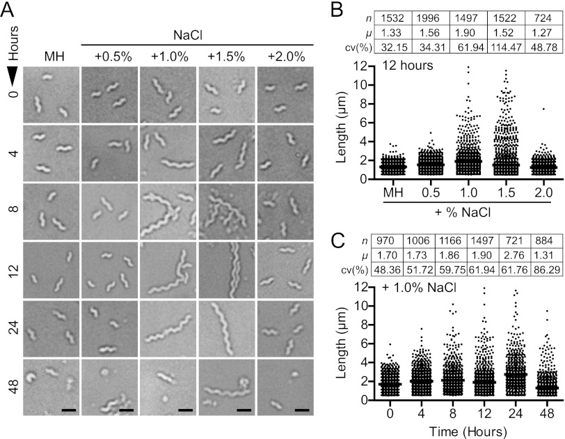
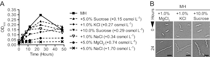
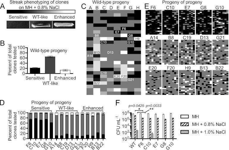
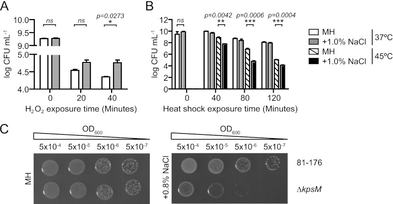
The diarrheal pathogen Campylobacter jejuni and other gastrointestinal bacteria encounter changes in osmolarity in the environment, through exposure to food processing, and upon entering host organisms, where osmotic adaptation can be associated with virulence. In this study, growth profiles, transcriptomics, and phenotypic, mutant, and single-cell analyses were used to explore the effects of hyperosmotic stress exposure on C. jejuni. Increased growth inhibition correlated with increased osmotic concentration, with both ionic and nonionic stressors inhibiting growth at 0.620 total osmol liter(-1). C. jejuni adaptation to a range of osmotic stressors and concentrations was accompanied by severe filamentation in subpopulations, with microscopy indicating septum formation and phenotypic diversity between individual cells in a filament. Population heterogeneity was also exemplified by the bifurcation of colony morphology into small and large variants on salt stress plates. Flow cytometry of C. jejuni harboring green fluorescent protein (GFP) fused to the ATP synthase promoter likewise revealed bimodal subpopulations under hyperosmotic stress. We also identified frequent hyperosmotic stress-sensitive variants within the clonal wild-type population propagated on standard laboratory medium. Microarray analysis following hyperosmotic upshift revealed enhanced expression of heat shock genes and genes encoding enzymes for synthesis of potential osmoprotectants and cross-protective induction of oxidative stress genes. The capsule export gene kpsM was also upregulated, and an acapsular mutant was defective for growth under hyperosmotic stress. For C. jejuni, an organism lacking most conventional osmotic response factors, these data suggest an unusual hyperosmotic stress response, including likely "bet-hedging" survival strategies relying on the presence of stress-fit individuals in a heterogeneous population.


Figures

Fig 1
Fig 1
Growth and survival of C. jejuni 81-176 in broth cultures containing increasing concentrations of NaCl in Mueller-Hinton (MH) medium. (A) Comparison of biologically relevant osmolarities and the conditions used in this experiment. (B and C) Optical density (OD600) readings (B) and CFU ml−1 counts (C) demonstrated that growth and survivability are inhibited at osmolarities exceeding +1.5% NaCl. Initial adaptation under the +1.0% NaCl condition (open triangles) was followed by logarithmic growth and late-stage growth defects; the dashed bracket and box in panel C show an expanded view of the 0- to 12-h time points for +1.0% NaCl versus unsupplemented medium. The MH+0.5% NaCl CFU ml−1 curve is identical to the MH curve but has been offset to enable viewing. The experiment is representative of three biological replicates; error bars for three technical replicates are present but in most cases are too small to see. *, P ≤ 0.05; **, P ≤ 0.01.
Fig 2
Fig 2
Hyperosmotic stress leads to C. jejuni cell length alteration. (A) Bright-field microscopy of bacteria from NaCl-supplemented cultures sampled over a 48-h period demonstrated that filamentation occurs in a narrow hyperosmotic range. Increased progression to the coccoid form was not observed, and not all cells are filamented. Scale bars, 1 μm. (B) Measurement of filament length from NaCl-supplemented cultures demonstrated that the proportion of filaments increased with osmolarity (the 12-h time point is shown) except under +2.0% NaCl conditions. (C) Filament length and abundance increased with time (the +1.0% NaCl condition is shown). Data tables include number of bacteria analyzed (n), mean length (μ) in μm, and coefficient of variation (cv%; standard deviation to mean ratio). All data are representative of at least 3 independent fields of view.
Fig 3
Fig 3
Growth inhibition and filamentation occur under multiple types of hyperosmotic stress. (A) OD600 readings of broth cultures of C. jejuni grown in MH medium supplemented with various concentrations of ionic (NaCl, MgCl2, or KCl) and nonionic (sucrose) osmotic stressors over 48 h showed that growth inhibition generally correlated with osmotic concentration. Error bars are present but in most cases are too small to see. (B) Bright-field microscopy illustrated that filamentation was an effect of both ionic (MgCl2 or KCl) and nonionic (sucrose) osmotic stressors. Scale bars, 1 μm; data from microscopy are representative of at least 3 independent fields of view.
Fig 4
Fig 4
Heterogeneity within hyperosmotically stressed populations and filaments and symmetrical staining with fluorescent vancomycin (Vanco-FL). Bacteria were grown for 12 h in unsupplemented MH broth or MH broth +1.0% NaCl and prepared for microscopy. (A) C. jejuni stained with propidium iodide (PI) (red) and Syto-9 (green) from the LIVE/DEAD BacLight kit. The wild type under standard conditions fluoresced with PI, while some cells under hyperosmotic conditions excluded PI, as did many of the cells within filaments (yellow arrows). (B) Filaments stained with Vanco-FL (white-gray) and PI (red) and analysis by fluorescence distribution. Fluorescence units are arbitrary. Scale bars, 1 μm.
Fig 5
Fig 5
Salt-sensitive isolates within the wild-type population. Two hundred single colonies were isolated from MH plates and patched on MH (control/passage) or MH+0.8% NaCl (test) plates. (A) Patches were assessed by microscopy and graded for growth by three categories; black represents minimal or no growth (“sensitive”), gray indicates characteristic (“WT-like”) growth, and white represents heavy growth (“enhanced”). (B) Percentages of 200 clones tested that were categorized as sensitive, WT-like, or enhanced. Error bars represent the fact that streaks were graded 3 times blind and that some streaks had intermediate phenotypes. (C) Visual heat map representation of the sensitivity profiles of the 200 clones. Intermediate phenotypes are represented by lighter or darker shades of gray, and numbered clones indicate progeny that were then selected from the MH (control/passage) plates, passed 2 times on MH, and then retested to examine the heritability of phenotypes. (D) Percentages of 200 clones of 15 progeny (5 sensitive, 5 WT-like, and 5 enhanced; “progeny of progeny”) falling into the same 3 categories. (E) Visual heat map representation of sensitivity profiles for 200 clones of the 15 progeny (“progeny of progeny”). For sensitive progeny, the majority of clones tested retained a sensitive phenotype, but WT-like or enhanced growth was also observed within those populations. Crossed-out boxes indicate tests where growth was not observed on the MH control plate. (F) CFU enumeration of sensitive clones F6, C10, E7, G8, and G10 in comparison to the wild type. Tenfold dilutions of an OD-standardized culture were plated on MH, MH+0.8%, or MH+1.0% NaCl plates and CFU enumerated; experiments were performed in triplicate. Most of the sensitive clones (F6, E7, G8, and G10) exhibited no growth on MH+1.0% NaCl (bars absent). All strains exhibited statistically significant (P ≤ 0.05) differences compared to the wild type for growth on the two NaCl concentrations and insignificant differences on MH alone. For space purposes, statistics are shown for wild type versus F6 only. *, P ≤ 0.05; **, P ≤ 0.01.
Fig 6
Fig 6
Bistability in colony size and expression of GFP from pAtpF′. (A) Heterogeneity in C. jejuni colony size when wild-type bacteria were plated on medium supplemented with 0.8% NaCl and grown for 48 h. Area measurements of colonies from plate images (upper panels) demonstrated a bimodal distribution of colony size on +0.8% NaCl plates (histograms, lower panels). (B) Bacteria harboring the pAtpF′-GFP plasmid were analyzed by flow cytometry. Following 12 h in MH plus 1.0% NaCl, the population bifurcated into GFPlow and GFPhigh populations (lower panel), while bacteria remaining in MH broth did not (upper panel). (C) Bifurcation was confirmed by fluorescence microscopy. Microscopy is representative of 3 independent fields of view. Scale bar, 3 μm. (D) FACS sorting of the GFP0, GFPlow, and GFPhigh populations and plating for CFU revealed that GFPhigh bacteria were as culturable as GFP0 bacteria and that GFPlow bacteria had ∼10× reduced culturability on MH medium (shown) and MH medium plus 0.8% NaCl (not shown). Error bars are derived from 3 independent experiments. **, P ≤ 0.01.
Fig 7
Fig 7
Hyperosmotic exposure cross-induces protective and detrimental effects on oxidative and heat shock stress responses, respectively, and the capsular polysaccharide protects against salt stress. (A and B) Bacteria were incubated in 1.0% NaCl for 2 h and then exposed to 5 mM H2O2 over 40 min (A) or 45°C conditions over 120 min (B). Cross-protection against oxidative stress but decreased tolerance to thermal stress occurred following hyperosmotic shock. Error bars are from three biological replicates. (C) Serial 10-fold dilutions of OD-standardized wild-type and ΔkpsM bacteria were spotted on MH agar with or without 0.8% NaCl. The ΔkpsM mutant exhibited increased sensitivity to hyperosmotic conditions. *, P ≤ 0.05; **, P ≤ 0.01; ***, P ≤ 0.001.

References

    1. Andersen MT, et al. 2005. Diverse roles for HspR in Campylobacter jejuni revealed by the proteome, transcriptome and phenotypic characterization of an hspR mutant. Microbiology 151:905–915 - PubMed
    1. Aspedon A, Palmer K, Whiteley M. 2006. Microarray analysis of the osmotic stress response in Pseudomonas aeruginosa. J. Bacteriol. 188:2721–2725 - PMC - PubMed
    1. Bacon DJ, et al. 2001. A phase-variable capsule is involved in virulence of Campylobacter jejuni 81-176. Mol. Microbiol. 40:769–777 - PubMed
    1. Bonis M, Ecobichon C, Guadagnini S, Prevost MC, Boneca IG. 2010. A M23B family metallopeptidase of Helicobacter pylori required for cell shape, pole formation and virulence. Mol. Microbiol. 78:809–819 - PubMed
    1. Boulos L, Prevost M, Barbeau B, Coallier J, Desjardins R. 1999. LIVE/DEAD BacLight: application of a new rapid staining method for direct enumeration of viable and total bacteria in drinking water. J. Microbiol. Methods 37:77–86 - PubMed

Publication types

LinkOut - more resources