. 2012 Nov;194(22):6105-15.
doi: 10.1128/JB.01366-12. Epub 2012 Sep 7.

Identification and characterization of five intramembrane metalloproteases in Anabaena variabilis

Affiliations

Identification and characterization of five intramembrane metalloproteases in Anabaena variabilis

Kangming Chen et al. J Bacteriol. 2012 Nov.

Abstract

Regulated intramembrane proteolysis (RIP) involves cleavage of a transmembrane segment of a protein, releasing the active form of a membrane-anchored transcription factor (MTF) or a membrane-tethered signaling protein in response to an extracellular or intracellular signal. RIP is conserved from bacteria to humans and governs many important signaling pathways in both prokaryotes and eukaryotes. Proteases that carry out these cleavages are named intramembrane cleaving proteases (I-CLips). To date, little is known about I-CLips in cyanobacteria. In this study, five putative site-2 type I-Clips (Ava_1070, Ava_1730, Ava_1797, Ava_3438, and Ava_4785) were identified through a genome-wide survey in Anabaena variabilis. Biochemical analysis demonstrated that these five putative A. variabilis site-2 proteases (S2Ps(Av)) have authentic protease activities toward an artificial substrate pro-σ(K), a Bacillus subtilis MTF, in our reconstituted Escherichia coli system. The enzymatic activities of processing pro-σ(K) differ among these five S2Ps(Av). Substitution of glutamic acid (E) by glutamine (Q) in the conserved HEXXH zinc-coordinated motif caused the loss of protease activities in these five S2Ps(Av), suggesting that they belonged to the metalloprotease family. Further mapping of the cleaved peptides of pro-σ(K) by Ava_4785 and Ava_1797 revealed that Ava_4785 and Ava_1797 recognized the same cleavage site in pro-σ(K) as SpoIVFB, a cognate S2P of pro-σ(K) from B. subtilis. Taking these results together, we report here for the first time the identification of five metallo-intramembrane cleaving proteases in Anabaena variabilis. The experimental system described herein should be applicable to studies of other RIP events and amenable to developing in vitro assays for I-CLips.


Figures

Fig 1
Fig 1
Diagram illustrates the typical regulated intramembrane proteolytic (RIP) activation of the membrane-anchored transcription factor in the cell. Inactive MTF (membrane portion shown in dark gray and cytosolic portion in light gray) is temporally membrane sequestered away from the transcription machinery. In quick response to an extracellular or intracellular signal, MTF undergoes an intramembrane proteolytic activation by an I-CLip, releasing an active MTF access to the transcription machinery to timely regulate target gene expressions.
Fig 2
Fig 2
Sequence alignment and phylogenetic tree of five putative A. variabilis site-2 proteases. (A) Sequence alignment of five S2PsAv with other homologs in diverse species including higher plants and humans. Secondary structural elements of S2PMj, a site-2 protease (S2P) from M. jannaschii (17), are shown at the top of the alignment. Three highly conserved motifs, HEXXH, AG(P/I), and N(X)7DG, are highlighted in red. The aligned sequences are M. jannaschii (GI 2499926), B. subtilis (GI 16079849), H. sapiens (GI 6016601); A. thaliana (GI 15238440), O. sativa (GI Os03g0792400), and A. variabilis (Ava_1070, Ava_1730, Ava_1797, Ava_3438, and Ava_4785). (B) Phylogenetic tree of five putative S2PsAv and the other five homologs in bacteria, archaea, higher plants, and human. The five S2PsAv are branched into three groups: Ava_1797 and Ava_4785 are close to archaeal M. jannaschii S2P; Ava_1070 and Ava_3438 are close to proteins of higher plants (A. thaliana and O. sativa); Ava_1730 is close to proteins of human and bacteria (H. sapiens and B. subtilis).
Fig 3
Fig 3
Five S2PsAv are membrane proteins. (A) Schematic representation of the predicted topology of five S2PsAv and SpoIVFB. Putative transmembrane helices were predicted using TMHMM (30). The length of the line is proportional to its amino acid sequence length. The C-terminal cystathionine-β-synthase (CBS) domain and PDZ domain are highlighted in blue and red, respectively. Ava_1070(201–417) and Ava_3438(184–407) are truncated versions of full-length proteins without the portions shown in gray. (B) Five S2PsAv were detected in the membrane fraction. E. coli cells bearing pZR632 to produce Ava_1730-F2-H6 (43 kDa; lanes 2 to 4), pZR796 to produce cytTM-Ava_1797-F2-H6 (50 kDa; lanes 5 to 7), pZR797 to produce Ava_4785-F2-H6 (43 kDa; lanes 8 to 10), pZR845 to produce truncated cytTM-Ava_3438(184–407)-F2-H6 (31 kDa; lanes 11 to 13), pZR874 to produce truncated cytTM-Ava_1070(201–417)-F2-H6 (31 kDa; lanes 14 to 16), pZR209 to produce cytTM-SpoIVFB-F2-H6 (42 kDa; lanes 18 to 20) as a control for membrane protein, and pZR173 to produce σK(21–126)-H6 (13 kDa; lanes 21 to 23) as a control for cytoplasmic protein were collected 3 h after IPTG induction. Extracts from equivalent cell amounts were fractionated as whole-cell lysate (W), the low-speed (12,000 × g) supernatant from centrifugation, the cytoplasmic fraction (C), and membrane fraction (M) after ultracentrifugation (200,000 × g) and then immunoblotted using antibodies against FLAG (left and top right panels) or His6 tag (bottom right panel).
Fig 4
Fig 4
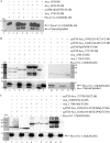
Five S2PsAv are site-2 metalloproteases. (A) Proteolytic activities of full-length Ava_1070, Ava_1730, and Ava_3438. E. coli cells bearing pZR632 (Ava_1730-F2-H6) (lane 3) or pZR633 (Ava_3438-F2-H6) (lane 6), pZR634 (Ava_1070-H6) (lane 9) alone or in combination with pZR327 [pro-σK(1–126)S20G-H6] (lanes 4, 7, and 8, respectively) were induced with IPTG for 3 h to express the indicated proteins; then whole-cell extracts were subjected to Western blot analysis using antibodies against pentahistidine. E. coli cells bearing pZR327 [pro-σK(1–126)S20G-H6] alone (lane 2) or in combination with pZR209 (cytTM-SpoIVFB-F2-H6) (lane 5) serve as negative and positive controls. (B) Proteolytic activities of five S2PsAv. E. coli cells bearing pZR632 (Ava_1730-F2-H6) (lane 3), pZR797 (Ava_4785-F2-H6) (lane 5), pZR796 (cytTM-Ava_1797-F2-H6) (lane 7), pZR845 [cytTM-Ava_3438(184–407)-F2-H6] (lane 11), or pZR874 [cytTM-Ava_1070(201–417)-F2-H6] (lane 13) alone or in combination with pZR327 [pro-σK(1–126)S20G-H6] (lanes 2, 4, 6, 10, and 12, respectively) were induced with IPTG for 3 h to express the indicated proteins; then whole-cell extracts were subjected to Western blot analysis using antibodies against FLAG (top panels) or pentahistidine (bottom panels). E. coli cells bearing pZR327 [pro-σK(1–126)S20G-H6] alone (lane 9) or in combination with pZR209 (cytTM-SpoIVFB-F2-H6) (lane 8) serve as negative and positive controls. (C) Proteolytic activities of the five S2PsAv were abolished by the E-to-Q substitution in the HEXXH motif, a hallmark of S2P metalloproteases. E. coli cells bearing pZR940 [cytTM-Ava_3438(184–407)E244Q-F2-H6] (lane 3), pZR922 [cytTM-Ava_1070(201–417)E266Q-F2-H6] (lane 5), pZR820 [Ava_1730(E18Q)-F2-H6] (lane 7), pZR819 [Ava_4785(E67Q)-F2-H6] (lane 9), or pZR817 [cytTM-Ava_1797(E63Q)-F2-H6] (lane 11) alone or in combination with pZR327 [pro-σK(1–126)S20G-H6] (lanes 2, 4, 6, 8, 10, respectively) were induced with IPTG for 3 h to express the indicated proteins; then whole-cell extracts were subjected to Western blot analysis using antibodies against FLAG (top panel) or pentahistidine (bottom panel). E. coli cells bearing pZR327 [pro-σK(1–126)S20G-H6] alone (lane 12) serve as a negative control.
Fig 5
Fig 5
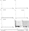
Mass spectrometric analysis for the peptides cleaved by S2PsAv. Q-TOF mass identification of peptides cleaved by Ava_4785-F2-H6 (A), cytTM-Ava_1797-F2-H6 (B), cytTM-SpoIVFB-F2-H6 (C), or cytTM-Ava_1797(E63Q)-F2-H6 (D) is shown. As a control, the m/z spectrum of intact pro-σK(1–126)S20G-H6 is also shown (E). (F) Purified pro-σK(1–126(S20G)-H6 or σK(21–126)-H6 detected by Coomassie staining (right panel) and Western blotting (left panel). Lanes 2 and 8, purified σK(21–126)-H6 cleaved by Ava_4785-F2-H6; lanes 3 and 9, purified peptide-H6 cleaved by cytTM-Ava_1797-F2-H6; lanes 4 and 10, purified peptide-H6 cleaved by cytTM-SpoIVFB-F2-H6; lanes 5 and 11, purified sample from cytTM-Ava_1797(E63Q)-F2-H6; lanes 6 and 12, purified pro-σK(1–126)S20G-H6. The calculated masses of σK(21–126)-H6 and pro-σK(21–126)S20G-H6 are 12,853 Da and 15,031 Da, respectively. (G) Partial amino acid sequence of pro-σK(1–126)S20G-H6 and σK(21–126)-H6. The cleavage site was mapped between residues Gly20 and Tyr21 by SpoIVFB. The italicized sequences represent transmembrane helix of pro-σK(1–126)S20G-H6.

Similar articles

Cited by

References

    1. Adams DG, Duggan PS. 1999. Tansley review no. 107. Heterocyst and akinete differentiation in cyanobacteria. New Phytol. 144:3–33
    1. Akiyama Y, Kanehara K, Ito K. 2004. RseP (YaeL), an Escherichia coli RIP protease, cleaves transmembrane sequences. EMBO J. 23:4434–4442 - PMC - PubMed
    1. Alba BM, Leeds JA, Onufryk C, Lu CZ, Gross CA. 2002. DegS and YaeL participate sequentially in the cleavage of RseA to activate the σE-dependent extracytoplasmic stress response. Genes Dev. 16:2156–2168 - PMC - PubMed
    1. Ali N, Knaüper V. 2007. Phorbol ester-induced shedding of the prostate cancer marker transmembrane protein with epidermal growth factor and two follistatin motifs 2 is mediated by the disintegrin and metalloproteinase-17. J. Biol. Chem. 282:37378–37388 - PubMed
    1. Blot G, et al. 2006. Luman, a new partner of HIV-1 TMgp41, interferes with Tat-mediated transcription of the HIV-1 LTR. J. Mol. Biol. 364:1034–1047 - PubMed

Publication types

MeSH terms

LinkOut - more resources