. 2013 Mar;101(3):809-18.
doi: 10.1002/jbm.a.34386. Epub 2012 Sep 8.

A novel suture-based method for efficient transplantation of stem cells

Affiliations

A novel suture-based method for efficient transplantation of stem cells

Jacques P Guyette et al. J Biomed Mater Res A. 2013 Mar.

Abstract

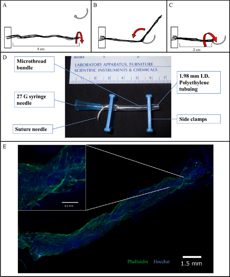
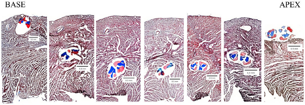
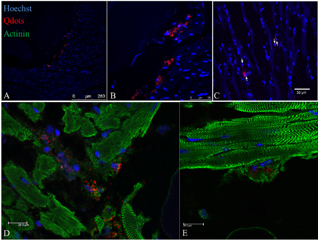
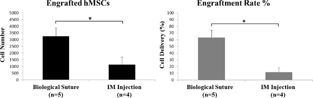
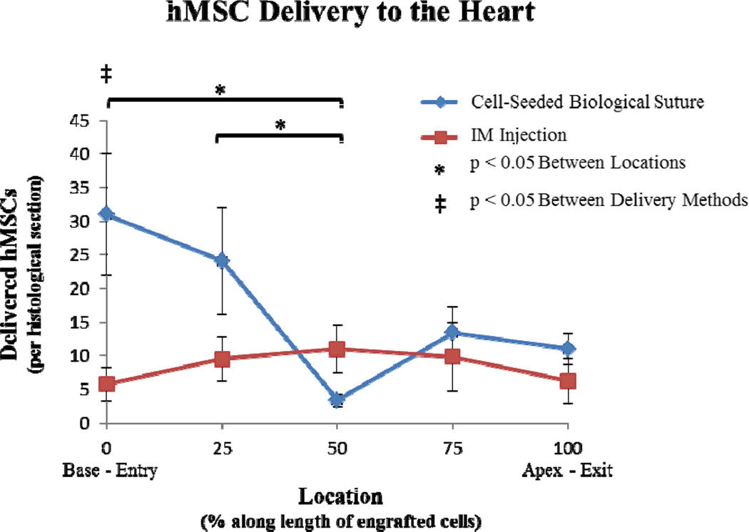
Advances in regenerative medicine have improved the potential of using cellular therapy for treating several diseases. However, the effectiveness of new cellular therapies is largely limited by low cell engraftment and inadequate localization. To improve on these limitations, we developed a novel delivery mechanism using cell-seeded biological sutures. We demonstrate the ability of cell-seeded biological sutures to efficiently implant human mesenchymal stem cells (hMSCs) to specific regions within the beating heart; a tissue known to have low cell retention and engraftment shortly after delivery. Cell-seeded biological sutures were developed by bundling discrete microthreads extruded from extracellular matrix proteins, attaching a surgical needle to the bundle and seeding the bundle with hMSCs. During cell preparation, hMSCs were loaded with quantum dot nanoparticles for cell tracking within the myocardium. Each biological suture contained an average of 5903 ± 1966 hMSCs/cm suture length. Delivery efficiency was evaluated by comparing cell-seeded biological suture implantation with intramyocardial (IM) cell injections (10,000 hMSCs in 35 μL) into the left ventricle of normal, noninfarcted rat hearts after 1 h. Delivery efficiency of hMSCs by biological sutures (63.6 ± 10.6%) was significantly higher than IM injection (11.8 ± 6.2%; p < 0.05). Cell-tracking analysis indicated suture-delivered hMSCs were found throughout the thickness of the ventricular myocardium: along the entire length of the biological suture track, localizing closely with native myocardium. These results suggest cell-seeded biological sutures can deliver cells to the heart more efficiently than conventional methods, demonstrating an effective delivery method for implanting cells in soft tissue.


Figures

Figure 1
Figure 1. Cell-seeded biological sutures
(A) Microthreads are anchored at one end and then twisted into a bundle. (B) The bundle is threaded through the eye of a 27 gauge needle and doubled over at the midpoint. (C) The thread is then twisted again to tighten the bundle, forming a biological suture. (D) The biological suture is placed in a bioreactor tube where the cell solution can be added via the needle. The bioreactor tube is then placed in a rotator and incubated for 24 hours. (E) A 2 cm length biological microthread bundle after 24 hours of seeding with hMSCs. (Inset) Nine 5× images merged; Hoechst-dyed nuclei are blue, Phalloidin stained f-actin filaments are green.
Figure 2
Figure 2. Cell-seeded biological suture implantation tracking
hMSC-seeded biological sutures were implanted from the base to the apex of the left ventricle. These images were used to determine the distance of the suture from the innermost section of the endocardium. Each section is 480 µm apart. (5× magnification, Masson’s Trichrome staining, collagen microthreads are blue, fibrin microthreads are pink).
Figure 3
Figure 3. Examples of quantum dot loaded hMSCs delivered to the rat heart
(A) Quantum dot loaded hMSCs injected into rat heart. (B) hMSCs delivered in close proximity to one of the biological microthreads that is part of the biological suture (20×). (C) hMSCs delivered in close proximity to biological microthread bundle (63×). (D) Quantum dot positive cells engrafted in the myocardium surrounding an implanted microthread. (E) Quantum dot positive cell in close proximity with a cardiac myofibers.
Figure 4
Figure 4. Biological sutures result in better cell engraftment than intramyocardial injection
(A) Total number of hMSCs engrafted in rat hearts, determined by counting QD-loaded cells in histological sections. (B) hMSC engraftment rate (%) was calculated based on the total number of cells engrafted in each heart and the starting cell number delivered. Asterisks indicate significance with a p-value less than 0.05.
Figure 5
Figure 5. Distribution of delivered hMSCs
Distributions of delivered hMSCs were compared between cell-seeded suture implantation and IM injection. The cell delivery areas were divided into 5 regions (0, 25, 50, 75, and 100%). For the cell-seeded sutures, 0% corresponded with the entry site and 100% corresponded with the exit site. For IM injections, 0% denoted the first QD-loaded cells identified at the base of the heart and 100% denoted the last QD-loaded cells found at the apex of the heart. Cells were found over 0.55 ± 0.06 cm for the biological suture group and 0.33 ± 0.13 cm for the IM injection group. For cell-seeded sutures, both the 0% and 25% regions contained significantly more cells than the 50% region. Comparing delivery methods at different locations, the cell-seeded suture modality delivered significantly more cells to the 0% entry-site region compared to IM injection.

References

    1. Beltrami AP, Barlucchi L, Torella D, Baker M, Limana F, Chimenti S, Kasahara H, Rota M, Musso E, Urbanek K, et al. Adult cardiac stem cells are multipotent and support myocardial regeneration. Cell. 2003;114(6):763–776. - PubMed
    1. Behfar A, Yamada S, Crespo-Diaz R, Nesbitt JJ, Rowe LA, Perez-Terzic C, Gaussin V, Homsy C, Bartunek J, Terzic A. Guided cardiopoiesis enhances therapeutic benefit of bone marrow human mesenchymal stem cells in chronic myocardial infarction. J Am Coll Cardiol. 2010;56(9):721–734. - PMC - PubMed
    1. He JQ, Ma Y, Lee Y, Thomson JA, Kamp TJ. Human embryonic stem cells develop into multiple types of cardiac myocytes: action potential characterization. Circ Res. 2003;93(1):32–39. - PubMed
    1. Kehat I, Kenyagin-Karsenti D, Snir M, Segev H, Amit M, Gepstein A, Livne E, Binah O, Itskovitz-Eldor J, Gepstein L. Human embryonic stem cells can differentiate into myocytes with structural and functional properties of cardiomyocytes. J Clin Invest. 2001;108(3):407–414. - PMC - PubMed
    1. Mauritz C, Schwanke K, Reppel M, Neef S, Katsirntaki K, Maier LS, Nguemo F, Menke S, Haustein M, Hescheler J, et al. Generation of functional murine cardiac myocytes from induced pluripotent stem cells. Circulation. 2008;118(5):507–517. - PubMed

Publication types

LinkOut - more resources