. 2012 Oct 19;287(43):36435-42.
doi: 10.1074/jbc.M112.397125. Epub 2012 Sep 7.

Snail represses the splicing regulator epithelial splicing regulatory protein 1 to promote epithelial-mesenchymal transition

Affiliations

Snail represses the splicing regulator epithelial splicing regulatory protein 1 to promote epithelial-mesenchymal transition

Lauren M Reinke et al. J Biol Chem. .

Abstract

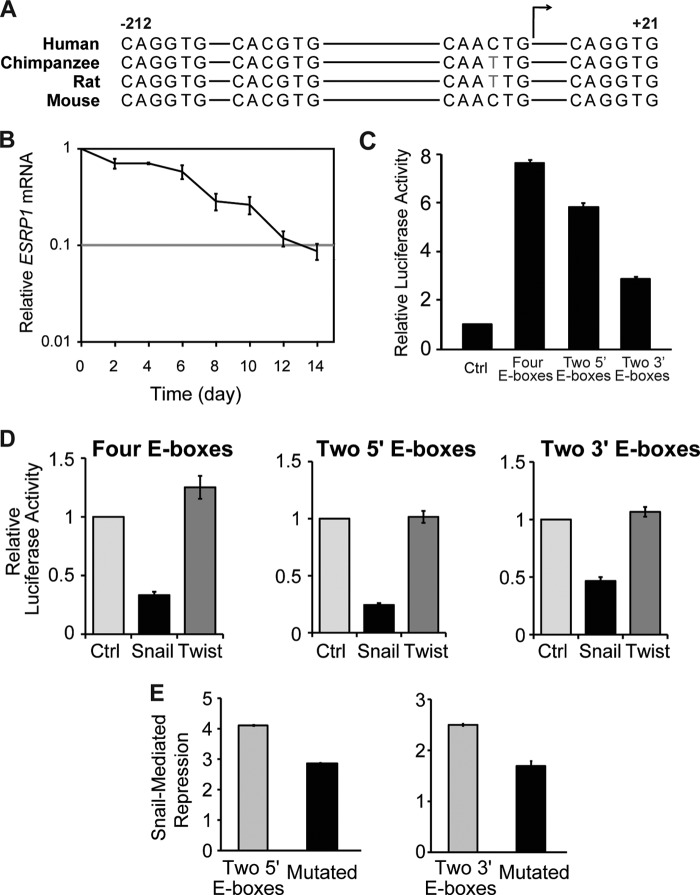
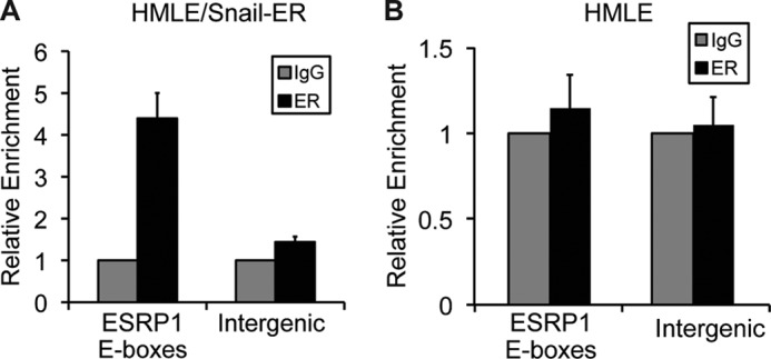
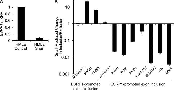
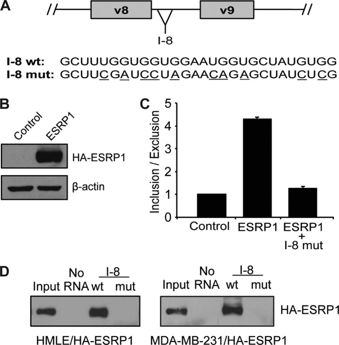
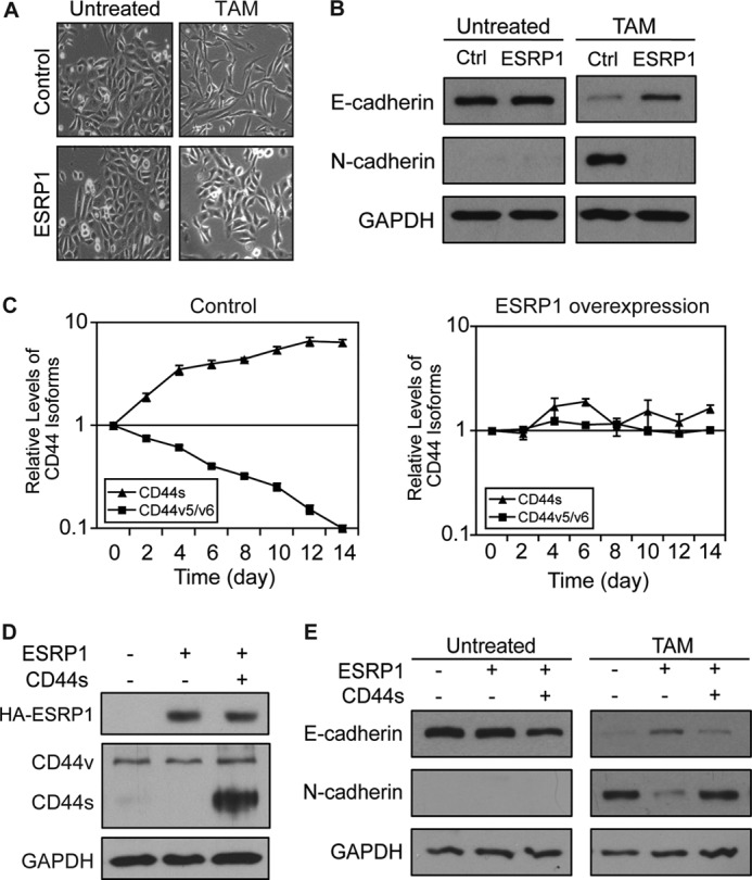
Epithelial-mesenchymal transition (EMT), a tightly regulated process that is critical for development, is frequently re-activated during cancer metastasis and recurrence. We reported previously that CD44 isoform switching is critical for EMT and showed that the splicing factor ESRP1 inhibits CD44 isoform switching during EMT. However, the mechanism by which ESRP1 is regulated during EMT has not been fully understood. Here we show that the transcription repressor Snail binds to E-boxes in the ESRP1 promoter, causing repression of the ESRP1 gene. Biochemically, we define the mechanism by which ESRP1 regulates CD44 alternative splicing: ESRP1 binds to the intronic region flanking a CD44 variable exon and causes increased variable exon inclusion. We further show that ectopically expressing ESRP1 inhibits Snail-induced EMT, suggesting that down-regulation of ESRP1 is required for function by Snail in EMT. Together, these data reveal how the transcription factor Snail mediates EMT through regulation of a splicing factor.


Figures

FIGURE 1.
FIGURE 1.
The transcriptional repressor Snail regulates ESRP1 expression. A, schematic of the ESRP1 promoter that contains four E-box sequences that are highly conserved among human, chimpanzee, rat, and mouse. The transcription start is indicated by a bent arrow. B, qRT-PCR analysis of ESRP1 levels in HMLE/Snail-ER cells during tamoxifen-induced EMT. Relative expression levels are normalized to TATA-binding protein at each time point, and the results are shown relative to day 0. Error bars indicate S.E.; n = 4. C, relative luciferase activity in HCT116 cells transfected with ESRP1 luciferase reporter constructs that include an E-box-containing fragment of the ESRP1 promoter upstream of luciferase. The ratios of Photinus to Renilla luciferase activities were normalized to cells transfected with a control luciferase reporter construct that lacks promoter and enhancer sequences. D, relative luciferase activities in HCT116 cells co-transfected with ESRP1 promoter luciferase reporter constructs and Snail. For each ESRP1 promoter construct, Photinus-to-Renilla luciferase activities were normalized to cells co-transfected with an empty vector control. E, relative luciferase activities in HCT116 cells co-transfected with a wild-type or mutant ESRP1 promoter luciferase reporter construct and Snail. C–E, bar graphs depict averages of at least three independent experiments; error bars indicate S.E.
FIGURE 2.
FIGURE 2.
Snail regulates ESRP1 expression by binding to E-box sequences located in the ESRP1 promoter. A and B, quantitative ChIP analysis of the relative occupancy of Snail at the ESRP1 promoter in HMLE/Snail-ER cells treated with tamoxifen for 6 days (A) and in HMLE cells (B). As a negative control, enrichment at an amplicon located in an intergenic region was also assayed. Additionally, ChIP assays were performed with preimmune IgG. Bar graphs show averages of three independent ChIP experiments.
FIGURE 3.
FIGURE 3.
Snail expression alters the ESRP1-associated splicing signature. A, qRT-PCR analysis of ESRP1 levels in control HMLE cells versus HMLE cells that stably express Snail (HMLE/Snail). B, qRT-PCR analysis indicating that Snail overexpression in HMLE cells altered the ESRP1-associated splicing signature. qRT-PCR assays were conducted using primer pairs that specifically detect exon inclusion or exclusion for each of the 11 ESRP1-regulated genes indicated. Exon inclusion-to-exclusion ratios were calculated for each gene in control and Snail-overexpressing cells. Bar graphs indicate -fold change in the inclusion-to-exclusion ratio in Snail-overexpressing cells compared with control cells. Error bars indicate S.E.; n = 4.
FIGURE 4.
FIGURE 4.
ESRP1 promotes CD44 exon inclusion by interacting with GU-rich elements in the CD44 pre-mRNA. A, schematic shows the location of the CD44 I-8 region. Sequences of the wild-type and mutated biotin-labeled RNA probes are shown; mutated nucleotides are underlined. B, immunoblot analysis shows overexpression of HA-tagged ESRP1 in 293 cells used in splicing reporter assays. C, quantitative RT-PCR analysis in 293 cells co-transfected with ESRP1 and a CD44v8 splicing reporter construct demonstrates that ESRP1-promoted inclusion of the CD44 v8 exon. When the putative ESRP1-binding motifs in the I-8 region are mutated, the ability of ESRP1 to promote inclusion of the CD44 v8 exon is diminished. Bar graphs depict averages of three independent experiments; error bars indicate S.E. D, immunoblot analysis using an HA antibody shows that ESRP1 interacted with the wild-type, but not the mutated, CD44 I-8 RNA probe (compare lane 3 with lane 4 in each panel). Assays were performed using whole cells lysates from HA-ESRP1-overexpressing HMLE cells (left panel) and HA-ESRP1-overexpressing MDA-MB-231 cells (right panel).
FIGURE 5.
FIGURE 5.
ESRP1 inhibits Snail-induced EMT by modulating CD44 alternative splicing. A, phase contrast images (10×) illustrate that ESRP1-overexpressing HMLE/Snail-ER cells exhibited impaired morphological changes following 14 days of tamoxifen (TAM) treatment compared with control cells. B, immunoblot analysis of E-cadherin and N-cadherin demonstrates that the control cells underwent a complete EMT, whereas the ESRP1-overexpressing cells maintained E-cadherin expression and failed to up-regulate N-cadherin following tamoxifen treatment. GAPDH served as a loading control. C, qRT-PCR analysis of mRNA levels of CD44 isoforms using primers that specifically detect either CD44s or CD44v that contain the v5 and v6 exons in TAM-treated control (left panel) or ESRP1-overexpressing (right panel) cells shows that ESRP1 overexpression prevents the CD44 isoform switch during EMT. Relative expression levels are normalized to TATA-binding protein at each time point, and the results are shown relative to day 0. Error bars indicate S.E.; n = 4. D, immunoblot analysis shows protein levels of HA-ESRP1 and CD44 isoforms in HMLE/Snail-ER cell lines that express ESRP1 or both ESRP1 and CD44s. GAPDH served as a loading control. E, immunoblot analysis of E-cadherin and N-cadherin shows that overexpression of the CD44s isoform in the ESRP1-overexpressing cells rescued the impaired EMT phenotype observed in the ESRP1-overexpressing cells. GAPDH served as a loading control.

References

    1. Moody S. E., Perez D., Pan T. C., Sarkisian C. J., Portocarrero C. P., Sterner C. J., Notorfrancesco K. L., Cardiff R. D., Chodosh L. A. (2005) The transcriptional repressor Snail promotes mammary tumor recurrence. Cancer Cell 8, 197–209 - PubMed
    1. Thiery J. P., Sleeman J. P. (2006) Complex networks orchestrate epithelial-mesenchymal transitions. Nat. Rev. Mol. Cell Biol. 7, 131–142 - PubMed
    1. Yang J., Mani S. A., Donaher J. L., Ramaswamy S., Itzykson R. A., Come C., Savagner P., Gitelman I., Richardson A., Weinberg R. A. (2004) Twist, a master regulator of morphogenesis, plays an essential role in tumor metastasis. Cell 117, 927–939 - PubMed
    1. Yang J., Weinberg R. A. (2008) Epithelial-mesenchymal transition: at the crossroads of development and tumor metastasis. Dev. Cell 14, 818–829 - PubMed
    1. Batlle E., Sancho E., Francí C., Domínguez D., Monfar M., Baulida J., García De Herreros A. (2000) The transcription factor Snail is a repressor of E-cadherin gene expression in epithelial tumor cells. Nat. Cell Biol. 2, 84–89 - PubMed

Publication types