. 2012 Dec;82(6):1136-49.
doi: 10.1124/mol.112.080507. Epub 2012 Sep 7.

Gβγ inhibits exocytosis via interaction with critical residues on soluble N-ethylmaleimide-sensitive factor attachment protein-25

Affiliations

Gβγ inhibits exocytosis via interaction with critical residues on soluble N-ethylmaleimide-sensitive factor attachment protein-25

Christopher A Wells et al. Mol Pharmacol. 2012 Dec.

Abstract

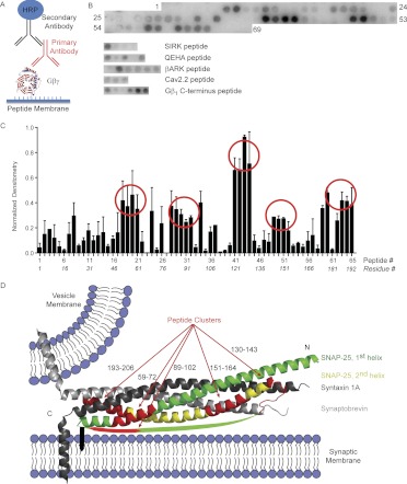
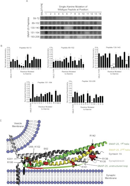
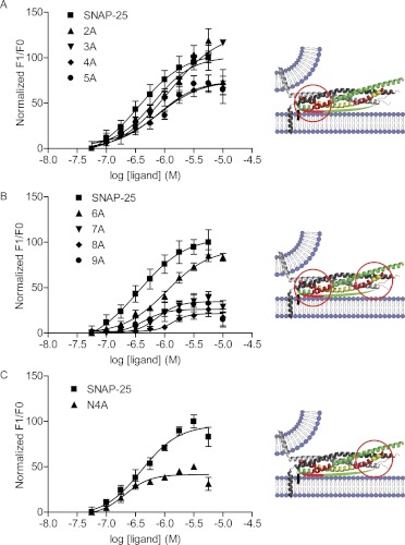
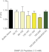
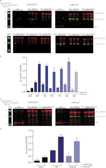
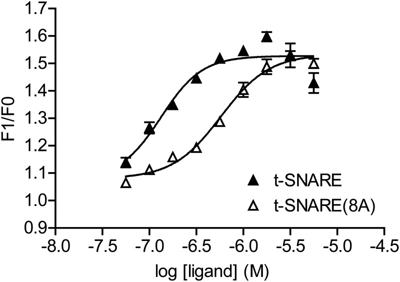
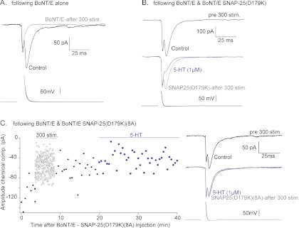
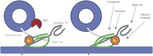
Spatial and temporal regulation of neurotransmitter release is a complex process accomplished by the exocytotic machinery working in tandem with numerous regulatory proteins. G-protein βγ dimers regulate the core process of exocytosis by interacting with the soluble N-ethylmaleimide-sensitive factor attachment protein receptor (SNARE) proteins soluble N-ethylmaleimide-sensitive factor attachment protein-25 (SNAP-25), syntaxin 1A, and synaptobrevin. Gβγ binding to ternary SNAREs overlaps with calcium-dependent binding of synaptotagmin, inhibiting synaptotagmin-1 binding and fusion of the synaptic vesicle. To further explore the binding sites of Gβγ on SNAP-25, peptides based on the sequence of SNAP-25 were screened for Gβγ binding. Peptides that bound Gβγ were subjected to alanine scanning mutagenesis to determine their relevance to the Gβγ-SNAP-25 interaction. Peptides from this screen were tested in protein-protein interaction assays for their ability to modulate the interaction of Gβγ with SNAP-25. A peptide from the C terminus, residues 193 to 206, significantly inhibited the interaction. In addition, Ala mutants of SNAP-25 residues from the C terminus of SNAP-25, as well as from the amino-terminal region decreased binding to Gβ₁γ₁. When SNAP-25 with eight residues mutated to alanine was assembled with syntaxin 1A, there was significantly reduced affinity of this mutated t-SNARE for Gβγ, but it still interacted with synaptotagmin-1 in a Ca²⁺ -dependent manner and reconstituted evoked exocytosis in botulinum neurotoxin E-treated neurons. However, the mutant SNAP-25 could no longer support 5-hydroxytryptamine-mediated inhibition of exocytosis.


Figures

Fig. 1.
Fig. 1.
Screening of SNAP-25 peptides for interaction with Gβ1γ1. A, the basic premise of the screening is a far-Western. The peptide synthesizer creates peptides on a derivatized membrane. With appropriate washes between steps, the membrane and peptides are sequentially exposed to Gβ1γ1, the primary antibody for Gβ, and horseradish peroxidase (HRP)-conjugated secondary antibody. B, representative image of a membrane exposed to Gβ1γ1. Numbering reflects spots with successive peptides 1 to 65. Spots 66 to 69 were left without peptide synthesized on them as negative controls. Shown separately are the peptide spots derived from the sequences for the SIRK peptide, QEHA peptide, βARK peptide, Gβγ binding domain of the calcium channel CaV2.2, and C terminus of Gβ1. C, densitometry was performed on the three membranes using ImageJ analysis of the image. Each membrane was normalized to the most intense spot on the membrane. The average of the three membranes was plotted for each set of 65 peptides that span the full length of SNAP-25. The x-axis reflects both the peptide number according to B as well as the residue number of the first residue in each respective peptide. Circles reflect clusters of peptides with the highest densitometric signal. D, representative sequences of the clusters of SNAP-25 peptides that were found are shown in red mapped onto the representation of the X-ray crystal structure of the core SNARE motifs (PDB 1SFC). The colors signify the following: light gray; synaptobrevin; dark gray, syntaxin 1A; green, first SNAP-25 helix; yellow, second SNAP-25 helix. Syntaxin 1A and synaptobrevin each have a transmembrane domain shown as α-helices. The black bar inserted into the membrane represents the palmitoylation sites on SNAP-25. The green arc represents the nonstructured sequence between the two α-helices of SNAP-25. This arc includes one of the peptide clusters (red) near the palmitoylation site. N signifies the N-terminal end of the helices within the SNARE complex; C signifies the C-terminal end of the helices within the SNARE complex.
Fig. 2.
Fig. 2.
Alanine mutagenesis screening of SNAP-25 peptides that bind Gβγ. A, representative image of the alanine screening for SNAP-25 peptides synthesized on a membrane. Five peptides are identified by their sequence number shown on the left. The first spot of each row contains wild-type peptide. The next 14 spots to the right are mutant peptides with a single alanine replacement of the residue at position 1, 2, 3. …, 14 for each wild-type peptide. B, densitometry was performed across three separate membranes for each respective peptide and its series of mutants. Means ± S.D. from the three membranes are shown for the five peptides in A (Student's t test; *, p < 0.05; **, p < 0.01; ***, p < 0.001). C, The residues (spheres, red) that had significantly reduced Gβγ binding when mutated to alanine are mapped onto the X-ray crystal structure of ternary snare (PDB 1SFC). The colors signify the following: dark gray, syntaxin 1A; light gray, synaptobrevin; green, first SNAP-25 helix; yellow, second SNAP-25 helix; green cartoon arc, unstructured domain between the two SNAP-25 α-helices.
Fig. 3.
Fig. 3.
Binding of SNAP-25 and its alanine mutants to MIANS-labeled Gβγ. A, a fixed concentration of MIANS-Gβ1γ1 (20 nM) was exposed to increasing concentrations of SNAP-25 with resulting increase in fluorescence. n = 4. F1/F0 is the ratio of fluorescence of Gβ1γ1 measured in the presence of SNAP-25 over the fluorescence of Gβ1γ1 in the absence of SNAP-25. The fluorescence was corrected for any intrinsic fluorescence of SNAP-25 at the various concentrations. Finally, all of the curves were normalized to the highest fluorescence achieved by binding of wild-type SNAP-25 to Gβ1γ1. A, dose-response curves for wild-type SNAP-25, SNAP-25 (2A), SNAP-25 (3A), SNAP-25 (4A), and SNAP-25 (5A). To the right of the dose-response curves is a cartoon modified from Fig. 2. The red circle denotes the area on the SNARE complex where these mutated residues are located together in the C terminus. B, the remaining alanine mutants of SNAP-25 were tested for binding to MIANS-Gβ1γ1. Compared with wild-type SNAP-25, increasing numbers of mutations resulted in decreased EC50 and then decreased maximum fluorescence enhancement of MIANS-Gβ1γ1 (A and B). C, a SNAP-25 mutant with residues in the amino-terminal region (R135A, R136A, R142A, and R161A) of the SNAP-25 protein termed N4A. When exposed to fluorescently labeled Gβ1γ1, this mutant (N4A) had a decreased maximal fluorescence, and its EC50 was 0.20 μM compared with 0.35 μM. The cartoon in the right portion of C shows the region with N-terminal mutated residues.
Fig. 4.
Fig. 4.
Inhibition of Gβγ-SNAP-25 binding by SNAP-25 peptides. Each peptide (1.5 mM) was added to the fluorescence assay detecting the interaction of 20 nM MIANS-Gβ1γ1with 0.3 μM SNAP-25. The C-terminal peptide (193–206) was the only peptide to significantly decrease the fluorescence enhancement (Student's t test, p < 0.01; n = 3). When the residues Arg198 and Lys201 were changed to alanine in that peptide, the peptide was no longer effective at reducing fluorescence enhancement by SNAP-25 (Student's t test, p > 0.05; n = 3).
Fig. 5.
Fig. 5.
Binding of SNAP-25 mutants to synaptotagmin-1 by GST pulldowns. A, GST, GST-SNAP-25 wild-type, and GST mutant SNAP-25 (6A) through SNAP-25 (9A) on glutathione beads were exposed to synaptotagmin-1 for 1 h in either 1 mM calcium or 2 mM EGTA, washed, and then eluted with sample buffer. Representative blots were imaged with Odyssey for simultaneous quantitation of synaptotagmin-1 (green) and GST (red) signal intensity. B, the ratio of normalized synaptotagmin-1: GST signals were averaged over three samples in the two conditions. The results are shown in the bar graph (**, p < 0.01 compared with WT in 2 mM EGTA; one-way analysis of variance, Tukey's multiple comparison post-test). C, GST, GST-SNAP-25 wild-type, and GST-fused mutant SNAP-25 (R161A) on glutathione beads were exposed to synaptotagmin-1 for 1 h in either 1 mM calcium or 2 mM EGTA, washed, and then eluted with sample buffer. Representative blots were imaged with Odyssey for simultaneous quantitation of synaptotagmin-1 (green) and GST (red) signal intensity. D, the ratio of normalized synaptotagmin-1:GST signals were averaged over three samples in the two conditions. The results are shown in the bar graph (**, p < 0.01 compared with WT in 2 mM EGTA; Student's t test). IB, immunoblot; LMW, low molecular weight.
Fig. 6.
Fig. 6.
Binding of wild-type t-SNARE and SNAP-25 (8A) t-SNARE to MIANS-labeled Gβγ. A fixed concentration of MIANS-Gβ1γ1 (20 nM) was exposed to increasing concentrations of wild-type t-SNARE with a resulting increase in fluorescence (▴, n = 4). The EC50 for t-SNARE binding to MIANS-Gβ1γ1 was 0.13 μM (95% CI 0.07–0.26 μM). Likewise, the t-SNARE complex of syntaxin 1A with SNAP-25 (8A) was exposed to MIANS-Gβ1γ1 with the resulting increase in fluorescence shown in the figure (▵, n = 4). The EC50 for this complex binding to Gβ1γ1 is 0.58 μM (95% CI, 0.47–0.70 μM).
Fig. 7.
Fig. 7.
Effect of SNAP-25 (8A) on presynaptic inhibition in lamprey with 5-HT. Paired cell recordings were made between lamprey giant reticulospinal axons and postsynaptic ventral horn target neurons. Each recording shown is the mean of at least 10 sequential responses. Overlaid presynaptic action potentials are shown below. A, in a recording in which BoNt/E was included in the presynaptic microelectrode, pressure injection of BoNt/E toxin left synaptic transmission intact (black). A period of 300 stimuli (1Hz) left no remaining chemical EPSC (early component is electrical) after loss of primed toxin-resistant vesicles. B, a similar recording in which BoNt/E and SNAP-25 (D179) were included in the presynaptic electrode. A period of 300 stimuli (1Hz) reduced but did not eliminate the EPSC (gray). Addition of 5-HT (1 μM) substantially reduced this remaining response (blue). C, with BoNt/E and SNAP-25 (D179) (8A) included in the presynaptic pipette. The graph shows peak chemical EPSC amplitudes recorded against time before (●) during (○) and after (●) 300 stimuli at 1Hz. Addition of 5-HT (1 μM, blue) failed to inhibit the synaptic response. EPSC examples are means of 10 from before (black), after 300 stimuli (1 Hz, gray), and after addition of 5-HT (1 μM, blue).
Fig. 8.
Fig. 8.
Gβγ-SNARE binding model. Based on the results of this study, not only does Gβγ appear to bind at or near the C terminus of SNAP-25, but also there are additional residues distal to the membrane-approximated portion of SNAP-25. Taken in the context of ternary SNARE and its proposed position at a docked synaptic vesicle, a single Gβγ dimer activated by a Gi/o-coupled GPCR that is bound to the C terminus of SNAP-25 would not be able to bind the distal portion of the SNARE complex at the same time. The additional residues appear to have implications in calcium-independent binding of synaptotagmin, but they may also have importance for Gβγ modulation of other interactions with SNARE proteins. These could include calcium channels, tomosyn, complexin, and Munc18.

Similar articles

Cited by

References

    1. Betke KM, Wells CA, Hamm HE. (2012) GPCR mediated regulation of synaptic transmission. Prog Neurobiol 96:304–321 - PMC - PubMed
    1. Binz T, Blasi J, Yamasaki S, Baumeister A, Link E, Südhof TC, Jahn R, Niemann H. (1994) Proteolysis of SNAP-25 by types E and A botulinal neurotoxins. J Biol Chem 269:1617–1620 - PubMed
    1. Blackmer T, Larsen EC, Bartleson C, Kowalchyk JA, Yoon EJ, Preininger AM, Alford S, Hamm HE, Martin TF. (2005) G protein βγ directly regulates SNARE protein fusion machinery for secretory granule exocytosis. Nat Neurosci 8:421–425 - PubMed
    1. Blackmer T, Larsen EC, Takahashi M, Martin TF, Alford S, Hamm HE. (2001) G protein βγ subunit-mediated presynaptic inhibition: regulation of exocytotic fusion downstream of Ca2+ entry. Science 292:293–297 - PubMed
    1. Brose N, Petrenko AG, Südhof TC, Jahn R. (1992) Synaptotagmin—a calcium sensor on the synaptic vesicle surface. Science 256(5059):1021–1025 - PubMed

Publication types

MeSH terms

LinkOut - more resources