. 2012 Nov 1;40(20):10203-14.
doi: 10.1093/nar/gks845. Epub 2012 Sep 8.

Excision of 5-hydroxymethyluracil and 5-carboxylcytosine by the thymine DNA glycosylase domain: its structural basis and implications for active DNA demethylation

Affiliations

Excision of 5-hydroxymethyluracil and 5-carboxylcytosine by the thymine DNA glycosylase domain: its structural basis and implications for active DNA demethylation

Hideharu Hashimoto et al. Nucleic Acids Res. .

Abstract

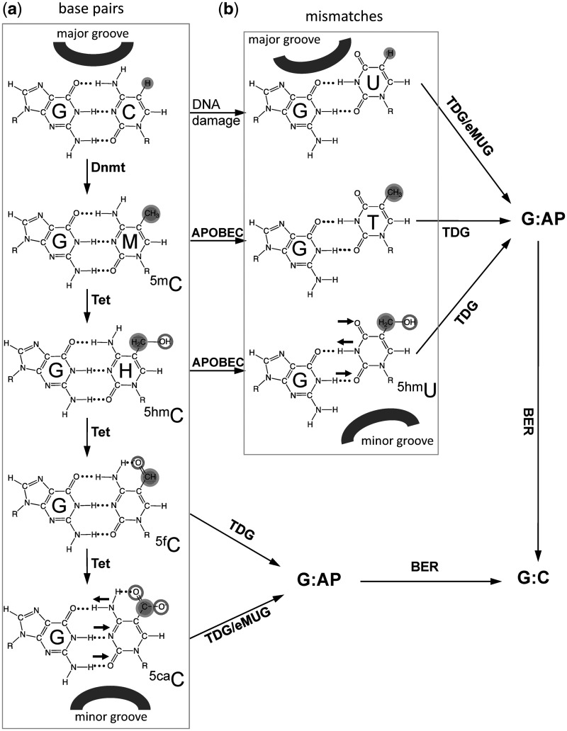
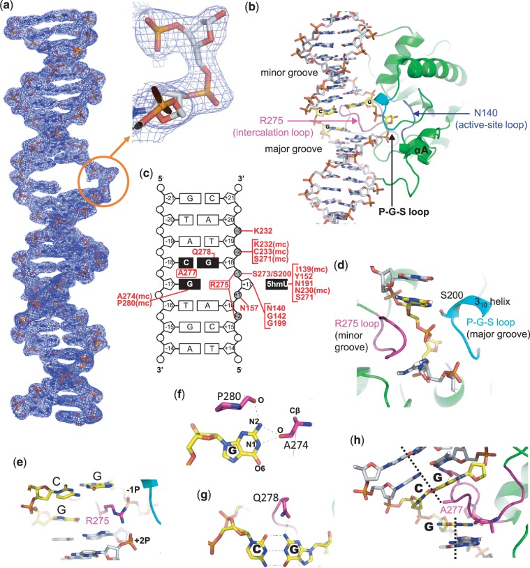
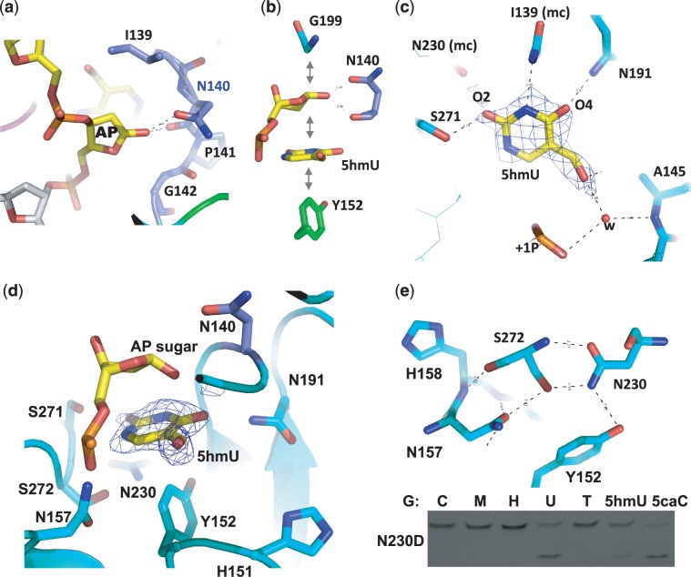
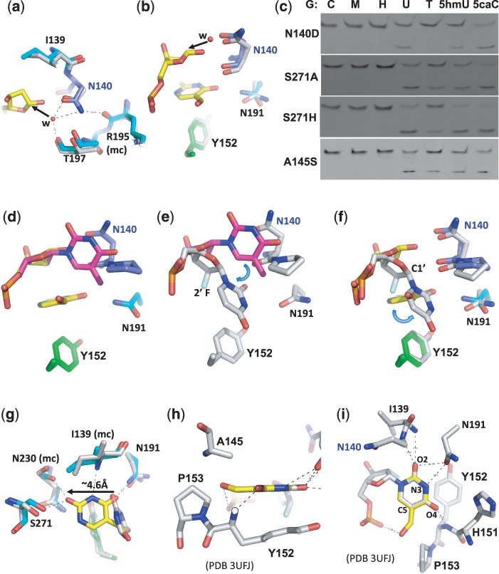
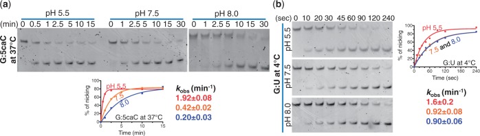
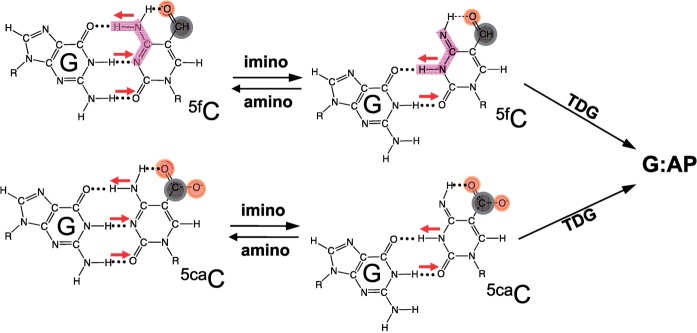
The mammalian thymine DNA glycosylase (TDG) is implicated in active DNA demethylation via the base excision repair pathway. TDG excises the mismatched base from G:X mismatches, where X is uracil, thymine or 5-hydroxymethyluracil (5hmU). These are, respectively, the deamination products of cytosine, 5-methylcytosine (5mC) and 5-hydroxymethylcytosine (5hmC). In addition, TDG excises the Tet protein products 5-formylcytosine (5fC) and 5-carboxylcytosine (5caC) but not 5hmC and 5mC, when paired with a guanine. Here we present a post-reactive complex structure of the human TDG domain with a 28-base pair DNA containing a G:5hmU mismatch. TDG flips the target nucleotide from the double-stranded DNA, cleaves the N-glycosidic bond and leaves the C1' hydrolyzed abasic sugar in the flipped state. The cleaved 5hmU base remains in a binding pocket of the enzyme. TDG allows hydrogen-bonding interactions to both T/U-based (5hmU) and C-based (5caC) modifications, thus enabling its activity on a wider range of substrates. We further show that the TDG catalytic domain has higher activity for 5caC at a lower pH (5.5) as compared to the activities at higher pH (7.5 and 8.0) and that the structurally related Escherichia coli mismatch uracil glycosylase can excise 5caC as well. We discuss several possible mechanisms, including the amino-imino tautomerization of the substrate base that may explain how TDG discriminates against 5hmC and 5mC.


Figures

Figure 1.
Figure 1.
A putative pathway of DNA demethylation involving DNA methylation by DNMTs, hydroxylation by Tet proteins, deamination by members of APOBEC superfamily, and base excision by TDG linked to base excision repair (BER). In addition, eMUG can excise 5caC as well (see Figure 2). DNA major groove and minor groove sides are indicated. Horizontal small arrows indicate the hydrogen bond donors and acceptors for 5caC and 5hmU bases. (a) C, 5mC and its oxidized derivatives (5hmC, 5fC and 5caC) form base pairs with an opposite G. (b) Deamination-linked mismatches.
Figure 2.
Figure 2.
Base excision and binding activities of TDG catalytic domain in the context of a double-stranded CpG dinucleotide. (a) Double-stranded 32-bp oligonucleotides bearing a single CpG dinucleotide were incubated with equal amount of the glycosylase domain of TDG or its noncatalytic mutant N140A at 37°C for 30 min. The oligonucleotide was labeled with FAM on the top strand, and the modification status was indicated (M = 5mC and H = 5hmC). The products of the reactions were separated on a denaturing polyacrylamide gel, and the FAM-labeled strand was excited by UV and photographed. (b) DNA binding assays were performed by incubating 0.5 µM FAM-labeled oligonucleotides with 1 µM of TDG at 37°C for 15 min. (c) Pairwise sequence alignment of human TDG domain (top line) and E. coli MUG (bottom line). Secondary structural elements are shown above or below the aligned sequences. White-on-black residues are invariant between the two sequences examined, while gray-highlighted positions are conserved (R and K, E and D, Q and N, T and S, F, Y and W, V, I, L and M, and G and P). Positions highlighted by * are active site residues responsible for catalysis (Asn140) and/or proposed for substrate base recognition (only two of them, Asn140 and Asn157, are invariant between human TDG and E. coli MUG). (d) eMUG is active on G:U and G:5caC substrates (top panel). Reactions were performed at room temperature (approximately 22°C) for 30 min with [EeMUG] = [SDNA] = 5 µM. The kinetic activities of eMUG on G:U substrates at 4°C (bottom left panel) and G:5caC at room temperature (approximately 22°C) (bottom right panel) were measured under single turnover condition ([EeMUG] = 2.5 µM and [SDNA] = 0.25 µM) at three different pH values (5.5 in red, 7.5 in orange and 8.0 in blue curves).
Figure 3.
Figure 3.
Structure of TDG in complex with G:5hmU containing DNA. (a) 2Fo-Fc electron density, contoured at 1σ above the mean, for the entire 28-bp DNA used in the TDG structure determination. The insert is an enlarged abasic sugar with a hydrolyzed C1′. (b) Overall structure of the WT TDG complex. DNA is in stick model, and TDG is in ribbon model. The Arg275-containing loop is colored in magenta, the P-G-S loop is in cyan and catalytic loop in blue. (c) Summary of the TDG–DNA interactions: mc, main-chain-atom-mediated contacts; black boxes represent the CpG sequence and extrahelical 5hmU. (d) The Arg275-containing intercalation loop (in magenta) and the P-G-S loop (in cyan) approach the modified DNA strand from opposite directions. (e) Arg275 penetrates into the DNA helix from the minor groove. (f) The three hydrogen bonds formed with the intrahelical orphaned guanine. (g) Gln278 forms a hydrogen bond from the minor groove side with Gua of the adjoining G:C base pair. (h) Ala277 intercalates between the central Cyt and Gua of the unmodified strand.
Figure 4.
Figure 4.
The binding of 5hmU in the active site. (a) In the WT structure, the N-glycosidic bond of the extrahelical nucleotide is cleaved, and a hydroxyl oxygen atom has been attached to the C1′ of the sugar ring (see Figure 3a inset). (b) Favorable face-to-face and edge-to-face hydrophobic interactions between the sugar, the cleaved 5hmU base and Tyr152. (c) Omit electron density, contoured at 3.5σ above the mean, is shown for omitting 5hmU. The hydrogen bond interactions (dashed lines) with the polar atoms of 5hmU are within 3.0 Å distance cutoff: mc, main-chain-atom-mediated contacts. (d) The 5hmU-binding pocket is rich in polar atoms. (e) A hydrogen-bonding network involves both side chain and main chain atoms of depicted residues. The activity of N230D is shown.
Figure 5.
Figure 5.
Comparison of post- and pre-reactive complex structures. (a, b) Superimposition of the post-reactive complex (in color) and the pre-reactive complex [in gray; PDB 3UFJ (20)] shows a putative nucleophilic water molecule, held in position by the side chain carbonyl oxygen atom of Asn140 in the pre-reactive complex, attacks the C1′ from the opposite position of the leaving base, generating the C1′-hydrolyzed abasic sugar as shown in the post-reactive complex (in yellow). (c) The activities of TDG mutants N140D, S271A, S271H and A145S. (d, e) Superimposition of a normal intrahelical thymine (colored in magenta) onto the post-reactive complex (panel d) or the pre-reactive complex (panel e) suggests a bent relative to the sugar ring and a rotation around the glycosidic bond. (f, g) Superimposition of the post-reactive complex (in color) and the pre-reactive complex (in gray) suggests the base undergoes another rotation after cleavage (panel f) and moves towards Ser271 (panel g). (h, i) Superimposition of a 5hmU base (in yellow) onto the flipped uracil in the pre-reactive complex (PDB 3UFJ).
Figure 6.
Figure 6.
The activity of TDG catalytic domain as a function of pH. The activity of TDG catalytic domain on (a) G:5caC at 37°C and (b) G:U substrates at 4°C under single turnover condition ([ETDG] = 2.5 µM and [SDNA] = 0.25 µM) at three different pH values. The reaction on G:U substrate was measured at 4°C because the reaction was complete within 1 min at 37°C or room temperature (∼22°C) (data not shown).
Figure 7.
Figure 7.
Amino-imino tautomerization. 5fC and 5caC exhibit an intramolecular hydrogen bond that could shift the amino/imino equilibrium toward the imino tautomeric form which would then base pair with guanine in a mismatch-like wobble pattern.

Similar articles

Cited by

References

    1. Globisch D, Munzel M, Muller M, Michalakis S, Wagner M, Koch S, Bruckl T, Biel M, Carell T. Tissue distribution of 5-hydroxymethylcytosine and search for active demethylation intermediates. PLoS One. 2010;5:e15367. - PMC - PubMed
    1. Tahiliani M, Koh KP, Shen Y, Pastor WA, Bandukwala H, Brudno Y, Agarwal S, Iyer LM, Liu DR, Aravind L, et al. Conversion of 5-methylcytosine to 5-hydroxymethylcytosine in mammalian DNA by MLL partner TET1. Science. 2009;324:930–935. - PMC - PubMed
    1. Kriaucionis S, Heintz N. The nuclear DNA base 5-hydroxymethylcytosine is present in Purkinje neurons and the brain. Science. 2009;324:929–930. - PMC - PubMed
    1. Booth MJ, Branco MR, Ficz G, Oxley D, Krueger F, Reik W, Balasubramanian S. Quantitative sequencing of 5-methylcytosine and 5-hydroxymethylcytosine at single-base resolution. Science. 2012;336:934–937. - PubMed
    1. Ito S, Shen L, Dai Q, Wu SC, Collins LB, Swenberg JA, He C, Zhang Y. Tet proteins can convert 5-methylcytosine to 5-formylcytosine and 5-carboxylcytosine. Science. 2011;333:1300–1303. - PMC - PubMed

Publication types

Associated data