. 2012;7(9):e44266.
doi: 10.1371/journal.pone.0044266. Epub 2012 Sep 4.

CD86 and IL-12p70 are key players for T helper 1 polarization and natural killer cell activation by Toll-like receptor-induced dendritic cells

Affiliations

CD86 and IL-12p70 are key players for T helper 1 polarization and natural killer cell activation by Toll-like receptor-induced dendritic cells

Felix S Lichtenegger et al. PLoS One. 2012.

Abstract

Background: Dendritic cells (DCs) determine the activation and polarization of T cells via expression of costimulatory molecules and secretion of cytokines. The function of DCs derived from monocytes ex vivo strongly depends on the composition of the maturation cocktail used.

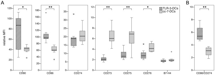
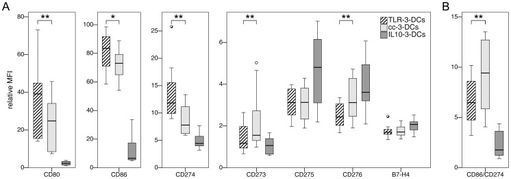
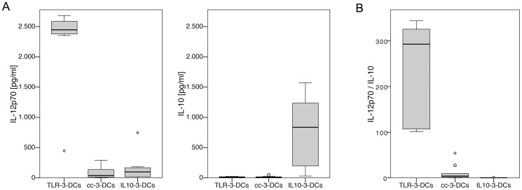
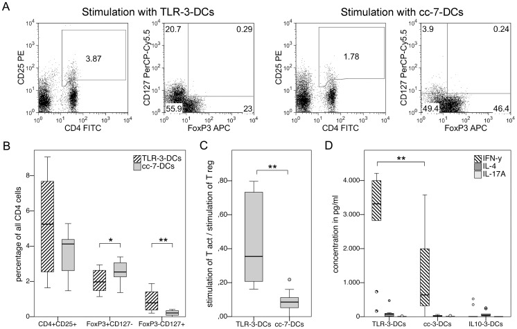
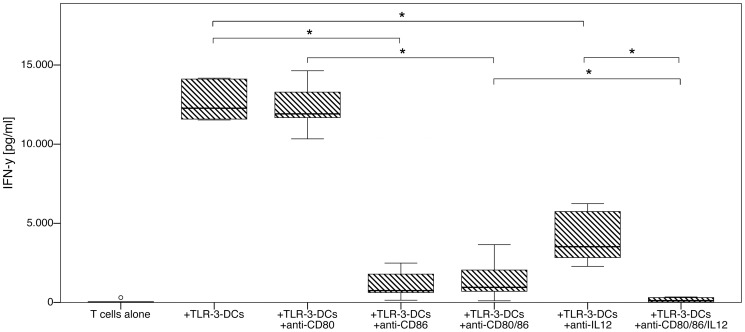
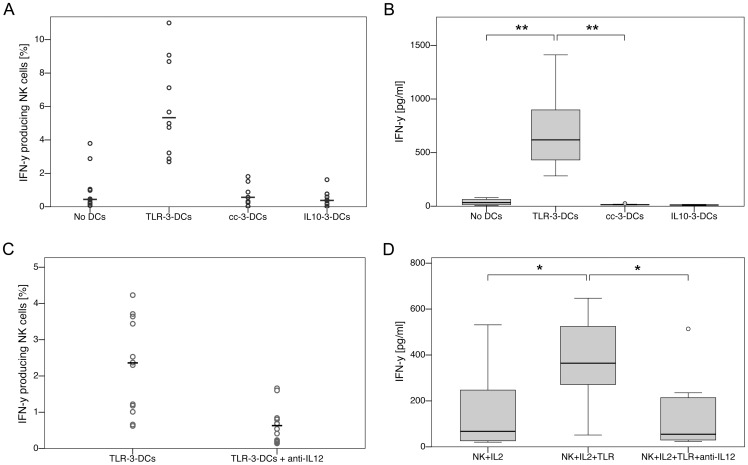
Methodology/principal findings: We analyzed the effect of costimulatory molecule expression and cytokine secretion by DCs on T and natural killer (NK) cell activation by conducting a head-to-head comparison of a Toll-like receptor (TLR) agonist-based cocktail with the standard combination of proinflammatory cytokines or IL-10 alone. We could show that TLR-induced DCs are characterized by a predominance of costimulatory over coinhibitory molecules and by high secretion of IL-12p70, but not IL-10. Functionally, these signals translated into an increase in IFN-γ secreting Th1 cells and a decrease in regulatory T cells. T cell activation and polarization were dependent on IL-12p70 and CD86, but remarkably not on CD80 signaling. By means of IL-12p70 secretion, only TLR-induced DCs activated NK cells.

Conclusions/significance: TLR-matured DCs are highly suitable for application in immunotherapeutic strategies that rely on strong type 1 polarization and NK cell activation. Their effects particularly depend on high CD86 expression and IL-12p70 secretion.


Conflict of interest statement

Competing Interests: Dolores J. Schendel declares a competing interest through pending patents submitted by the Helmholtz Zentrum München, German Research Center for Environmental Health (Munich, Germany). These pending patents relate to “Composition for the Preparation of Mature Dendritic Cells”. This does not alter the authors' adherence to all the PLoS ONE policies on sharing data and materials.

Figures

Figure 1
Figure 1. Costimulatory profiles of TLR-3-DCs and cc-7-DCs.
TLR-3-DCs and cc-7-DCs were generated from peripheral blood of healthy donors, and expression of various costimulatory markers was analyzed by flow cytometry. *, p<0.05; **, p<0.01. (A) Expression of the cell surface antigens on both DC populations (n = 7 for B7-H4, n = 8 for CD276, n = 10 for all other markers). (B) Comparison of the CD86/CD274 ratio for TLR-3-DCs and cc-7-DCs (n = 10).
Figure 2
Figure 2. Costimulatory profiles of TLR-3-DCs, cc-3-DCs and IL10-3-DCs.
TLR-3-DCs, cc-3-DCs and IL10-3-DCs were generated from peripheral blood of healthy donors, and expression of various costimulatory markers was analyzed by flow cytometry. Differences between TLR-3-DCs and cc-3-DCs were tested. *, p<0.05; **, p<0.01. (A) Expression of the cell surface antigens on all three DC populations (n = 10). (B) Comparison of the CD86/CD274 ratio for all three DC populations (n = 10).
Figure 3
Figure 3. Cytokine secretion patterns of TLR-3-DCs, cc-3-DCs and IL10-3-DCs.
DCs generated from peripheral blood of healthy donors were analyzed for their cytokine secretion patterns (n = 9). (A) Mature DCs were cocultured with CD40L expressing mouse fibroblasts for 24 hours, the concentration of IL-12p70 and IL-10 in the supernatants was measured by CBA, and the difference to the basal secretion of the same cell populations without CD40 ligation was calculated. (B) Comparison of the IL-12p70/IL-10 ratio for TLR-3-DCs, cc-3-DCs and IL10-3-DCs.
Figure 4
Figure 4. Preferential induction of activated, IFN-γ secreting T cells by TLR-3-DCs.
(A to C) TLR-3-DCs and cc-7-DCs generated from peripheral blood of healthy donors were cocultured with autologous monocyte-depleted (non-adherent) PBMCs for 24 hours, and antigen-independent stimulatory capacity of the DCs on regulatory and activated T cells was quantified using flow cytometry with the markers CD4, CD25, CD127 and FoxP3. *, p<0.05; **, p<0.01. (A) Data from one representative donor. Gating strategies used to determine different T cell subsets. (B) Capacity of TLR-3-DCs and cc-7-DCs to stimulate CD4+CD25+ T cells and their regulatory (FoxP3+CD127-) and non-regulatory activated (FoxP3-CD127+) subsets. Differences between stimulated and unstimulated cells are shown (n = 10). (C) Ratio of activated and regulatory T cells induced by coculture. (D) TLR-3-DCs, cc-3-DCs and IL10-3-DCs were cocultured with CD3-selected autologous T cells for 4 days. Supernatants were analyzed for secretion of IFN-γ, IL-4 and IL-17A by CBA. Differences between stimulated and unstimulated cells are shown (n = 10).
Figure 5
Figure 5. Effect of costimulation and IL-12p70 blockade on Th1 polarization by TLR-3-DCs.
TLR-3-DCs generated from peripheral blood of 6 healthy donors were cocultured with CD3-selected autologous T cells for 4 days either alone or with addition of various combinations of blocking antibodies. Supernatants were analyzed for secretion of IFN-γ by ELISA. T cells without DC stimulation were used as a negative control. Box-and-whisker plots for the different conditions are shown. *, p<0.05.
Figure 6
Figure 6. IL-12p70 dependency of NK cell activation by TLR-3-DCs.
(A/B) DCs generated from peripheral blood of 10 healthy donors were cocultured with autologous monocyte-depleted (non-adherent) PBMCs for 24 hours with addition of IL-2. Activation of NK cells was analyzed by intracellular IFN-γ staining of CD3CD56+ cells (A; circles represent single experiments, the mean is displayed as horizontal line) and by ELISA measurement of IFN-γ in the supernatant (B; box-and-whisker plots). (C/D) In a similar set of experiments, cocultures of TLR-3-DCs and autologous monocyte-depleted PBMCs were compared with or without addition of IL-12p70 blocking antibody. Activation of NK cells was analyzed by intracellular IFN-γ staining of CD3CD56+ cells (C; n = 13) and by ELISA measurement of IFN-γ in the supernatant (D; n = 8). *, p<0.05; **, p<0.01.

Similar articles

Cited by

References

    1. Steinman RM, Cohn ZA (1973) Identification of a novel cell type in peripheral lymphoid organs of mice. I. Morphology, quantitation, tissue distribution. J Exp Med 137: 1142–1162. - PMC - PubMed
    1. Banchereau J, Steinman RM (1998) Dendritic cells and the control of immunity. Nature 392: 245–252. - PubMed
    1. Schuler G, Schuler-Thurner B, Steinman RM (2003) The use of dendritic cells in cancer immunotherapy. Curr Opin Immunol 15: 138–147. - PubMed
    1. Banchereau J, Palucka AK (2005) Dendritic cells as therapeutic vaccines against cancer. Nat Rev Immunol 5: 296–306. - PubMed
    1. Melief CJ (2008) Cancer immunotherapy by dendritic cells. Immunity 29: 372–383. - PubMed

Publication types

MeSH terms