

doi: 10.1080/07391102.2012.718528.
Epub 2012 Sep 11.
Possible origin of life between mica sheets: does life imitate mica?
Affiliations
- PMID: 22963072
- PMCID: PMC3725661
- DOI: 10.1080/07391102.2012.718528
Item in Clipboard
Possible origin of life between mica sheets: does life imitate mica?
J Biomol Struct Dyn.
2013.
Free PMC article
Abstract
The mica hypothesis for the origin of life proposes that life originated between the sheets of muscovite mica. This paper elaborates on two ways that life resembles what might have originated between mica sheets. First, enzymes: The configurations and dynamics of enzymes, with their substrates, cofactors, and sometimes transition metal ions, often resemble mica sheets, with their open-and-shut motions, acting on small molecules between them, sometimes assisted by transition metal ions. Second, organisms: Mica world had the potential to be a community or ecosystem of prebiotic organisms in a way unlike other models for the origin of life.
Figures
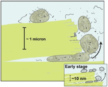
A mica world: diagrams of the possible origin of life between mica sheets. Protocells, the large gray structures, have a proto-cytoplasm that is distinct from the aqueous environment. Inset: at an early stage in the mica world, macromolecules and lipid vesicles are seen. The vesicles are filled with water and a few macromolecules. Note the scale change between early and later stages. Mica sheets are green. Green lines in the inset are individual mica sheets; white spaces between the green lines contain K+ bridging adjacent mica sheets. Blue is the aqueous environment. Adapted from Hansma (2010)
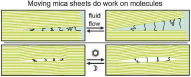
Mechanical energy from moving mica sheets may have provided an energy source for the origin of life. With mechanical energy doing work on molecules, no energy transduction is needed. Top panel: Mica sheets move open and shut in response to water movements, stretching and compressing polymer strands attached to them. Lower panel: Bubble between mica sheets functions as a heat pump in which hot and cold cycles cause the bubble to expand and contract, exerting forces on polymer molecules within the bubble. Adapted from Hansma (2010)
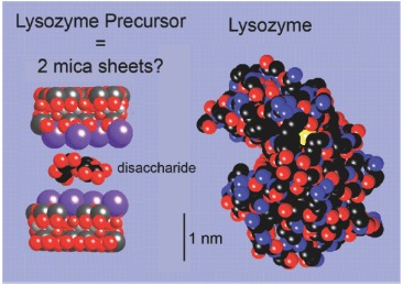
Mica sheets with a disaccharide (left) and a molecule of the enzyme, lysozyme (right). Enzyme motion may have originated from the motions of mica sheets. These models show the size of lysozyme's cleft in relationship to the disaccharide mannose between muscovite mica sheets. The enzyme lysozyme cleaves polysaccharides. The disaccharide, mannose, is a dimer of glucose. Potassium ions are divided equally between the mica sheets. Atoms are colored as follows: Yellow = Sulfur, S; Purple-blue = Potassium ions, K+; Black = Carbon, C; Reds = Oxygen, O, and Hydroxyl, OH; Grays = Silicon, Si, and Aluminum, Al; and Light blue = Aqueous environment. Models were prepared with Crystal Maker Software 2.3.6, using CPK van der Waals radii to define element sizes
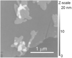
Lipids form bilayer and multilayer membranes on mica under water. This image shows a polymerized lipid film, but biological lipids form bilayers on mica when deposited as vesicles. Image adapted from Hansma et al. (1991). This image is an example of the ease with which biomolecules adhere to mica, as seen by atomic force microscopy
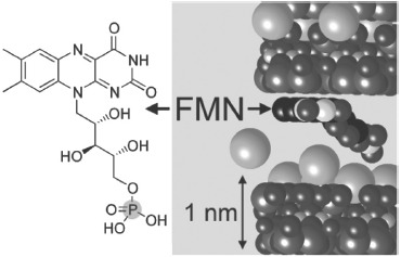
A cofactor, Flavin mononucleotide, FMN, between sheets of mica. This model of a cofactor between muscovite mica sheets bears a strong resemblance to enzyme–cofactor complexes and may be a precursor of what are now enzyme–cofactor complexes. The flavin rings are oriented on a sheet of mica without counterions, while the yellow phosphate on the end of the polymer chain is oriented toward a sheet of mica with potassium counterions, K+. For other atoms’ colors, and other details, see Figure 3 caption
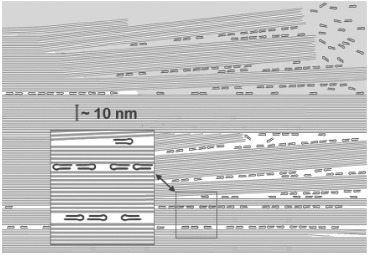
Mica provides massive redundancy and high error tolerance. Error tolerance is high in the spaces between mica sheets, as compared with prebiotic molecules in solution or on surfaces. Therefore, almost everything can go wrong. Error tolerance is one of the main requirements for the origin of life. This diagram envisages a self-replicating ribozyme inhabiting many spaces between mica sheets and diffusing in water to other spaces between mica sheets. Much of this mica world could be destroyed without eliminating all the molecules contained within it
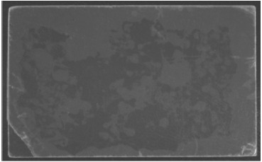
This mica-water ‘sandwich’ shows the way in which mica provides an optimum ‘Goldilocks’ solution to the problem of too much water vs. too much dryness during the process of life's origin. Mica is 50 × 75 mm. Dark areas are wet; light areas are dry. For other experimental details, see Hansma (2010)
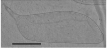
Did biopolymers evolve in a bubble defect in mica such as this one? High-grade muscovite mica in a dissecting microscope (Edmund Scientific Company, Barrington, NJ, USA) was photographed through a microscope objective with a Canon SDH camera. The fringes on the right side are interference patterns near the edge of the piece of mica. Scale bar = 1 mm
References
-
- Adamala K., Luisi P.L. Experimental systems to explore life origin: Perspectives for understanding primitive mechanisms of cell division. In: Kubiak J.Z., editor. Results and problems cell differentiation. 2011/06/02 ed. Vol. 53. Berlin: Springer-Verlag; 2011. pp. 1–9. - PubMed
-
- Bernal J.D. The physical basis of life. London: Routledge & Kegan Paul; 1951.
-
- Berrisford D.J., Bolm C., Sharpless K.B. Ligand-accelerated catalysis. Angewandte Chemie International Edition in English. 1995;34(10):1059–1070. doi: 10.1002/anie.199510591.
MeSH terms
Substances
LinkOut - more resources
Full Text Sources
Other Literature Sources