. 2013 Feb;25(2):206-15.
doi: 10.1111/j.1365-2826.2012.02381.x.

A novel reproductive peptide, phoenixin

Affiliations

A novel reproductive peptide, phoenixin

G L C Yosten et al. J Neuroendocrinol. 2013 Feb.

Abstract

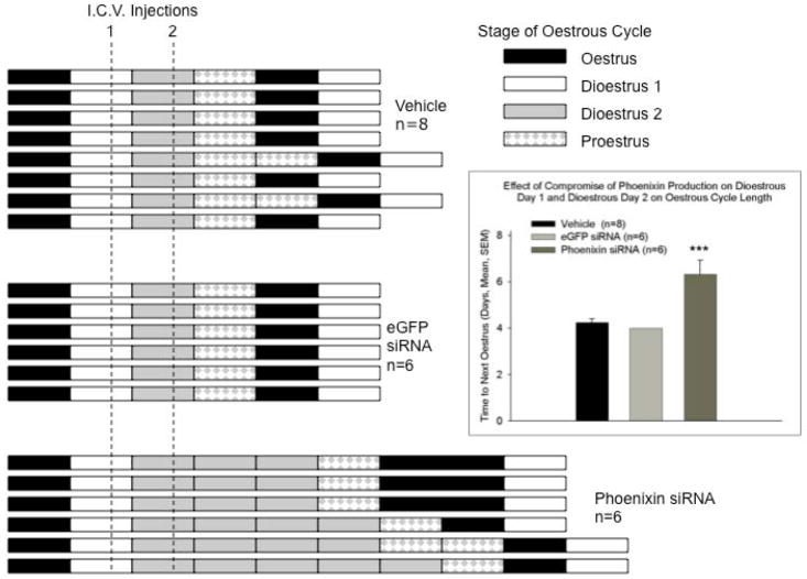
Normal anterior pituitary function is essential for fertility. Release from the gland of the reproductive hormones luteinising hormone and follicle-stimulating hormone is regulated primarily by hypothalamically-derived gonadotrophin-releasing hormone (GnRH), although other releasing factors (RF) have been postulated to exist. Using a bioinformatic approach, we have identified a novel peptide, phoenixin, that regulates pituitary gonadotrophin secretion by modulating the expression of the GnRH receptor, an action with physiologically relevant consequences. Compromise of phoenixin in vivo using small interfering RNA resulted in the delayed appearance of oestrus and a reduction in GnRH receptor expression in the pituitary. Phoenixin may represent a new class of hypothalamically-derived pituitary priming factors that sensitise the pituitary to the action of other RFs, rather than directly stimulating the fusion of secretary vesicles to pituitary membranes.


Conflict of interest statement

AUTHOR INFORMATION

The authors declare no competing financial interests. R.M.L. and J.K.C. are employees of Phoenix Pharmaceuticals.

Figures

Figure 1
Figure 1. Isolation of endogenous phoenixin
(A) Peptide sequence of phoenixin. Potential cleavage sites are indicated by red lettering on the consensus (top) sequence. Phoenixin is highly conserved across species. (B) Phoenixin immunoreactivity can be detected in various tissues in adult rats, as indicated by EIA. Mass spectrometry analysis of endogenous phoenixin isolated from rat hypothalamus (C) and bovine heart (D) revealed peptides of 20 and 14 amino acids, respectively.
Figure 2
Figure 2. Localization of phoenixin in brain
Male and female Sprague-Dawley rats were anaesthetised with urethane and perfused intracardially. Brains were removed and coronal sections of 40 um were prepared using a vibratome. Tissues were blocked with 10% goat serum, and then incubated in PNX antiserum (1:750 dilution). Sections were developed using DAB (see Supplemental Methods). PNX immunoreactivity was observed in both hypothalamus (A–I) and brainstem (J–O). (A) Staining was observed in both PVN and SON. Enlargements of these areas are presented in (D) and (G), respectively. (B) Staining was detected also in the ZI (enlargements presented in E and H). 3V, third ventricle; AP, area postrema; Arc, arcuate nucleus; cc, central canal; DA, dorsal hypothalamus; EW, Edinger-Westphal nucleus; ME, median eminence; opt, optic chiasm; SO, supraoptic nucleus; Pe, periventricular nucleus; PeF, perifornical area; PVN, paraventricular nucleus; Sol, nucleus tractus solitarius; VMH, ventromedial hypothalamus; ZI, zona incerta. (P) A hypothalamic section through the median eminence (ME) where numerous small irPNX cells are seen in the arculate nucleus (Arc). (Q) a section of anterior pituitary lobe, where small irPNX cells are detected. (R) a section of posterior pituitary lobe, where irPNX cell processes are noted. Abbreviations: f, fornix; opt, optic tract, 3V, 3rd ventricle. Scale bar: A–I, 100 μm; J–O, 250 μm; P–R, 50 μm.
Figure 3
Figure 3. Phoenixin regulates pituitary gonadotropin secretion by modulating expression of the GnRH receptor
Anterior pituitary cell cultures from random cycle female rats were incubated overnight with either phoenixin-14 (A) or phoenixin-20 (B), and then exposed to GnRH for one hour. Both phoenixin-14 and -20 potentiated GnRH-stimulated LH release (A and B). To evaluate the time course of the effect of phoenixin on LH release, female anterior pituitary cells were incubated with either vehicle or 1000nM phoenixin-20 for 24 hours prior to loading onto BioGel P-2 columns and exposure to the perifusate media alone (Fractions 0–10), media containing 10 nM GnRH (Fractions 11–25), and media containing 60 mM KCl (Fractions 30–40) (C). Pretreatment with phoenixin enhanced GnRH-stimulated LH release. Overnight pretreatment with phoenixin-20 also potentiated the ability of GnRH to upregulate the expression GnRH receptor in dispersed male anterior pituitary cell cultures (D). In the immortalised mouse pituitary gonadotroph cell line, alphaT3-1, treatment with phoenixin-20 increased GnRH receptor mRNA (E), as determined by RT-PCR. Phoenixin-20 and phoenixin-14 bound with similar affinity to pituitary adenoma cell membrane preparations (F). In primary pituitary cell cultures collected from female donors, phoenixin, in the absence of GnRH, significantly increased GnRH receptor expression (G). *p<0.05, **p<0.01, ***p<0.001 versus non-pretreated control cells (A and B) or vehicle treated cells (C, D, E, and G). #p<0.05 vs. control-pretreated, GnRH-treated cultures (D). For A and B, a one-way ANOVA was utilised, and data in C, D, E, and G was analyzed using a t test.
Figure 4
Figure 4. Compromise of endogenous hypothalamic phoenixin delays the appearance of the subsequent oestrus in cycling female rats
Following verification of a four-day oestrous cycle in rats bearing an indwelling cerebroventricular cannula, test substances [vehicle (2 ul sterile saline), or eGFP siRNA or phoenxin siRNA (2 ug in 2 ul sterile saline)] were administered i.c.v. on the afternoon of dioestrous day 1 and again on dioestrous day 2. Vaginal cytology was employed to monitor oestrous cycle progression and detect the next day of oestrus. (A) The bars indicate stage of the oestrous cycle for individual animals. Animals were then sacrificed on the first day of dioestrus following the verified appearance of oestrus and brains harvested for determination of phoenixin mRNA levels. The inset demonstrates the significant interruption of the four-day oestrous cycle in phoenixin siRNA treated animals, due mainly to the extension of the time in dioestrus. *** p < 0.001 versus vehicle and eGFP siRNA treated animals (ANOVA). In cycling female rats that were treated with siRNA directed against phoenixin on dioestrus days 1 and 2, and sacrificed the following day, hypothalamic content of phoenixin was significantly (p<0.01 vs. eGFP siRNA-injected controls, t test) reduced, as determined by radioimmunoassay (B). However, treatment with siRNA did not alter body weight (C).

Similar articles

Cited by

References

    1. McCann SM, Lumpkin MD, Mizunuma H, Khorram O, Samson WK. Brain peptides in control of anterior pituitary hormone secretion. Peptides. 1984;5:3–7. - PubMed
    1. Daniel PM. The blood supply of the hypothalamus and pituitary gland. Br Med Bull. 1966;22:202–208. - PubMed
    1. Gahete MD, Duran-Rado M, Luque RM, Martinez-Fuentes AJ, Quintero A, Gutierrez-Pascual E, Cordoba-Chacon J, Malagon MM, Gracia-Navarro F, Castano JP. Understanding the multifactorial control of growth hormone release by somatotropes: Lessons from comparative endocrinology. Ann NY Acad Sci. 2009;1163:137–153. - PubMed
    1. Burger LL, Haisenleder DJ, Dalkin AC, Marshall JC. Regulation of gonadotropin subunit gene transcription. J Mol Endo. 2004;33:559–584. - PubMed
    1. Clarke IJ, Cummins JT. The temporal relationship between gonadotropin releasing hormone (GnRH) and luteinizing hormone (LH) secretion in ovariectomized ewes. Endocrinology. 1982;111:1737–1739. - PubMed

Publication types

MeSH terms