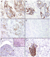
Gross cystic disease fluid protein-15 and mammaglobin A expression determined by immunohistochemistry is of limited utility in triple-negative breast cancer
- PMID: 22963676
- PMCID: PMC3881372
- DOI: 10.1111/j.1365-2559.2012.04344.x
Gross cystic disease fluid protein-15 and mammaglobin A expression determined by immunohistochemistry is of limited utility in triple-negative breast cancer
Abstract
Aims: In addition to oestrogen and progesterone receptors, gross cystic disease fluid protein-15 (GCDFP-15) and mammaglobin A (MAM) are the most common markers used to identify breast origin by immunohistochemistry. GCDFP-15 expression has been reported in approximately 60% of breast carcinomas and MAM expression in approximately 80%. Data on their expression in triple-negative breast cancer (TNBC) are very limited. The aim of this study was to examine the expression of these markers in TNBC to determine their utility in pathological diagnosis.
Methods and results: We studied the immunohistochemical (IHC) expression of GCDFP-15 and MAM in 63 primary and 118 metastatic TNBCs. GCDFP-15 staining was present in 14% of primary and 21% of metastatic TNBCs. MAM staining was present in 25% of primary and 41% of metastatic TNBCs. The frequency of expression of GCDFP-15 and/or MAM was 30% in primary and 43% in metastatic TNBCs, and many positive tumours had only focal staining.
Conclusions: Staining for GCDFP-15 and/or MAM in triple-negative carcinomas helps to confirm breast origin, but most tumours in this subgroup of breast carcinomas lack expression of either marker.
© 2012 Blackwell Publishing Limited.
Figures

Similar articles
-
GATA-binding protein 3 enhances the utility of gross cystic disease fluid protein-15 and mammaglobin A in triple-negative breast cancer by immunohistochemistry.Histopathology. 2015 Aug;67(2):245-54. doi: 10.1111/his.12645. Epub 2015 Feb 23. Histopathology. 2015. PMID: 25564996
-
Utility of different immunostains for diagnosis of metastatic breast carcinomas in both surgical and cytological specimens.Ann Diagn Pathol. 2017 Oct;30:21-27. doi: 10.1016/j.anndiagpath.2017.05.006. Epub 2017 May 17. Ann Diagn Pathol. 2017. PMID: 28965624
-
Differential expression of immunohistochemical markers in primary lung and breast cancers enriched for triple-negative tumours.Histopathology. 2016 Feb;68(3):367-77. doi: 10.1111/his.12765. Epub 2015 Aug 7. Histopathology. 2016. PMID: 26118394
-
Markers of metastatic carcinoma of breast origin.Histopathology. 2016 Jan;68(1):86-95. doi: 10.1111/his.12877. Histopathology. 2016. PMID: 26768031 Review.
-
The Novel Marker GATA3 is Significantly More Sensitive Than Traditional Markers Mammaglobin and GCDFP15 for Identifying Breast Cancer in Surgical and Cytology Specimens of Metastatic and Matched Primary Tumors.Appl Immunohistochem Mol Morphol. 2016 Apr;24(4):229-37. doi: 10.1097/PAI.0000000000000186. Appl Immunohistochem Mol Morphol. 2016. PMID: 25906123 Free PMC article. Review.
Cited by
-
Breast carcinoma metastasising to the gastric wall and the peritoneum: what physicians need to know.BMJ Case Rep. 2021 May 12;14(5):e241467. doi: 10.1136/bcr-2020-241467. BMJ Case Rep. 2021. PMID: 33980555 Free PMC article.
-
Prolactin-induced protein as a potential therapy response marker of adjuvant chemotherapy in breast cancer patients.Am J Cancer Res. 2016 May 1;6(5):878-93. eCollection 2016. Am J Cancer Res. 2016. PMID: 27293986 Free PMC article.
-
Gross cystic disease fluid protein 15 (GCDFP-15) expression in breast cancer subtypes.BMC Cancer. 2014 Jul 28;14:546. doi: 10.1186/1471-2407-14-546. BMC Cancer. 2014. PMID: 25070172 Free PMC article. Clinical Trial.
-
Characterization of GATA3 and Mammaglobin in breast tumors from African American women.Res Sq [Preprint]. 2023 Jan 25:rs.3.rs-2463961. doi: 10.21203/rs.3.rs-2463961/v1. Res Sq. 2023. Update in: Arch Microbiol Immunol. 2023;7(1):18-28. doi: 10.26502/ami.936500101. PMID: 36747860 Free PMC article. Updated. Preprint.
-
Immunohistochemical Characterization of a Large Cohort of Triple Negative Breast Cancer.Int J Surg Pathol. 2024 Apr;32(2):239-251. doi: 10.1177/10668969231171936. Epub 2023 Jun 12. Int J Surg Pathol. 2024. PMID: 37306115 Free PMC article.
References
-
- Chaignaud B, Hall TJ, Powers C, Subramony C, Scott-Conner CE. Diagnosis and natural history of extramammary tumors metastatic to the breast. J Am Coll Surg. 1994;179:49–53. - PubMed
-
- Hadju SI, Urban JA. Cancers metastatic to the breast. Cancer. 1972;29:1691–1696. - PubMed
-
- McCrea ES, Johnston C, Haney PJ. Metastases to the breast. AJR Am J Roentgenol. 1983;141:685–690. - PubMed
-
- Sanderson AT. Metastatic tumours in the breast. Br J Surg. 1959;47:54–58. - PubMed
-
- Lee BH, Hecht JL, Pinkus JL, Pinkus GS. WT1, estrogen receptor, and progesterone receptor as markers for breast or ovarian primary sites in metastatic adenocarcinoma to body fluids. Am J Clin Pathol. 2002;117:745–750. - PubMed
Publication types
MeSH terms
Substances
Grants and funding
LinkOut - more resources
Full Text Sources
Other Literature Sources
Medical
Research Materials
Miscellaneous

