

Motion correction of multi-b-value diffusion-weighted imaging in the liver
- PMID: 22963726
- PMCID: PMC4786013
- DOI: 10.1016/j.acra.2012.07.005
Motion correction of multi-b-value diffusion-weighted imaging in the liver
Abstract
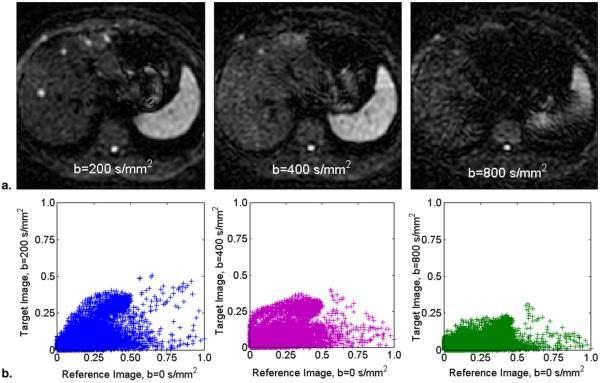
Rationale and objectives: Motion artifacts are a significant source of error in the acquisition and quantification of parameters from multi-b-value diffusion-weighted imaging (DWI). The objective of this article is to present a reliable method to reduce motion-related artifacts during free-breathing at higher b-values when signal levels are low.
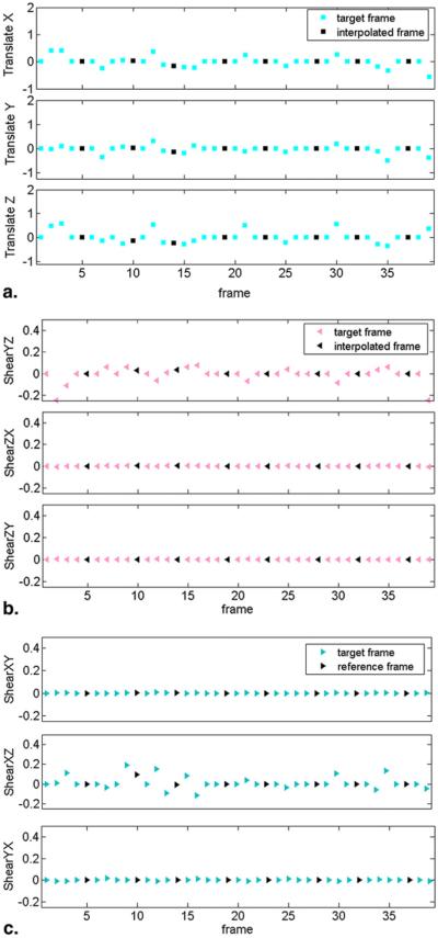
Materials and methods: Twelve patients referred for magnetic resonance imaging of the liver underwent a clinical magnetic resonance imaging examination of the abdominal region that included DWI. Conventional single-shot spin-echo echo planar imaging acquisitions of the liver during free breathing were repeated in a "time-resolved" manner during a single acquisition to obtain data for multi-b-value analysis, alternating between low and high b-values. Image registration using a normalized mutual information similarity measure was used to correct for spatial misalignment of diffusion-weighted volumes caused by motion. Registration error was estimated indirectly by comparing the normalized root-mean-square error (NRMSE) values of data fitted to the biexponential intra-voxel incoherent motion model before and after motion correction. Regions of interest (ROIs) were selected in the liver close to the surface of the liver and close to internal structures such as large bile ducts and blood vessels.
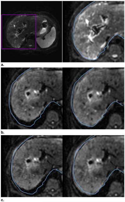
Results: For the 12 patient datasets, the mean NRMSE value for the motion-corrected ROIs (0.38 ± 0.16) was significantly lower than the mean NRMSE values for the non-motion-corrected ROIs (0.41 ± 0.13) (P < .05). In cases where there was substantial respiratory motion during the acquisition, visual inspection verified that the algorithm markedly improved alignment of the liver contours between frames.
Conclusions: The proposed method addresses motion-related artifacts to increase robustness in multi-b-value acquisitions.
Copyright © 2012 AUR. Published by Elsevier Inc. All rights reserved.
Figures


References
-
- Le Bihan D, Turner R, MacFall JR. Effects of intravoxel incoherent motions (IVIM) in steady-state free precession (SSFP) imaging: application to molecular diffusion imaging. Magn Reson Med. 1989;10:324–337. - PubMed
-
- Le Bihan D, Breton E, Lallemand D, et al. MR imaging of intravoxel incoherent motions: application to diffusion and perfusion in neurologic disorders. Radiology. 1986;161:401–407. - PubMed
-
- Le Bihan D, Breton E, Lallemand D, et al. Separation of diffusion and perfusion in intravoxel incoherent motion MR imaging. Radiology. 1988;168:497–505. - PubMed
-
- Yamada I, Aung W, Himeno Y, et al. Diffusion coefficients in abdominal organs and hepatic lesions: evaluation with intravoxel incoherent motion echo-planar MR imaging. Radiology. 1999;210:617–623. - PubMed
-
- Luciani A, Vignaud A, Cavet M, et al. Liver cirrhosis: intravoxel incoherent motion MR imaging—pilot study. Radiology. 2008;249:891–899. - PubMed
MeSH terms
Grants and funding
LinkOut - more resources
Full Text Sources
