. 2013 Aug 22;32(34):4017-27.
doi: 10.1038/onc.2012.408. Epub 2012 Sep 10.

CIB1 prevents nuclear GAPDH accumulation and non-apoptotic tumor cell death via AKT and ERK signaling

Affiliations

CIB1 prevents nuclear GAPDH accumulation and non-apoptotic tumor cell death via AKT and ERK signaling

T M Leisner et al. Oncogene. .

Abstract

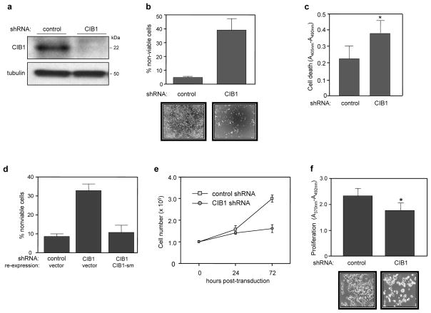
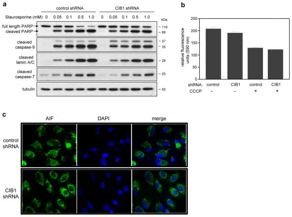
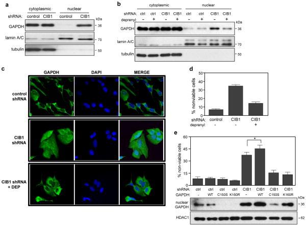
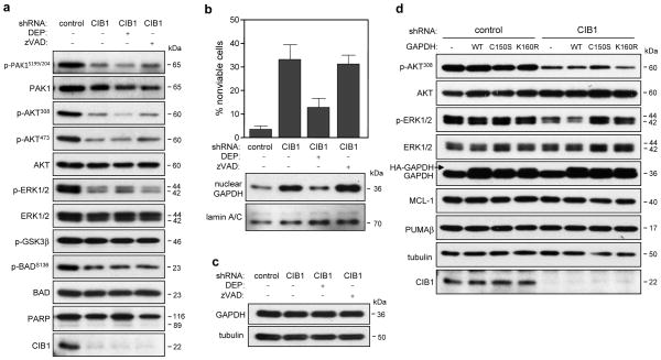
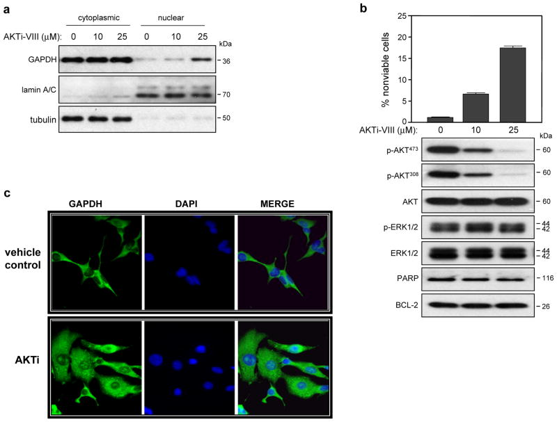
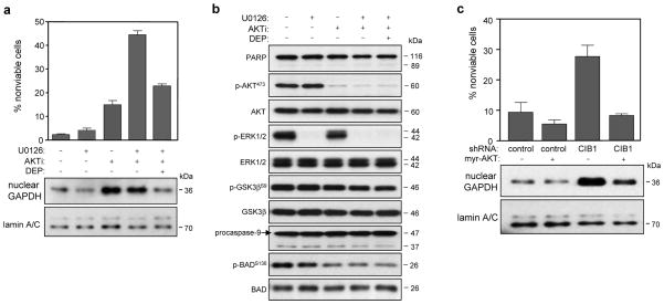
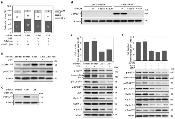
CIB1 is a 22-kDa regulatory protein previously implicated in cell survival and proliferation. However, the mechanism by which CIB1 regulates these processes is poorly defined. Here, we report that CIB1 depletion in SK-N-SH neuroblastoma and MDA-MB-468 breast cancer cells promotes non-apoptotic, caspase-independent cell death that is not initiated by increased outer mitochondrial membrane permeability or translocation of apoptosis-inducing factor to the nucleus. Instead, cell death requires nuclear GAPDH accumulation. Furthermore, CIB1 depletion disrupts two commonly dysregulated, oncogenic pathways-PI3K/AKT and Ras/MEK/ERK, resulting in a synergistic mechanism of cell death, which was mimicked by simultaneous pharmacological inhibition of both pathways, but not either pathway alone. In defining each pathway's contributions, we found that AKT inhibition alone maximally induced GAPDH nuclear accumulation, whereas MEK/ERK inhibition alone had no effect on GAPDH localization. Concurrent GAPDH nuclear accumulation and ERK inhibition were required, however, to induce a significant DNA damage response, which was critical to subsequent cell death. Collectively, our results indicate that CIB1 is uniquely positioned to regulate PI3K/AKT and MEK/ERK signaling and that simultaneous disruption of these pathways synergistically induces a nuclear GAPDH-dependent cell death. The mechanistic insights into cell death induced by CIB1 interference suggest novel molecular targets for cancer therapy.


Figures

Figure 1
Figure 1. CIB1 depletion induces cell death and slows proliferation
(a) Immunoblot showing efficient shRNA-induced CIB1 depletion from SK-N-SH cells. (b) Increased cell death in CIB1-depleted SK-N-SH cells as determined by trypan blue dye exclusion. Results are expressed as the mean percentage of dead cells (i.e., trypan blue positive cells) from both adherent and floating cell populations, data represent means +/− SEM, n=5. Representative phase contrast images show significant loss of adherent CIB1-depleted cells (bottom panels). (c) Analysis of control and CIB1-depleted SK-N-SH cells by immunochemical ELISA assays that detect histone-complexed DNA fragments as a marker of cell death (*p<0.05). (d) Ectopic expression of a shRNA-resistant CIB1 silent mutant (CIB1-sm) in CIB1-depleted SK-N-SH cells prevents cell death. Cell death was quantified as in (a), n=4. (e) Cell proliferation over 72 h, quantifed as total cell numbers at the indicated times post-transduction, n=2. (f) CIB1 depleted SK-N-SH cells show decreased cell proliferation as determined by BrdU proliferation assays (*p<0.05). Representative phase contrast images show significant loss of cell number from wells containing CIB1-depleted cells (bottom panels).
Figure 2
Figure 2. Effect of CIB1 depletion on caspase activation and mitochondrial function
(a) Control and CIB1 depleted SK-N-SH cells were incubated in the absence (0) and presence of increasing concentrations (0.05–1.0 mM) of staurosporine (STS); whole cell lysates were immunoblotted using the indicated antibodies, n=3. (b) CIB1-depletion does not affect mitochondrial membrane potential (Δψm). The mitochondrial dye JC-10 was used to quantify Δψm in adherent control and CIB1-depleted cells. In non-compromised mitochondria, JC-10 accumulates as J-aggregates which fluoresce at 590 nm (data expressed in relative fluorescence units). As positive control, cells were treated with 4 μM of the mitochondrial depolarizing agent carbonyl cyanide 3-chlorophenylhydrazone (CCCP), resulting in the release of JC-10 monomers, as indicated by decreased fluorescence at 590 nm. Data represent means +/− SEM, n=2. (c) Control and CIB1-depleted SK-N-SH cells were immunostained with an anti-AIF antibody (green fluorescence) and nuclei were visualized by counterstaining with DAPI (blue fluorescence). Images are representative of at least 3 separate experiments. Scale bar = 20 μm
Figure 3
Figure 3. Cell death induced by CIB1 depletion requires GAPDH nuclear accumulation and GAPDH S-nitrosylation and acetylation
(a) Western blot analysis of cytoplasmic and nuclear fractions shows increased nuclear GAPDH in CIB1-depleted SK-N-SH cells. αTubulin and lamin A/C antibodies were used to confirm cytoplasmic and nuclear fractions, respectively. (b) Deprenyl blocks nuclear accumulation of GAPDH. Control (ctrl) and CIB1-depleted cells were treated with or without 25 nM deprenyl (DEP) and analyzed as in (a), n=4. (c) Immunohistochemical analysis of GAPDH localization. Control and CIB1-depleted cells were treated with and without deprenyl as in (b) and immunostained with anti-GAPDH (green) and nuclei were visualized with DAPI (blue). Bar = 20 μm. (d) Deprenyl protects CIB1-depleted cells from cell death. Cells were treated with or without DEP as in (b). Cell death was determined as in Figure 1b. Data represent means ± SEM, n=8. (e) Cell death in control and CIB1-depleted cells expressing empty vector, HA-GAPDH-WT, -C150S or -K160R was quantified as in Figure 1b. Data represent means +/− SEM, n=4 (t-test; * p<0.05 versus CIB1 shRNA). Effect of GAPDH-WT, -C150, or -K160R expression on GAPDH nuclear accumulation. Nuclear lysates were immunoblotted with anti-GAPDH or -histone deacetylase-1 (HDAC1) antibodies, lower panels.
Figure 4
Figure 4. Effect of CIB1 depletion on AKT, ERK and PAK1 signaling pathways in the absence and presence of deprenyl or zVAD-FMK
(a) Immunoblots of whole cell lysates prepared from control and CIB1-depleted SK-N-SH cells, n ≥ 3. (b) Deprenyl (DEP), but not caspase inhibition with zVAD-FMK (zVAD) blocks nuclear GAPDH accumulation and cell death. (c) GAPDH expression is unchanged in the absence and presence of either DEP or zVAD. (d) Whole cell lysates from SK-N-SH cells expressing emtpy vector, HA-GAPDH-WT, -CI50S or -K160R were immunoblotted with the indicated antibodies.
Figure 5
Figure 5. AKT inhibition induces cell death and GAPDH nuclear accumulation
Untransduced SK-N-SH cells were treated in the absence and presence of either 10 or 25 μM AKT inhibitor VIII (AKTi-VIII) for 48 h. (a) Analysis of cytoplasmic and nuclear fractions was performed as in Figure 3a, n=4. (b) AKT inhibition induces SK-N-SH cell death. Cell death was determined as in Figure 1b. Data represent means ± SEM, n=4. Immunoblot analysis of whole cell lysates, n=3. (c) SK-N-SH cells were treated as in (a) and GAPDH cellular localization was visualized as in Figure 3c.
Figure 6
Figure 6. Effects of combined AKT and ERK inhibition on SK-N-SH cell death and GAPDH nuclear accumulation
Untransduced SK-N-SH cells were treated in the absence and presence of either 10 μM U0126 (MEK inhibitor) and/or 20 μM AKTi. Cells exposed to both inhibitors were also treated with 25 nM deprenyl. (a) Concurrent inhibition of AKT and ERK significantly increases cell death but not GAPDH nuclear accumulation. Cell death was quantified as in Figure 1b (upper graph). Data represent means ± SEM, n=3. Analysis of cytoplasmic and nuclear fractions was performed as in Figure 3a (bottom panels). (b) Immunoblotting of whole cell lysates with the indicated antibodies, prepared from cells treated with or without AKTi and/or U0126. (c) Overexpression of a constitutively active AKT mutant (myr-AKT) blocks cell death in CIB1-depleted cells. Cell viability was quantified as Figure 1b. Data represent +/- SEM, n=3. Expression of myr-AKT blocks GAPDH nuclear accumulation (lower panels). Nuclear lysates were immunobloted with anti-GAPDH or -lamin A/C antibodies.
Figure 7
Figure 7. Increased DNA damage and disruption of cell cycle regulation by CIB1 depletion is recapitulated by concurrent AKT and ERK inhibition
(a) Cell cycle analysis of propidium iodide-stained control and CIB1-depleted SK-N-SH cells treated with 25 nM deprenyl or expressing CIB1-sm (CIB1-silent mutation). (b) CIB1-depleted cells show induction of DNA damage markers. Analysis of whole cell lysates prepared from control and CIB1-depleted cells in the absence and presence of deprenyl (DEP). Analysis of adherent CIB1-depleted (CIB1-Adh) cells (excluding floating cell populations) shows induction of both γH2AX and p-CHK1. (c) Ectopic expression of CIB1-sm in CIB1-depleted cells blocks induction of γH2AX. (d) Ectopic expression of either HA-GAPDH-C150S or -K160R blocks induction of γH2AX. (e) Deprenyl does not significantly rescue the cell proliferation defect in CIB1-depleted cells. Proliferation was quantified as in Figure 1c. Data represent means ± SEM, n=7. Effect of DEP on the activation and expression of cell cycle proteins (blots, lower panels). (f) Effect of AKTi and/or U0126 on proliferation and cell cycle regulatory proteins. Proliferation of SK-N-SH cells treated with and without inhibitors expressed as the fold increase over the cell number at time of cell plating (time 0) (upper graph). Data represent means ± SEM, n=3. Effect of AKT and/or ERK inhibition on the activation and expression of cell cycle regulatory proteins. Whole cell lysates were analyzed as in (b) (lower panels).

References

    1. McCubrey JA, Steelman LS, Abrams SL, Lee JT, Chang F, Bertrand FE, et al. Roles of the RAF/MEK/ERK and PI3K/PTEN/AKT pathways in malignant transformation and drug resistance. Adv Enzyme Regul. 2006;46:249–79. - PubMed
    1. Slack-Davis JK, Eblen ST, Zecevic M, Boerner SA, Tarcsafalvi A, Diaz HB, et al. PAK1 phosphorylation of MEK1 regulates fibronectin-stimulated MAPK activation. J Cell Biol. 2003;162(2):281–91. - PMC - PubMed
    1. Huynh N, Liu KH, Baldwin GS, He H. P21-activated kinase 1 stimulates colon cancer cell growth and migration/invasion via ERK- and AKT-dependent pathways. Biochim Biophys Acta. 2010;1803(9):1106–13. - PubMed
    1. Grant S. Cotargeting survival signaling pathways in cancer. The Journal of clinical investigation. 2008;118(9):3003–6. Epub 2008/08/30. - PMC - PubMed
    1. Datta SR, Dudek H, Tao X, Masters S, Fu H, Gotoh Y, et al. Akt phosphorylation of BAD couples survival signals to the cell-intrinsic death machinery. Cell. 1997;91(2):231–41. - PubMed

Publication types

MeSH terms