Review
. 2012 Sep;192(1):33-53.
doi: 10.1534/genetics.112.141390.

The selfish Segregation Distorter gene complex of Drosophila melanogaster

Affiliations
Review

The selfish Segregation Distorter gene complex of Drosophila melanogaster

Amanda M Larracuente et al. Genetics. 2012 Sep.

Abstract

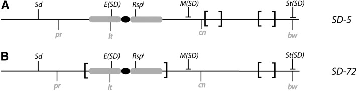
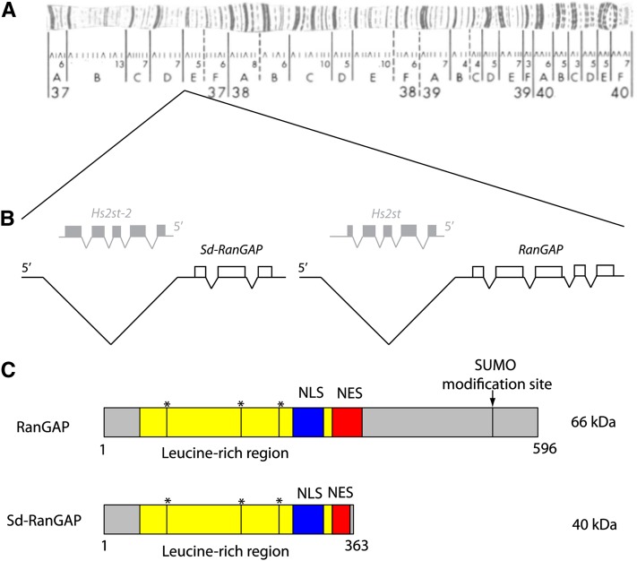
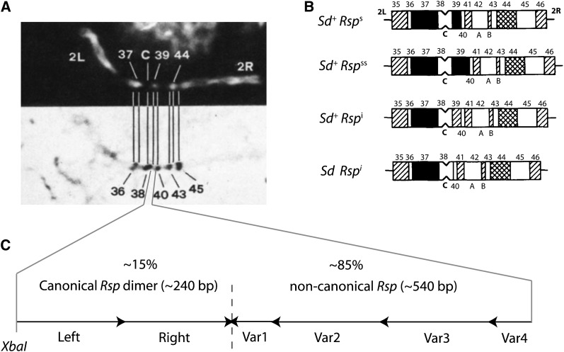
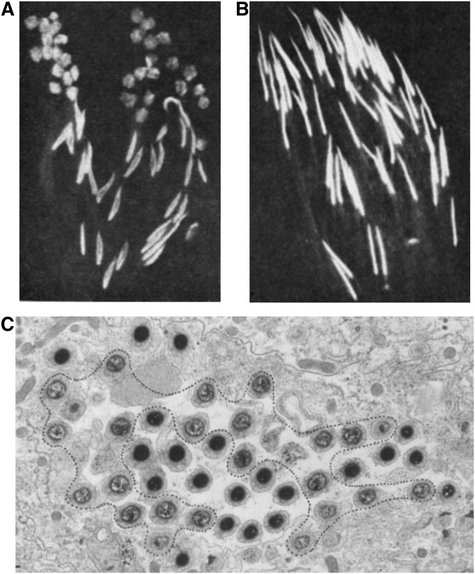
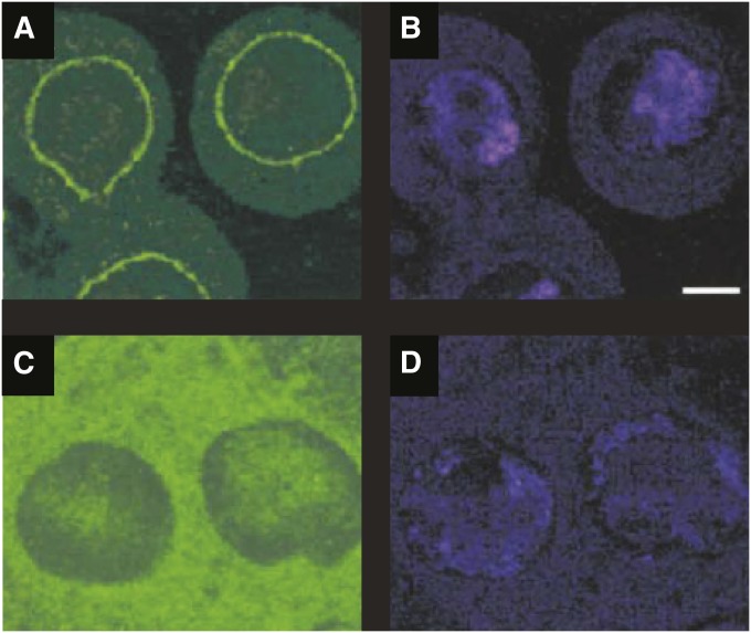
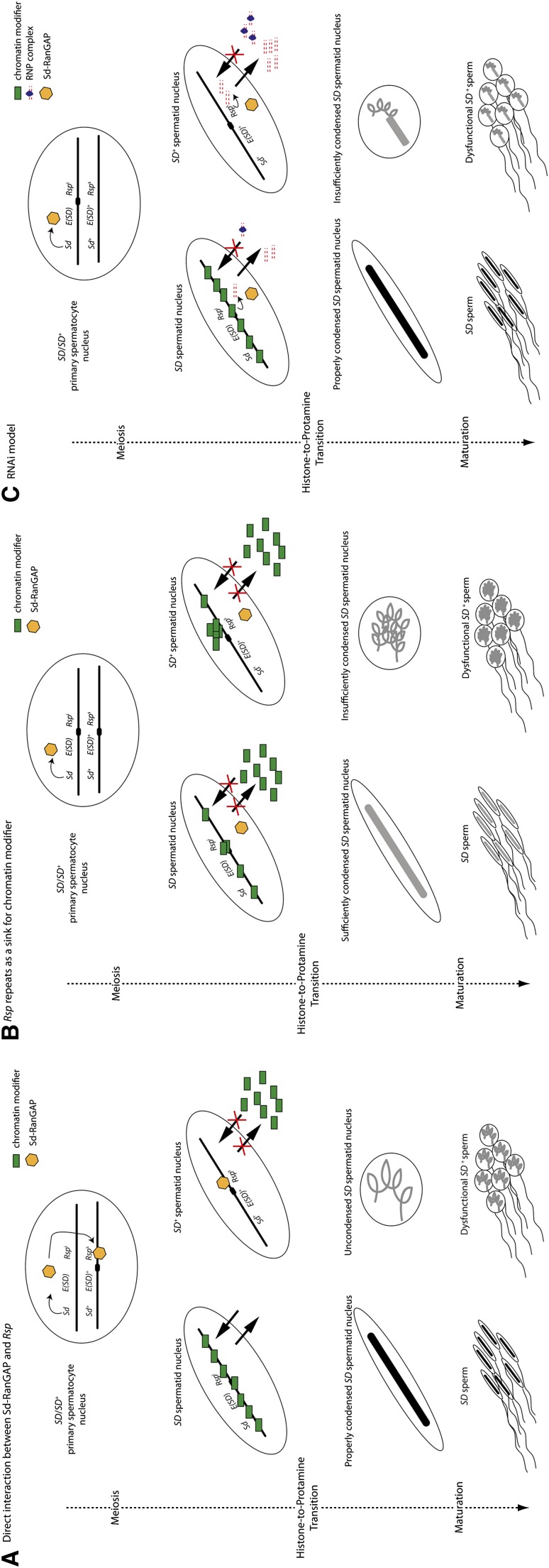
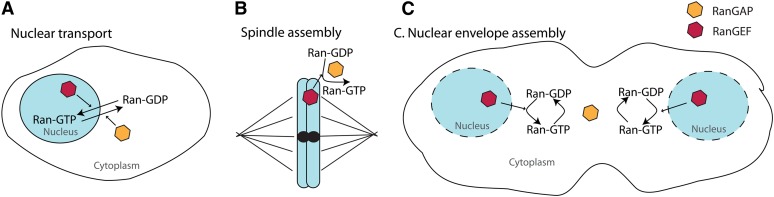
Segregation Distorter (SD) is an autosomal meiotic drive gene complex found worldwide in natural populations of Drosophila melanogaster. During spermatogenesis, SD induces dysfunction of SD(+) spermatids so that SD/SD(+) males sire almost exclusively SD-bearing progeny rather than the expected 1:1 Mendelian ratio. SD is thus evolutionarily "selfish," enhancing its own transmission at the expense of its bearers. Here we review the molecular and evolutionary genetics of SD. Genetic analyses show that the SD is a multilocus gene complex involving two key loci--the driver, Segregation distorter (Sd), and the target of drive, Responder (Rsp)--and at least three upward modifiers of distortion. Molecular analyses show that Sd encodes a truncated duplication of the gene RanGAP, whereas Rsp is a large pericentromeric block of satellite DNA. The Sd-RanGAP protein is enzymatically wild type but mislocalized within cells and, for reasons that remain unclear, appears to disrupt the histone-to-protamine transition in drive-sensitive spermatids bearing many Rsp satellite repeats but not drive-insensitive spermatids bearing few or no Rsp satellite repeats. Evolutionary analyses show that the Sd-RanGAP duplication arose recently within the D. melanogaster lineage, exploiting the preexisting and considerably older Rsp satellite locus. Once established, the SD haplotype collected enhancers of distortion and suppressors of recombination. Further dissection of the molecular genetic and cellular basis of SD-mediated distortion seems likely to provide insights into several important areas currently understudied, including the genetic control of spermatogenesis, the maintenance and evolution of satellite DNAs, the possible roles of small interfering RNAs in the germline, and the molecular population genetics of the interaction of genetic linkage and natural selection.


Figures

Figure 1
Figure 1
The SD complex. (A) Schematic of the SD-5 chromosome showing the location of the distorting gene, Sd, the major enhancer of distortion, E(SD) on 2L (the black dot is the centromere), the target locus, Rspi, and the strong upward modifiers M(SD) and St(SD), on 2R above the chromosome. Markers commonly used to dissect the genetics of SD are diagramed below the chromosome: pr (2-54.5), lt (2-55), cn (2-57.5) and bw (2-104.5). SD-5 has two paracentric inversions on 2R (brackets). (B) Same as (A) except for the SD-72 chromosome. SD-72 has a pericentric inversion (brackets) and a paracentric inversion on 2R.
Figure 2
Figure 2
Structure of the SD region. (A) A polytene map showing the location of the Sd locus at band 37D2-6. (B) The structure of the Sd locus showing that Sd–RanGAP is a partial duplication of the RanGAP locus. Hs2st (shaded in gray) is a gene occurring in the intron of RanGAP that is also duplicated in Sd–RanGAP (Hs2st-2). (C) A schematic of RanGAP (66 kD) and Sd–RanGAP (40 kD) proteins. RanGAP contains a large leucine-rich domain with a nuclear localization signal (NLS, blue) and two adjacent nuclear export signals (NES, in red). The * denotes some of the sites required for RanGAP activity. RanGAP contains a SUMO modification site at its C terminus that is required for tethering RanGAP to the cytoplasmic side of the nuclear pore. Sd–RanGAP is missing 234 amino acids at its C terminus including a NES and the SUMO modification site. Sd–RanGAP retains the NLS and sites required for activity.
Figure 3
Figure 3
Structural organization of the Rsp locus. (A) Hoeschst fluoresence (above) and N-banding (below) of mitotic chromosome 2 from larval neuroblasts (modified from Figure 1B of Pimpinelli and Dimitri 1989). (B) Thick bars represent chromosome 2 heterochromatin and thin lines represent euchromatin. The shading represents the intensity of Hoeschst fluorescence in mitotic chromosomes of larval neuroblasts (modified from Figure 5, A, C–E of Pimpinelli and Dimitri 1989, with permission from the Genetics Society of America). (C) Schematic of a canonical Rsp dimer and noncanonical Rsp repeats (Houtchens and Lyttle 2003) comprising ∼15% and ∼85% of the ∼600-kb cn bw Rsp locus, respectively. Canonical Rsp dimers consist of related left and right repeats and a characteristic XbaI restriction site.
Figure 4
Figure 4
Stages of spermatogenesis in D. melanogaster. (A) Cell divisions are represented with a solid arrow and differentiation events without cell divisions are represented by a dotted arrow. Germline stem cells (GSC) are surrounded by cyst progenitor cells (CPC), which give rise to the cyst cells encapsulating developing germ cells. Each germline stem cell divides to produce another stem cell or a daughter spermatogonial cell. The spermatogonia undergo four rounds of mitotic divisions to create 16 primary spermatocytes. Only 1 of the 16 primary spermatocytes is shown. Most of the transcription during spermatogenesis occurs in primary spermatocytes prior to entering meiosis. Primary spermatocytes undergo two rounds of meiotic divisions to create 64 spermatids. Spermatids differentiate and individualize as mature sperm, which are coiled and deposited in the seminal vesicle. All divisions have incomplete cytokinesis so that cells in a cyst are connected through cytoplasmic bridges. Prior to individualization, all cells develop within a cyst (Fuller 1993). (B) Spermiogenesis showing the histone-to-protamine transition. The arrow corresponds to the first visible difference between SD and SD+ spermatids in SD/SD+ heterozygotes. Beneath the images of nuclei during spermiogenesis is a schematic showing the primary chromatin component during spermiogenesis (modified from Figure 7 of Rathke et al. 2007 (DOI: 10.1242/jcs.004663), with permission from the Journal of Cell Science).
Figure 5
Figure 5
SD+ spermatid dysfunction is due to a failure to proper condense chromatin after meiosis. (A) A fluorescent image of a cyst of elongating spermatids in an SD/SD+ heterozygote. About half of the spermatids (those corresponding to SD+) are not elongating. (B) A comparable cyst in a wild-type testis showing a cyst of elongating spermatids. (Images in A and B are from figure 3, B and C of Hauschteck-Jungen and Hartl 1978; reprinted with permission from the Genetics Society of America.) (C) An ultrastructure image of SD/SD+ testes at the coiling stage showing that approximately half of the spermatids (again corresponding to SD+) have abnormal condensation (spermatids within dotted line). (Image in part C is reprinted from figure 2 of Tokuyasu et al. 1977, with permission from Elsevier.)
Figure 6
Figure 6
Sd–RanGAP is mislocalized in some primary spermatocytes. (A) Immunolocalization of RanGAP (green) in primary spermatocytes of SD+/SD+ testes. (B) Propidium Iodide staining showing the location of DNA (blue) in the nucleus. RanGAP normally localizes to the cytolplasmic side of the nuclear envelope. (C and D) The same as A and B except showing the localization of Sd–RanGAP (with an anti-HA antibody in a Sd–RanGAP-HA transgenic fly showing segregation distortion). Sd–RanGAP-HA diffusely stains the cytoplasm and shows aberrant expression in the nucleus (A–D are reprinted from Kusano et al. 2001, with permission from Elsevier).
Figure 7
Figure 7
Current models of SD action. (A) Sd–RanGAP directly binds Rsp repeats, disrupting chromatin condensation in Rsps-bearing spermatids causing spermatid dysfunction either as a consequence of disrupted nuclear transport or some other cellular function of RanGAP. (B) Sd–RanGAP disrupts nuclear transport globally, but Rsps-bearing spermatids are disproportionately sensitive to this disruption because large blocks of Rsp act as a sink for chromatin modifiers when their access to the nucleus is limited. (C) Rsp rasiRNAs, presumably required for postmeiotic chromatin condensation, are exported from the nucleus, where they form ribonucleoprotein (RNP) complexes; however, the RNP complexes fail to target chromatin modifiers to the genomic Rsp satellite because of some disrupted RanGAP, or ran-like, function (see text). Although the disruption is shown as a failure to reenter the nucleus due to disrupted transport, a disrupted Ran-GTP/Ran-GDP (or ran-like-GTP/ran-like-GDP) gradient could affect chromatin condensation more directly.
Figure 8
Figure 8
The role of the Ran cycle throughout the cell cycle. (A) The Ran cycle during interphase aids in nuclear transport. A gradient of Ran-GTP/Ran-GDP is established by RanGAP and RanGEF located in the cytoplasm and nucleus, respectively. (B) The Ran cycle during mitosis, prior to metaphase. A gradient of Ran-GTP/Ran-GDP aids in spindle assembly. (C) The Ran cycle at telophase during cell division. After mitosis, the Ran cycle is involved in reassembling the nuclear envelope.

References

    1. Aravin A. A., Lagos-Quintana M., Yalcin A., Zavolan M., Marks D., et al. , 2003. The small RNA profile during Drosophila melanogaster development. Dev. Cell 5: 337–350 - PubMed
    1. Aravin A. A., Klenov M. S., Vagin V. V., Bantignies F., Cavalli G., et al. , 2004. Dissection of a natural RNA silencing process in the Drosophila melanogaster germ line. Mol. Cell. Biol. 24: 6742–6750 - PMC - PubMed
    1. Baudry E., Viginier B., Veuille M., 2004. Non-African populations of Drosophila melanogaster have a unique origin. Mol. Biol. Evol. 21: 1482–1491 - PubMed
    1. Brennecke J., Aravin A. A., Stark A., Dus M., Kellis M., et al. , 2007. Discrete small RNA-generating loci as master regulators of transposon activity in Drosophila. Cell 128: 1089–1103 - PubMed
    1. Brittnacher J. G., Ganetzky B., 1983. On the components of segregation distortion in Drosophila melanogaster. II. Deletion mapping and dosage analysis of the SD locus. Genetics 103: 659–673 - PMC - PubMed

Publication types

MeSH terms

Substances

LinkOut - more resources