. 2012 Nov;92(11):1541-52.
doi: 10.1038/labinvest.2012.125. Epub 2012 Sep 10.

Heme oxygenase-1 promotes granuloma development and protects against dissemination of mycobacteria

Affiliations

Heme oxygenase-1 promotes granuloma development and protects against dissemination of mycobacteria

Doron Regev et al. Lab Invest. 2012 Nov.

Abstract

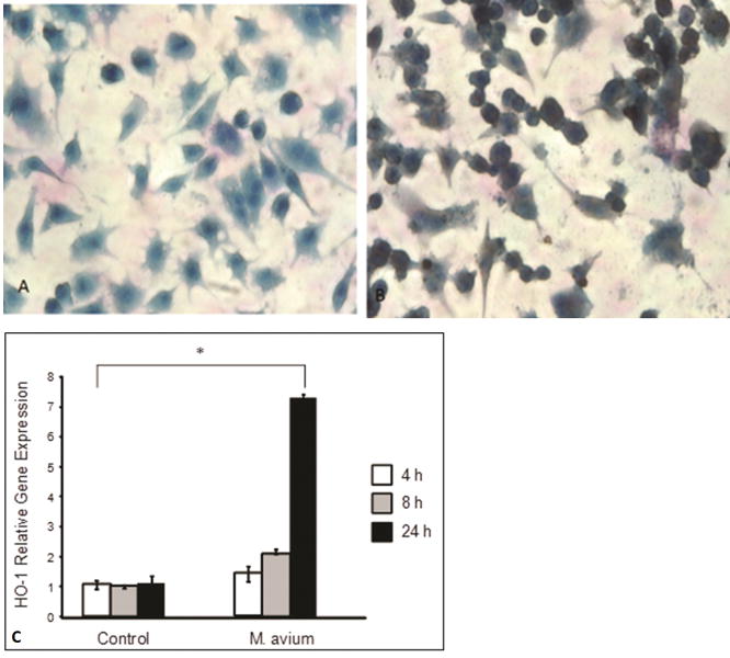
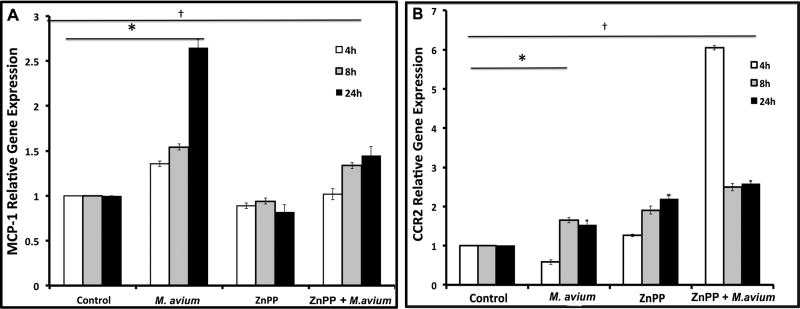
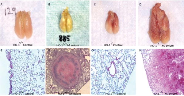
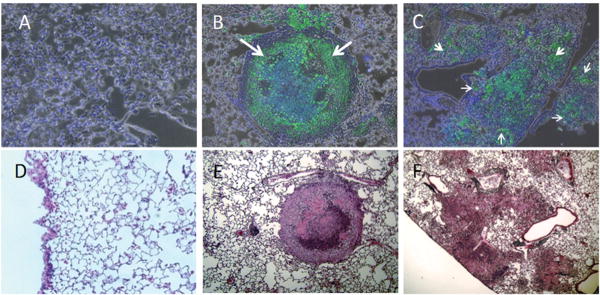
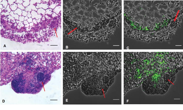
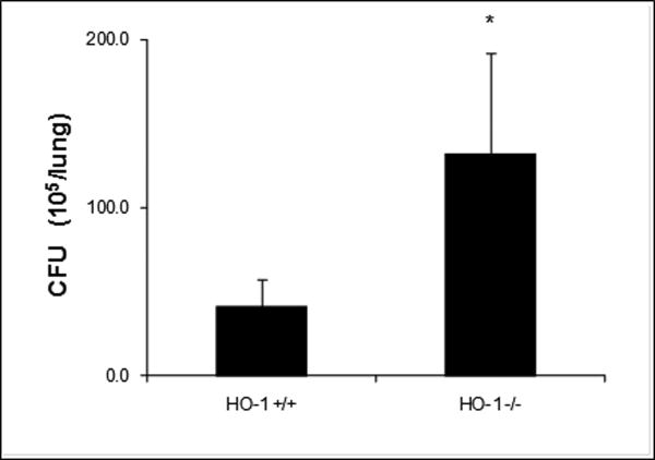
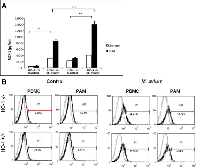
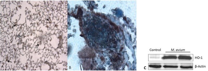
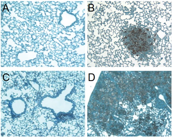
Non-tuberculous mycobacterial (NTM) infections occur in both immunocompromised and immunocompetent hosts and are an increasingly recognized cause of morbidity and mortality. The hallmark of pulmonary mycobacterial infections is the formation of granuloma in the lung. Our study focuses on the role of heme oxygenase-1 (HO-1), a cytoprotective enzyme, in the regulation of granuloma development and maturation following infection with Mycobacterium avium. We examined the role of HO-1 in regulating monocyte chemoattractant protein-1 (MCP-1) and chemokine receptor 2 (CCR2), two molecules involved in monocyte-macrophage cell trafficking after infection. We showed that RAW 264.7 mouse monocytes exposed to M. avium expressed HO-1 and MCP-1. Inhibition of HO by zinc protoporphyrin-IX led to inhibition of MCP-1 and increased expression of CCR2, its cognate receptor. HO-1⁻/⁻ mice did not develop organized granuloma in their lungs, had higher lung colony forming unit of M. avium when infected with intratracheal M. avium, and had loose collections of inflammatory cells in the lung parenchyma. Mycobacteria were found only inside defined granulomas but not outside granuloma in the lungs of HO-1⁺/⁺ mice. In HO-1⁻/⁻ mice, mycobacteria were also found in the liver and spleen and showed increased mortality. Peripheral blood monocytes isolated from GFP⁺ mice and given intravenously to HO-1⁺/⁺ mice localized into tight granulomas, while in HO-1⁻/⁻ mice they remained diffusely scattered in areas of parenchymal inflammation. Higher MCP-1 levels were found in bronchoalveolar lavage fluid of M. avium infected HO-1(-/-) mice and CCR2 expression was higher in HO-1⁻/⁻ alveolar macrophages when compared with HO-1⁺/⁺ mice. CCR2 expression localized to granuloma in HO-1⁺/⁺ mice but not in the HO-1⁻/⁻ mice. These findings strongly suggest that HO-1 plays a protective role in the control of M. avium infection.


Figures

Figure 1
Figure 1. M. avium infection induces HO-1 in RAW 264.7 monocyte-macrophages
(A, B) Representative light micrograph showing immunocytochemical detection of HO-1 in RAW 264.7. Panel A shows the control cells were incubated in SFM while panel B shows the cells infected with M. avium (50 MOI) for 24 hours. Cells were fixed and stained with anti-HO-1 antibody and visualized by optical microscopy at 400× magnification. The infected cells stained for HO-1 expression (brown) after 24 hours as compared to control cells. Blue color shows counter-stain with hematoxylin. The photomicrograph is the representative of three individual staining. (C) HO-1 relative mRNA expression significantly increases in RAW 264.7 cells after infection with M. avium. Control cells were incubated in SFM and treated cells were incubated with M. avium. For 4, 8 and 24 hours. HO-1 relative gene expression was analyzed by RT-PCR. HO-1 levels significantly increased in cells treated with M. avium as compared to nontreated cells in a time dependent manner (*P < 0.001). Values represent the mean of three different experiments.
Figure 2
Figure 2. HO inhibition decreases MCP-1 while increase CCR2 relative gene expression in vitro following the infection by M. avium
RAW 264.7 cells were pretreated with SFM (control) or ZnPP-IX (HO inhibitor) for 30 minutes and then incubated with M. avium for 4, 8 and 24 hours. MCP-1 and CCR2 relative gene expression was analyzed by RT-PCR. (A) MCP-1 levels were higher in M. avium treated cells as compared to controls (* P<0.001) and were significantly down-regulated († P<0.001) when cells were pretreated with ZnPP-IX, for 8 and 24 hours as compared to M. avium alone for the same time points. No significant differences were observed at the 4 hour time-point. Values represent the mean of three different experiments. (B) CCR2 gene expression was significantly increased as compared with controls at all time points (*P<0.05). CCR2 relative gene expression was higher in cells pre-treated with ZnPP-IX as compared with cells treated with M. avium alone († P<0.05). Values represent the mean of three different experiments.
Figure 3
Figure 3. Mouse lung tissue showing differential granuloma formation in HO-1+/+ and HO-1-/- mice after 6 month M. avium infection or saline inoculation
Mouse lungs were harvested following 6 month M. avium inoculation and visually inspected for presence of granuloma. Alternatively, lung tissue was fixed and stained with H&E to confirm granuloma formation. (A) Absence of granuloma in HO-1+/+ control lung. (B) Presence of organized, mature granulomas in HO-1+/+ lung inoculated with M. avium for 6 months. (C) Absence of granuloma in HO-1-/- control lung. (D) Diffuse infection and unorganized granuloma formation in HO-1-/- lung inoculated with M. avium for 6 months. (E) 200× H&E stain of normal tissue from HO-1+/+ control lung. (F) 200× H&E stain showing organized granuloma formation in HO-1+/+ lung inoculated with M. avium for 6 months. (G) 200× H&E stain of normal tissue from HO-1-/- control lung. (H) 200× H&E stain of diffuse infection and dysfunctional granuloma formation in HO-1-/- lung inoculated with M. avium for 6 months. Five mice were included in each treatment group (lungs are not shown to scale).
Figure 4
Figure 4. HO-1-/- mice showed diffuse M. avium infection in lung issue as opposed to HO-1+/+ mice which contained the mycobacteria in mature, wellformed granulomas
Lung sections were stained with fluorescent auramine stain (BD Biosciences, Sparks, MD, USA) to show M. avium (green color) in lung tissue. (A) Auramine stain of HO-1+/+ control lung. (B) Auramine stain of HO-1+/+ lung infected with M. avium for 6 months. (C) Auramine stain of HO-1-/- lung after 6 months M. avium infection. White arrows show the presence of M. avium. (D-F) H&E stain corresponding to panels A-C.
Figure 5
Figure 5. HO-1 modulates development and recruitment of monocytes recruitment into granuloma
HO-1-/- and HO-1+/+ mice were infected with M. avium intratracheallyas described in material and methods. At 6 weeks control and experimental mice were injected with PBMCs from GFP positive mice. The mice were euthanized 24 hrs post PBMC injection; the lungs were perfused with heparinised normal saline and fixed in 4% paraformaldehyde. The lungs were embedded in paraffin and 4 micron sections were prepared, stained with H&E (A, D). HO-1-/- mice demonstrated poorly organized granuloma formation (A, B). The HO-1+/+ mice demonstrated highly organized granuloma (D, E). The red arrows points to the granuloma. No organized granuloma formation was present in the HO-1-/- mice (A, B). This is a single representative image of H&E stained images of three mice from each strain. (C, F) are the transmission microscopy images of pulmonary granulomas from HO-1-/- and HO-1+/+ mice respectively. The HO-1 positive GFP labeled monocytes given intravenously tracked into the granuloma of HO-1+/+ mice (F) but they remain dispersed in HO-1-/- mice (C). This is a single representative image of three similar images observed in six mice from each strain. The white arrows point to the granulomas. The green fluorescence indicated GFP positive monocytes. Magnification= 200×. Scale bar = 100 μm.
Figure 6
Figure 6. M. avium CFU counts are higher in lung tissue of infected HO-1 -/- mice as compared to lung tissue form infected HO-1+/+ mice
Mouse lung tissue was homogenized in 2ml of 7H9 broth medium. Then, 100 μl of 10-fold serial dilutions were plated on 7H10 agar plates and incubated for 10-21 days in 37°C after which colony forming units were counted. CFU counts were significantly higher (* P<0.05) in lung tissue of infected HO-1-/- mice as compared to HO-1+/+ mouse tissue. Values represent the average CFU counts from four mice in either non-infected or M. avium infected groups.
Figure 7
Figure 7. (A) MCP-1 levels are up-regulated in BALF and serum samples of M. avium infected HO-1+/+ and HO-1-/- mice
BALF and serum were collected and MCP-1 protein levels were measured by ELISA. MCP-1 levels were significantly increased in BALF of infected HO-1+/+ mice as compared to HO-1+/+ saline controls (*P <0.001) and HO-1-/- mice as compared to HO-1-/- saline controls (** P <0.001). MCP-1 levels are increased in mouse serum after 6 months M. avium infection and are significantly higher in infected HO-1+/+ as compared with saline controls (* P < 0.0001) and HO-1-/- mice (** P< .0001) as compared with saline controls. MCP-1 levels were significantly higher in M. avium infected HO-1-/- mice then in M. avium infected HO-1+/+ mice (*** P< 0.05) Samples from five mice in each group were assayed in triplicate. (B) CCR2 expression in PBMC and PAM of HO-1+/+ and HO-1-/- mice post M. avium infection. CCR2 receptor PAM in HO-1-/- mice with M. avium infection is significantly increased compared to M. avium infected HO-1+/+ mice. CCR2 expression in PBMC and PAM in HO-1-/- mice with M. avium infection were comparable. This is a representative histogram of three experiments with similar observation. Control mice received saline.
Figure 8
Figure 8. HO-1 protein expression is up-regulated and co-localizes to granuloma after M. avium infection in mouse lung tissue
Paraffin fixed lung sections were stained using primary antibody against HO-1. (A) HO-1+/+ mouse lung showing basal levels of HO-1 (brown) at 100× magnification. (B) HO-1+/+ M. avium infected mouse lung showing increased expression of HO-1 (brown) in granuloma at 200× magnification. Nuclei were counterstained with hematoxylin (blue). Experiments were done in triplicate. (C) HO-1 protein levels were assessed in lung tissue of saline control mice (lane 1) and M. avium infected mice (lanes 2, 3) by western blot using primary antibody against HO-1. β-Actin was probed as an internal loading control. Blotted antibody was developed by horseradish peroxidase-conjugated secondary antibody and enhanced chemiluminescence detection system.
Figure 9
Figure 9. CCR2 co-localizes to granuloma in HO-1+/+ mouse lung tissue but shows a diffuse pattern of expression in HO-1-/- mouse lungs following M. avium infection
Lung sections were stained for CCR2 receptor using anti-CCR2 antibody. (A) HO-1+/+ lung tissue from saline inoculated mouse showing basal levels of CCR2 (brown). (B) HO-1+/+ lung tissue from mouse inoculated with M. avium for 6 months. CCR2 (brown) expression was upregulated and co-localized to granuloma. (C) HO-1-/- saline inoculated mouse lung section showing basal levels of CCR2 expression (brown). (D) Increased and diffuse CCR2 expression in HO-1-/- mouse lung tissue following 6 months M. avium infection. Blue color shows counter-stain with hematoxylin. All pictures were taken at a magnification of 100×. Lungs were harvested from five mice in each treatment group.

References

    1. Falkinham JO., 3rd Epidemiology of infection by nontuberculous mycobacteria. Clinical microbiology reviews. 1996;9:177–215. - PMC - PubMed
    1. Falkinham JO., 3rd Surrounded by mycobacteria: nontuberculous mycobacteria in the human environment. Journal of applied microbiology. 2009;107:356–67. - PubMed
    1. Horsburgh R, Nelson AM, editors. Mycobacterium avium: Amerian Society for Microbiology. 1998.
    1. Kahana LM, Kay JM, Yakrus MA, Waserman S. Mycobacterium avium complex infection in an immunocompetent young adult related to hot tub exposure. Chest. 1997;111:242–5. - PubMed
    1. Ahn CH, Lowell JR, Onstad GD, Shuford EH, Hurst GA. A demographic study of disease due to Mycobacterium kansasii or M intracellulare-avium in Texas. Chest. 1979;75:120–5. - PubMed

Publication types