. 2012 Nov;40(21):10937-49.
doi: 10.1093/nar/gks832. Epub 2012 Sep 10.

Small RNA deep sequencing reveals a distinct miRNA signature released in exosomes from prion-infected neuronal cells

Affiliations

Small RNA deep sequencing reveals a distinct miRNA signature released in exosomes from prion-infected neuronal cells

Shayne A Bellingham et al. Nucleic Acids Res. 2012 Nov.

Abstract

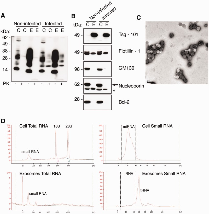
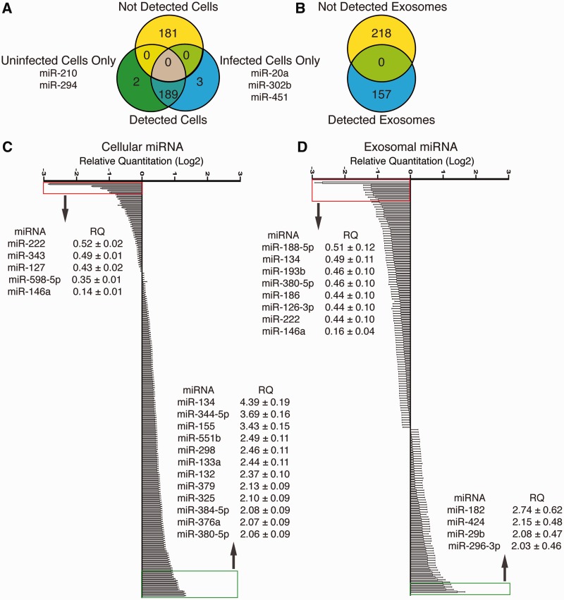
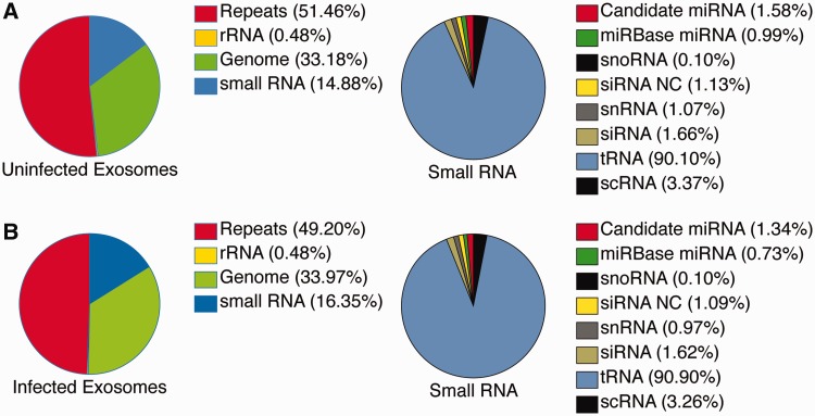
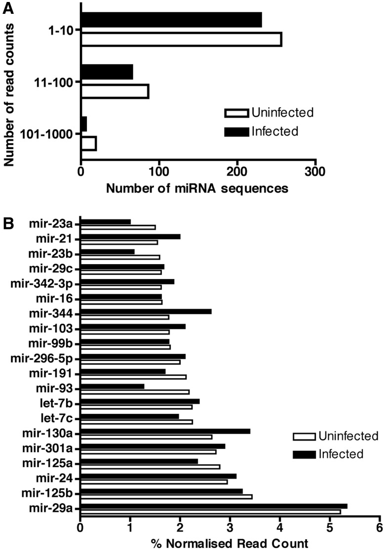
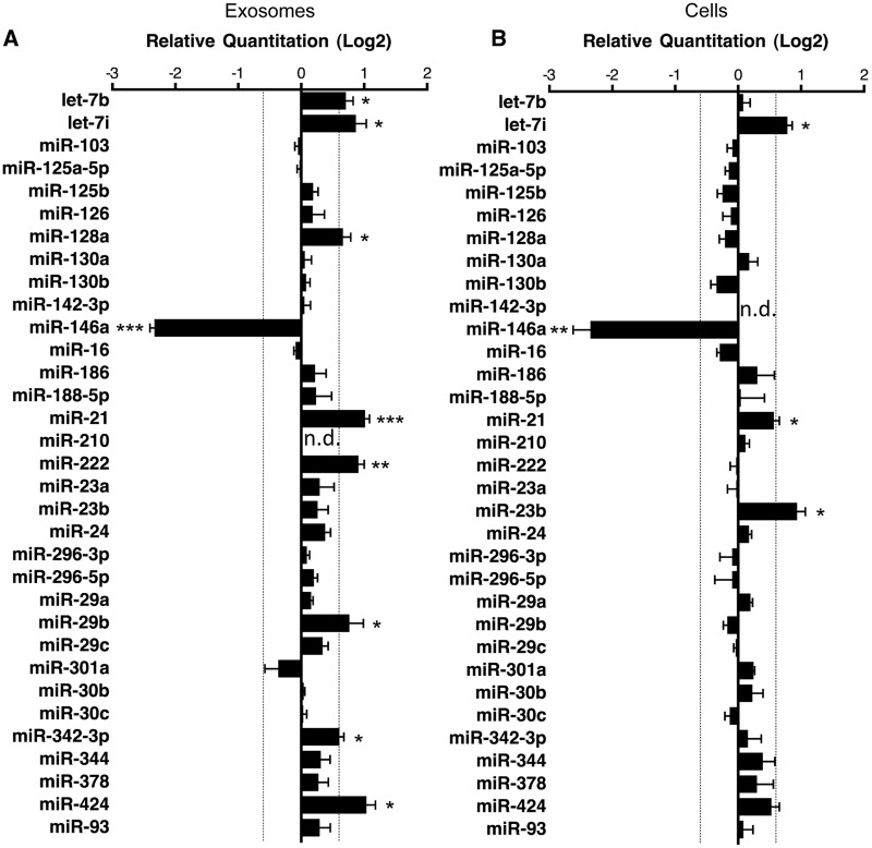
Prion diseases are transmissible neurodegenerative disorders affecting both humans and animals. The cellular prion protein, PrP(C), and the abnormal infectious form, PrP(Sc), are found associated with exosomes, which are small 50-130 nm vesicles released from cells. Exosomes also contain microRNAs (miRNAs), a class of non-coding RNA, and have been utilized to identify miRNA signatures for diagnosis of disease. While some miRNAs are deregulated in prion-infected brain tissue, the role of miRNA in circulating exosomes released during prion disease is unknown. Here, we investigated the miRNA profile in exosomes released from prion-infected neuronal cells. We performed the first small RNA deep sequencing study of exosomes and demonstrated that neuronal exosomes contain a diverse range of RNA species including retroviral RNA repeat regions, messenger RNA fragments, transfer RNA fragments, non-coding RNA, small nuclear RNA, small nucleolar RNA, small cytoplasmic RNA, silencing RNA as well as known and novel candidate miRNA. Significantly, we show that exosomes released by prion-infected neuronal cells have increased let-7b, let-7i, miR-128a, miR-21, miR-222, miR-29b, miR-342-3p and miR-424 levels with decreased miR-146 a levels compared to non-infected exosomes. Overall, these results demonstrate that circulating exosomes released during prion infection have a distinct miRNA signature that can be utilized for diagnosis and understanding pathogenic mechanisms in prion disease.


Figures

Figure 1.
Figure 1.
Exosomes isolated from prion-infected cells contain small RNA. (A) Western blot of PrPC and PrPSc in GT1-7 cell lysates and GT1-7 exosomes. Anti-PrP antibody ICSM-18 reveals the presence of PrP in GT1-7 cell (C) lysates and GT1-7 exosome (E) lysates (40 µg total protein loaded) from both non-infected and infected samples, and following PK-digested (100 µg total protein digested with 25 µg/ml PK) PrPSc is indicated by presence PK-resistant protein in infected (C) cell lysates and exosomes (E). (B) Isolated exosomes were confirmed by enrichment of exosome markers tsg-101 and flotillin-1, and negative for Golgi marker GM130, nuclear marker nucleoporin and mitochondrial marker Bcl-2. Asterisks indicate non-specific band for anti-nucleoporin, p62. (C) Representative TEM of non-infected GT1-7 exosomes revealed homogenous populations of extracellular vesicles of ∼100 nm in diameter that are characteristic of exosomes. Scale bar 200 nm. (D) Bioanalyser analysis of RNA isolated from cells and exosomes on the total RNA and small RNA Chips. Exosomes lack detectable 18 S and 28S rRNA bands compared to total cell RNA. Cells and exosomes both contain miRNA between 4 and 40 nt, with exosomes enriched in tRNA at ∼60 nt.
Figure 2.
Figure 2.
Profiling of miRNA Isolated from prion-infected cells and exosomes. (A) Venn diagram of detected miRNA in uninfected and prion-infected cells with TLDA cards that detect 375 known miRNA (n = 2). (B) Venn diagram of detected miRNA in uninfected and prion-infected exosomes with TLDA cards that detect 375 known miRNA (n = 2). (C) Relative quantitation of 189 miRNA is detected in uninfected and infected cells. Twelve miRNAs were detected >2-fold up-regulated and five miRNAs were detected as <2-fold down-regulated. Data represent mean ± SEM normalized to sno135 and sno202 endogenous controls using ΔΔCt method (n = 2). (D) Relative quantitation of 157 miRNAs detected in uninfected and infected exosomes. Four miRNAs were detected >2-fold up-regulated and eight miRNA were detected as <2-fold down-regulated. Data represent mean ± SEM normalized to sno135 and sno202 endogenous controls using ΔΔCt method (n = 2).
Figure 3.
Figure 3.
Small RNA library composition from deep sequencing of neuronal exosomes. (A and B) Percentage reads mapping in uninfected exosome (A) and infected exosome (B) libraries to RNA repeats, rRNA, genomic regions and small RNA. Small RNA mapping is further categorized to tRNA, siRNA and siRNA derived from ncRNA, snoRNA, scRNA, snRNA, mirBase miRNA and candidate miRNA sequences (n = 2).
Figure 4.
Figure 4.
Known miRNAs sequenced in uninfected and prion-infected exosomes. (A) Distribution of known miRNA sequences in uninfected and infected exosomes based on the total read counts. Mature miRNA sequences are categorized by number of read counts corresponding to each individual miRNA (n = 2). (B) Top 20 known miRNAs expressed in uninfected and infected exosomes. The percent contribution of each miRNA sequence in the total pool of known miRNA sequences was calculated by dividing the individual miRNA read count by the total number of known miRNAs sequenced in the corresponding library (n = 2).
Figure 5.
Figure 5.
Validation of miRNA ‘hits’ in infected exosomes and cells. (A) Exosomes miRNA detection by TaqMan qPCR miRNA assay, miR-210 was used as a negative control for exosomal miRNA. Relative quantitation data represent mean ±SEM normalized to sno135 and sno202 using ΔΔCt method. Fold change >1.5 or <1.5 is indicated with significance determined by Mann–Whitney U-test; *P < 0.05; **P < 0.01; ***P < 0.001 (n = 6–7 independent experiments, n.d. = not detected). (B) Cellular miRNA detection by TaqMan qPCR miRNA assay, miR-142-3 p was used as a negative control for cellular miRNA. Relative quantitation data represent mean ± SEM of normalized to sno135 and sno202 using ΔΔCt method. Fold change >1.5 or <1.5 is indicated by the dashed line with significance determined by Mann–Whitney U-test; *P < 0.05; **P < 0.01 (n = 6–7 independent experiments, n.d. = not detected).

References

    1. Prusiner SB. Novel proteinaceous infectious particles cause scrapie. Science. 1982;216:136–144. - PubMed
    1. Aguzzi A, Heikenwalder M. Pathogenesis of prion diseases: current status and future outlook. Nat. Rev. Microbiol. 2006;4:765–775. - PubMed
    1. Thery C, Zitvogel L, Amigorena S. Exosomes: composition, biogenesis and function. Nat. Rev. Immunol. 2002;2:569–579. - PubMed
    1. Fevrier B, Vilette D, Archer F, Loew D, Faigle W, Vidal M, Laude H, Raposo G. Cells release prions in association with exosomes. Proc. Natl Acad. Sci. USA. 2004;101:9683–9688. - PMC - PubMed
    1. Vella LJ, Sharples RA, Lawson VA, Masters CL, Cappai R, Hill AF. Packaging of prions into exosomes is associated with a novel pathway of PrP processing. J. Pathol. 2007;211:582–590. - PubMed

Publication types