. 2012 Nov;40(21):11009-22.
doi: 10.1093/nar/gks792. Epub 2012 Sep 10.

The Ighmbp2 helicase structure reveals the molecular basis for disease-causing mutations in DMSA1

Affiliations

The Ighmbp2 helicase structure reveals the molecular basis for disease-causing mutations in DMSA1

Siew Choo Lim et al. Nucleic Acids Res. 2012 Nov.

Abstract

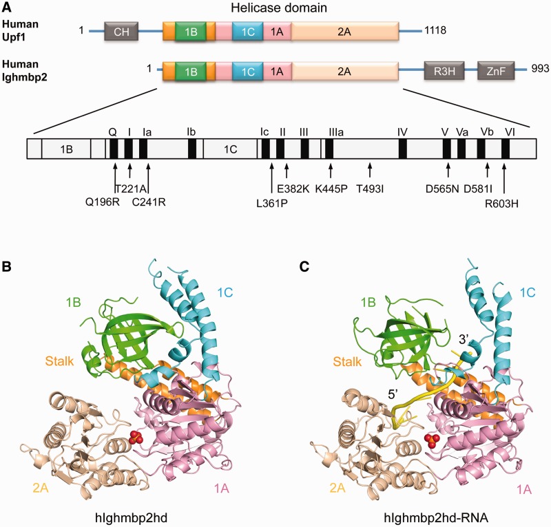
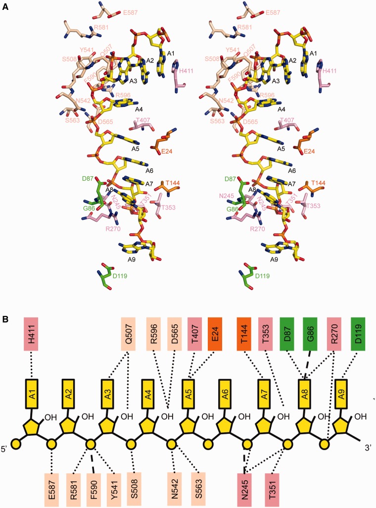
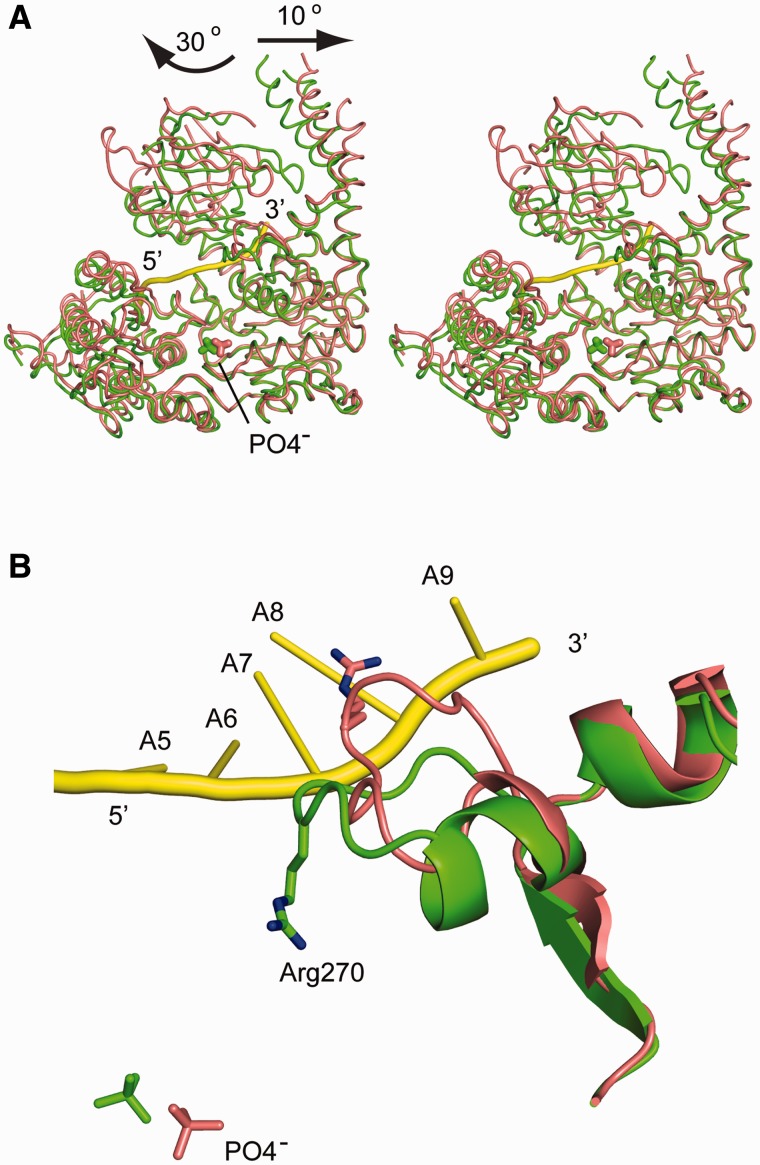
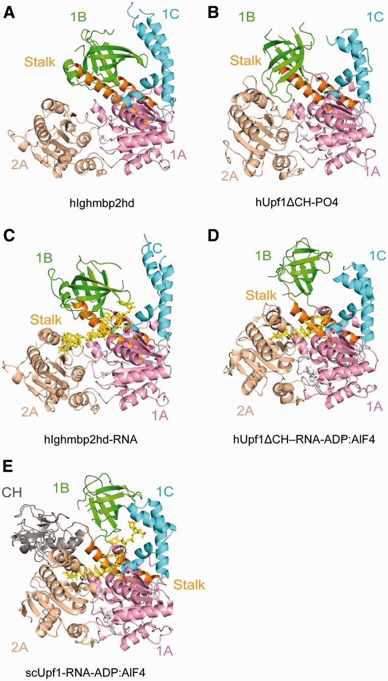
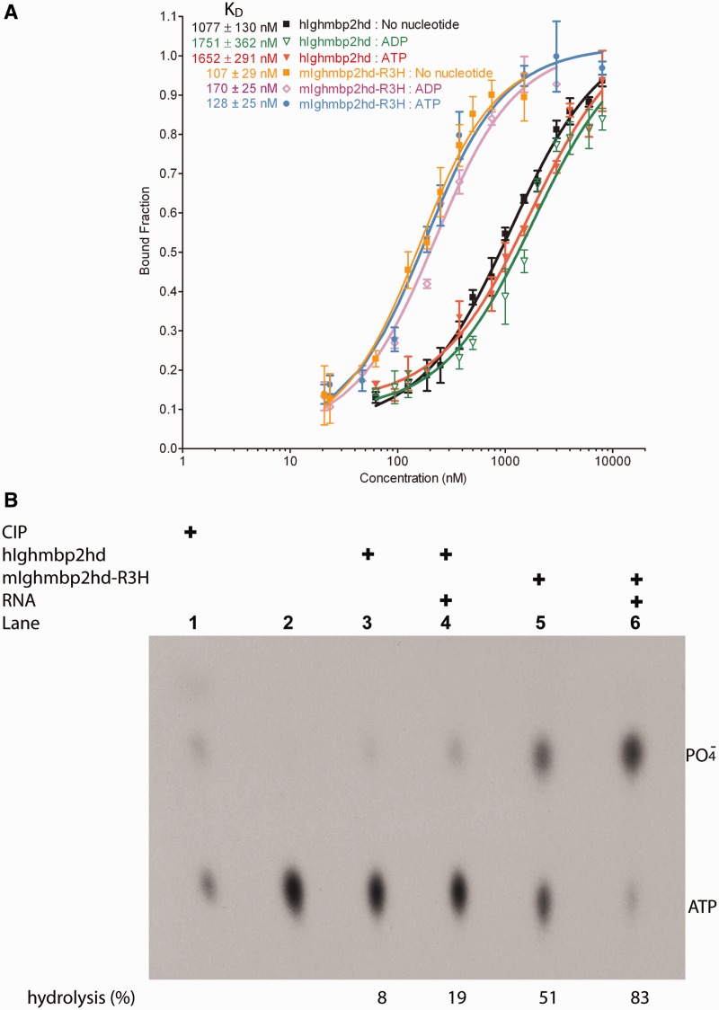
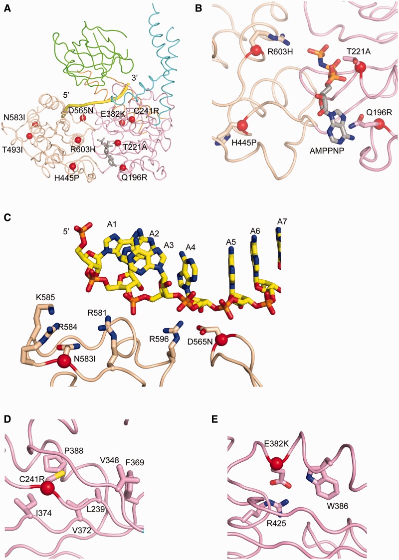
Mutations in immunoglobulin µ-binding protein 2 (Ighmbp2) cause distal spinal muscular atrophy type 1 (DSMA1), an autosomal recessive disease that is clinically characterized by distal limb weakness and respiratory distress. However, despite extensive studies, the mechanism of disease-causing mutations remains elusive. Here we report the crystal structures of the Ighmbp2 helicase core with and without bound RNA. The structures show that the overall fold of Ighmbp2 is very similar to that of Upf1, a key helicase involved in nonsense-mediated mRNA decay. Similar to Upf1, domains 1B and 1C of Ighmbp2 undergo large conformational changes in response to RNA binding, rotating 30° and 10°, respectively. The RNA binding and ATPase activities of Ighmbp2 are further enhanced by the R3H domain, located just downstream of the helicase core. Mapping of the pathogenic mutations of DSMA1 onto the helicase core structure provides a molecular basis for understanding the disease-causing consequences of Ighmbp2 mutations.


Figures

Figure 1.
Figure 1.
Structural overview of hIghmbp2hd and hIghmbp2hd–RNA. (A) Schematic representation of the domain arrangement in Upf1 and Ighmbp2. The helicase core region contains two RecA-like domains: domain 1A (pink) and domain 2A (wheat) and additional regulatory domains: domain 1B (green), domain 1C (cyan) and the ‘stalk’ (orange). Upf1 contains another regulatory domain, the CH domain (gray) located upstream of the helicase region while Ighmbp2 features two more another regulatory domains: a R3H domain (gray) and an AN-1 type zinc-finger domain (gray) downstream of the helicase region. Missense mutations and canonical motifs in Ighmbp2 are labeled. (B) Crystal structures of hIghmbp2hd. (C) Crystal structure of hIghmbp2hd–RNA. The coloring schemes for domains are as in (A). The bound phosphate ion and ssRNA are shown as red sphere and yellow cartoon tube, respectively.
Figure 2.
Figure 2.
Protein–RNA interactions. (A) Stereo view showing the ssRNA-binding site in hIghmbp2hd–RNA. The bound ssRNA and the residues involved protein–RNA interactions are shown in ball-stick models. The color scheme is as in Figure 1. (B) Schematic representation of the contacts between the hIghmbp2hd protein and RNA.
Figure 3.
Figure 3.
Conformational changes of hIghmbp2hd upon RNA binding. (A) Stereo view showing the superposition of hIghmbp2hd–RNA (pink) with hIghmbp2hd (green). Phosphate ions are shown as sticks and the bound ssRNA is shown in yellow cartoon. The arrows indicate the movement of domain 1B and 1C in the RNA-bound state compared to the RNA-free state. (B) Conformational change of a loop region (residues 264–273) and the reorientation of residue Arg270 in response to ssRNA binding. The position of the phosphate ions indicates the location of the nucleotide-binding site.
Figure 4.
Figure 4.
Structural comparison of Ighmbp2 with Upf1. The ribbon diagrams are drawn with domains 1A (pink) and 2A (wheat) in the same orientation. The bound phosphate ion and nucleotide are shown in gray sticks. ssRNA is show in yellow sticks. The coloring scheme for domains are as in Figure 1. (A) hIghmbp2hd. (B) hUpf1ΔCH-PO4 (PDB code: 2GK7). (C) hIghmbp2hd–RNA. (D) hUpf1ΔCH-RNA-ADP:AlF4 (PDB code 2XZO). (E) Yeast Upf1-RNA-ADP:AlF4 (PDB code: 2XZL).
Figure 5.
Figure 5.
RNA-binding and ATPase activities of Ighmbp2. (A) Quantitative measurements of RNA-binding affinities of hIghmbp2hd and mIghmbp2hd-R3H in solution by fluorescence anisotropy. Dissociation constants (KD) values were determined by fitting the experimental data to a binding equation describing a single-site binding model. The KD values and their corresponding errors are the mean and standard deviation of three independent experiments. (B) ATPase activities of hIghmbp2hd and hIghmbp2hd-R3H in the absence or presence of ssRNA. The position of phosphate was determined by treating the substrate with CIP.
Figure 6.
Figure 6.
Structural roles of residues associated with disease-causing missense mutations in SMARD1. (A) Disease-causing missense mutations in DSMA1 are mapped in the structure of hIghmbp2hd–RNA with the Cα atoms of the mutated residues shown as red spheres. AMPPNP (in stick model) is modeled by superposition of the structure of hIghmbp2hd–RNA with that of human Upf1ΔCH-AMPPNP (PBD code: 2GJK). (B) Class I mutations residing in or close to the nucleotide-binding pocket. (C) Class II mutations located in the RNA-binding channel. (D) and (E) Class III mutations located far away from either the nucleotide-binding pocket or the RNA-binding channel.

Similar articles

Cited by

References

    1. Mellins RB. Pulmonary physiotherapy in the pediatric age group. Am. Rev. Respir. Dis. 1974;110:137–142. - PubMed
    1. Lefebvre S, Burglen L, Reboullet S, Clermont O, Burlet P, Viollet L, Benichou B, Cruaud C, Millasseau P, Zeviani M, et al. Identification and characterization of a spinal muscular atrophy-determining gene. Cell. 1995;80:155–165. - PubMed
    1. Gubitz AK, Feng W, Dreyfuss G. The SMN complex. Exp. Cell Res. 2004;296:51–56. - PubMed
    1. Paushkin S, Gubitz AK, Massenet S, Dreyfuss G. The SMN complex, an assemblyosome of ribonucleoproteins. Curr. Opin. Cell Biol. 2002;14:305–312. - PubMed
    1. Meister G, Eggert C, Fischer U. SMN-mediated assembly of RNPs: a complex story. Trends Cell. Biol. 2002;12:472–478. - PubMed

Publication types

MeSH terms

Supplementary concepts