. 2012 Nov;40(21):10851-65.
doi: 10.1093/nar/gks833. Epub 2012 Sep 10.

Common chaperone activity in the G-domain of trGTPase protects L11-L12 interaction on the ribosome

Affiliations

Common chaperone activity in the G-domain of trGTPase protects L11-L12 interaction on the ribosome

Dandan Zhang et al. Nucleic Acids Res. 2012 Nov.

Abstract

Translational GTPases (trGTPases) regulate all phases of protein synthesis. An early event in the interaction of a trGTPase with the ribosome is the contact of the G-domain with the C-terminal domain (CTD) of ribosomal protein L12 (L12-CTD) and subsequently interacts with the N-terminal domain of L11 (L11-NTD). However, the structural and functional relationships between L12-CTD and L11-NTD remain unclear. Here, we performed mutagenesis, biochemical and structural studies to identify the interactions between L11-NTD and L12-CTD. Mutagenesis of conserved residues in the interaction site revealed their role in the docking of trGTPases. During docking, loop62 of L11-NTD protrudes into a cleft in L12-CTD, leading to an open conformation of this domain and exposure of hydrophobic core. This unfavorable situation for L12-CTD stability is resolved by a chaperone-like activity of the contacting G-domain. Our results suggest that all trGTPases-regardless of their different specific functions-use a common mechanism for stabilizing the L11-NTD•L12-CTD interactions.


Figures

Figure 1.
Figure 1.
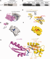
Functional significance of loop62 in translation. (A) The crystal structure of 70S•EF-G•FA in POST state (PDB 2WRL) (22) shows that EF-G is located between L12-CTD and L11-NTD. MD simulations reveal details of the L11-NTD•L12-CTD interaction. Loop62 of L11-NTD (magenta) inserts into the cleft between α-helixes 4 and 6 of L12-CTD (yellow). Four residues on loop62, Tyr61 (Y61), Ala62 (A62), Asp63 (D63) and Arg64 (R64), interact directly with L12-CTDMD. (B) Multiple alignments show that residues 61, 63 and 64 are highly conserved. Red residues show 100% identity, while blue residues show 80% identity. (C and D) In vivo analyses of loop62 mutations on cell growth. (C) The doubling time of rplK knock-out E. coli cells which are rescued by WT L11 or its mutant (L11-Y61A, A62K, D63L or R64A). (D) Equal amount of the rplK knock-out E. coli cells rescued by WT L11 or its mutant were grown as indicated and the proportion of mutant cells (%) was determined. (E–G) Importance of loop62 for protein synthesis. WT L11 (deep blue), L11-Y61A (pink), L11-A62K (light pink), L11-D63L (red) or L11-R64A (pink) L11 mutants were reconstituted into ribosomes lacking L11. Ribosomes reconstituted with WT L11 were incubated with Thio (blue) or micrococcin (ice blue). (E) Quantification of the total effect of each mutant on GFP synthesis in the RTS system. (F) Chemical footprinting of E. coli 70S ribosome·EF-G·GDPNP complexes. G and T, sequencing lanes; Co, control without DMS modification; 70SWT, WT ribosome; 70S−L11, the ribosome depleted of L11; 70S−L11 + L11, the ribosome depleted of L11 was reconstituted with WT L11 or the L11 mutant as indicated. The EF-G protection pattern of A2660 and A1607 of 23S rRNA is indicated by arrows. (G) GTP hydrolysis by EF-G is given as the amount of GTP hydrolyzed in 5 min per ribosome. Error bars represent the mean ± SEM of data from at least three separate experiments. P-values were calculated with unpaired t-tests using Welch’s correction by comparing to WT samples (*P < 0.05, **P < 0.01, ***P < 0.001). See also Supplementary Figure S1.
Figure 2.
Figure 2.
Direct interaction between L11 and L12 requires L12-CTD to be in an open conformation with the hydrophobic core exposed. (A and B) Interactions between 6× His-tagged L12 and L11 WT (A) or mutants (B) were detected by in vitro pull-down assay coupled to immunoblotting against the L11 antibody (αL11). Purified L11 protein was loaded in the first lane (L11) as a marker. (A) Pull-down assays show that L11 binds with L12. No L11 was detected in the control (the third lane) when L11 was incubated with empty beads. (B) The bait-prey pair was the L12 + L11 mutant. The D63L mutant did not bind with L12 at all. Y61A and R64A mutants bound a lower intensity, whereas the A62K mutant bound with intensity similar to WT L11. (C–F) Structural comparison of L12-CTD in the isolated state (gray) and on the ribosome (yellow). (C) When L12-CTD on the ribosome complex (PDB 2WRL) (22) was replaced by its isolated structure (PDB 1CTF) (29), the interplay between L11-NTD (magenta) and L12-CTD was hindered by a steric clash at the connection site (circled region). (D) Structural alignment shows L12-CTD on the ribosome with a more open conformation. When the two structures were aligned to helix 6 (α6), the tip of helix 4, i.e. the Cα of G74, moved 6.4 Å and the helices opened 32.4° more, as measured by the angle of A113:Cα to G74:Cα in the two states. (E, F) Surface display of L12-CTD with its hydrophobic core in blue (E) or orange (F) in the closed and open states, respectively. (G) The interface between L11-NTD and L12-CTD in the open state. The interface is composed of Y61 (orange), A62 (orange), D63 (red, negative charged) and R64 (blue, positive charged) from L11 and K95 (blue, positive charged) from L12. The hydrophobic core of L2-CTD includes L58, L94, L106 and L110 (orange). See also Supplementary Figures S2 and S3.
Figure 3.
Figure 3.
Both electrostatic and hydrophobic forces contribute to the L11–L12 connection. (A) An interaction between L11 (WT or mutant) and L12 (WT or mutant) was detected by pull-down assays as described in Figure 2A and B. The bait–prey pair is indicated as L12 + L11 (L12 = WT or K95A or L106A; L11 = WT or R64K). When Arg64 of loop62 was mutated to Lys, the interaction between L11 and L12 was enhanced. When Lys95 of L12 was replaced by Ala, the interaction vanished. Similarly, the L106A mutation abolished the interaction as well. (B) Chemical footprinting of E. coli 70S ribosome•EF-G•GDPNP complexes. G and T, sequencing lanes; Co, without DMS modification. 70S reconstituted with L11 or L12 are indicated as in (A). The EF-G protection pattern of A2660 and A1607 of 23S rRNA is indicated by arrows. (C and D) Importance of the L11–L12 interaction for EF-G-dependent GTP hydrolysis and protein synthesis, respectively. WT L12 and WT L11 are shown in deep blue and all other combinations in pink. The corresponding proteins L12 and L11 were reconstituted into ribosomes lacking L11 and L12. (C) GTP hydrolysis by EF-G is given as the amount of GTP hydrolyzed in 5 min per ribosome. (D) The total effect of each mutant on GFP synthesis was quantified in the RTS system. Error bars represent the mean ± SEM of data from at least three separate experiments. P-values were calculated with unpaired t-test using Welch’s correction by comparing to WT + WT samples (**P < 0.01, ***P < 0.001). See also Supplementary Figure S4.
Figure 4.
Figure 4.
Identification of EF4’s chaperone activity. Denatured protein in the absence (empty column) and presence of EF-G (gray) or EF4 (black) was studied in the refolding assay. (A) 0.1 μM denatured CS was incubated in the absence and presence of EF-G or EF4 at the indicated concentrations. (B) Influence of guanosine phosphates on the chaperone activity of EF4. A total of 0.1 μM denatured CS and 5 μM EF-G or EF4 were incubated in the absence (−) and presence of 2 mM GDP, GTP or its non-hydrolysable analogue GDPNP. (C) Influence of the ribosome on the chaperone activity of EF4. A total of 0.1 μM denatured CS and 5 μM EF-G or EF4 were incubated in the absence (−) and presence of 0.2 μM 70S ribosomes. (D) Influence of EF4 on the refolding of denatured α-glucosidase. Denatured α-glucosidase (0.1 μM) in the absence and presence of 5 μM EF-G or EF4, with 2 mM GDP, GTP or GDPNP, or with 0.2 μM 70S ribosomes. (E) Influence of EF4 on the thermal aggregation of CS. The aggregation of CS at 43°C was monitored by light scattering at 320 nm. Light gray: no chaperone added; gray: EF-G; black: EF4. Error bars represent the mean ± SEM of data from at least three separate experiments. P-values were, calculated with unpaired t-test with Welch’s correction by comparing to EF-G or EF4 samples (*P < 0.05, ** P < 0.01, ***P < 0.001).
Figure 5.
Figure 5.
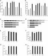
Common chaperone activity on the G-domains of trGTPase. (A and B) Chaperone activity of trGTPases. Denatured CS (A) or α-glucosidase (B) in the absence (−) and presence of translational factors was studied in a refolding assay. Denatured protein (0.1 μM) was incubated in the absence and presence of 5 μM translational factor. Similar to EF4, all trGTPases (black) showed chaperone activity, while all other translational factors (gray) exhibited much lower values, close to the negative control (−). (C–H) The G-domain of trGTPases is a chaperone activity center. To localize the enzymatic center, EF-G (C) or EF4 (D) was truncated as indicated. Full length and truncated proteins were studied in the CS (E, F) and α-glucosidase (G, H) refolding assays. Denatured protein (0.1 μM) was incubated in the absence (−) and presence of 5 μM of the factor constructs. Error bars represent the mean ± SEM of data from at least three separate experiments. P-values were calculated with unpaired t-test with Welch’s correction by comparing to full length EF-G or EF4 samples, (*P < 0.05, ***P < 0.001). See also Supplementary Figure S5 and Supplementary Table S1.
Figure 6.
Figure 6.
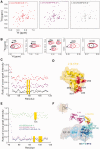
TrGTPase facilitates L11–L12 interaction in vitro. (A–C) NMR analysis of L12-CTD·EF4 interaction. (A) 1H-15N HSQC spectra of the 15N-labeled L12-CTD (E. coli) titrated with EF4 (T. thermophilus) at different ratios. (B) A close-up view of the cross-peaks showing distinct changes in chemical shifts. The black arrow indicates the direction of the chemical shift. The K108 diagram is from a sample of the less affected cross-peak obtained by EF4 titration. (C) Cross-peak intensities of residues in L12-CTD plotted versus residue number. Varying molar ratios of EF4 to L12-CTD are colored as in A. Cross-peak intensities were evaluated as peak heights. Data were normalized using the intensities before EF4 titration. The highlighted regions (residues 78–86) exhibit comparably strong decrease in intensity. (D) Spatial locations of the residues on L12-CTD that are responsible for interacting with EF4. (E) L12-CTD was titrated by L11 or EF-G or both proteins. Scales of axes are the same as in C. Data were normalized using the intensity before titration. The highlighted regions exhibited comparably strong decreases in intensity. (F) EF4 from the cryo-EM structure of 70S·EF4·GDPNP in the PRE state (PDB 3DEG) (33) aligned with the EF-G in the crystal structure in (2WRL) (22). The G-domains of EF4 (blue) and EF-G (cyan) are represented as cartoons. The G′ domain of EF-G contacts L12-CTD from the lower right-hand side. Red residues in L12-CTD are the same as in D, namely those interacting EF4. It suggests the G-domain of EF4 might interact with the L12-CTD in a similar way as EF-G. See also Supplementary Figure S6 and Supplementary Table S2.
Figure 7.
Figure 7.
A ‘Support-and-Protect’ model of the L11–L12 interaction protected by the G-domain chaperone. (A) The ribosome is in the “Factor-Free” state that L12-CTD (yellow) protruding from the body of the ribosome has no interaction with L11-NTD (magenta). (B) Upon trGTPase loading, i.e. EF-G (dotted), L12-CTD initiates the interaction of the factor with the ribosome— “With Factor” state. (C) After EF-G fully docked and triggered cis-trans isomerization of PS22, L11-NTD (from magenta to red) inserts into L12-CTD, which has now an open conformation. Here, L12-CTD is in the state of “With Factor and L11”. EF-G from the lower right-hand side supports and protects L12-CTD. For details, see the text and Supplementary Movie S1.

Similar articles

Cited by

References

    1. Diaconu M, Kothe U, Schlunzen F, Fischer N, Harms JM, Tonevitsky AG, Stark H, Rodnina MV, Wahl MC. Structural basis for the function of the ribosomal L7/12 stalk in factor binding and GTPase activation. Cell. 2005;121:991–1004. - PubMed
    1. Traut RR, Dey D, Bochkariov DE, Oleinikov AV, Jokhadze GG, Hamman B, Jameson D. Location and domain structure of Escherichia coli ribosomal protein L7/L12: site specific cysteine cross-linking and attachment of fluorescent probes. Biochem. Cell Biol. 1995;73:949–958. - PubMed
    1. Agrawal RK, Linde J, Sengupta J, Nierhaus KH, Frank J. Localization of L11 protein on the ribosome and elucidation of its involvement in EF-G-dependent translocation. J. Mol. Biol. 2001;311:777–787. - PubMed
    1. Christodoulou J, Larsson G, Fucini P, Connell SR, Pertinhez TA, Hanson CL, Redfield C, Nierhaus KH, Robinson CV, Schleucher J, et al. Heteronuclear NMR investigations of dynamic regions of intact Escherichia coli ribosomes. Proc. Natl Acad. Sci. USA. 2004;101:10949–10954. - PMC - PubMed
    1. Kavran JM, Steitz TA. Structure of the base of the L7/L12 stalk of the Haloarcula marismortui large ribosomal subunit: analysis of L11 movements. J. Mol. Biol. 2007;371:1047–1059. - PubMed

Publication types

MeSH terms