. 2012 Nov 1;40(20):10334-44.
doi: 10.1093/nar/gks802. Epub 2012 Sep 10.

A hybrid G-quadruplex structure formed between RNA and DNA explains the extraordinary stability of the mitochondrial R-loop

Affiliations

A hybrid G-quadruplex structure formed between RNA and DNA explains the extraordinary stability of the mitochondrial R-loop

Paulina H Wanrooij et al. Nucleic Acids Res. .

Abstract

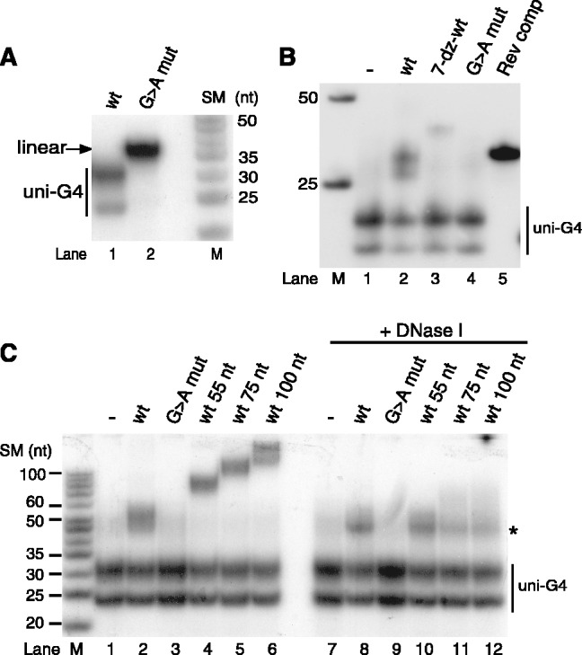
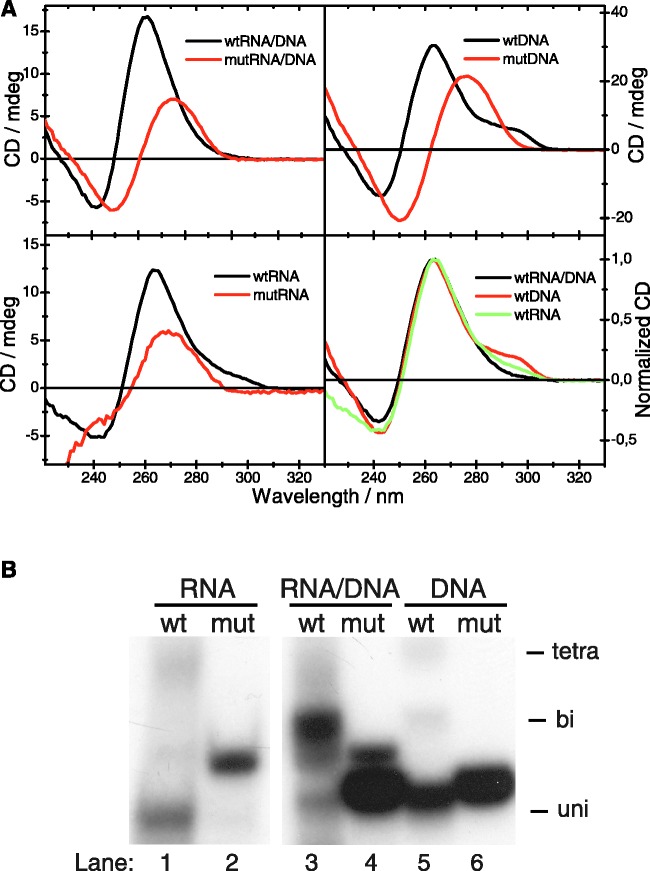
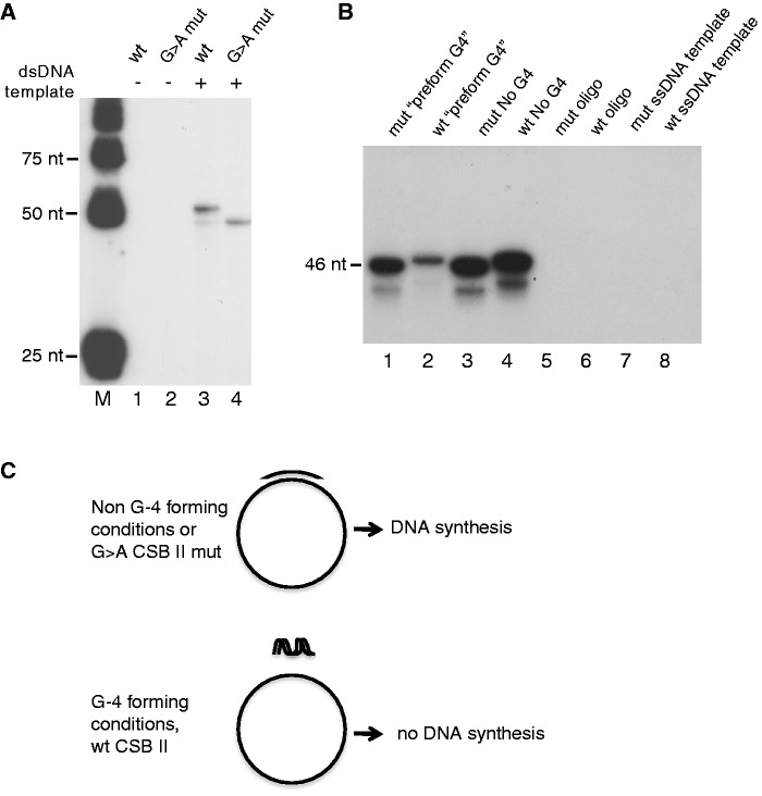
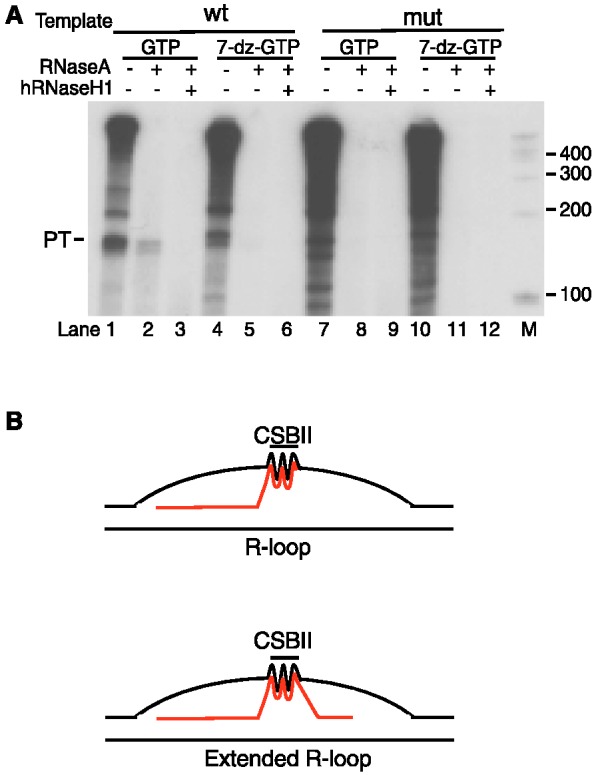
In human mitochondria the transcription machinery generates the RNA primers needed for initiation of DNA replication. A critical feature of the leading-strand origin of mitochondrial DNA replication is a CG-rich element denoted conserved sequence block II (CSB II). During transcription of CSB II, a G-quadruplex structure forms in the nascent RNA, which stimulates transcription termination and primer formation. Previous studies have shown that the newly synthesized primers form a stable and persistent RNA-DNA hybrid, a R-loop, near the leading-strand origin of DNA replication. We here demonstrate that the unusual behavior of the RNA primer is explained by the formation of a stable G-quadruplex structure, involving the CSB II region in both the nascent RNA and the non-template DNA strand. Based on our data, we suggest that G-quadruplex formation between nascent RNA and the non-template DNA strand may be a regulated event, which decides the fate of RNA primers and ultimately the rate of initiation of DNA synthesis in human mitochondria.


Figures

Figure 1.
Figure 1.
G-quadruplex formation in nascent RNA, but not in template DNA, causes premature termination of transcription. In vitro transcription with the mitochondrial transcription apparatus was carried out in the presence or absence of 7-deaza-GTP to monitor effects of G-quadruplex formation in RNA. To address the effects of G-quadruplex formation in DNA, the DNA templates used were produced by PCR amplification of positions 1–512 of human mtDNA in the presence or absence of 7-deaza-dGTP.
Figure 2.
Figure 2.
Native gel analysis reveals hybrid G-quadruplex formation between RNA and DNA oligonucleotides spanning CSB II. (A) An RNA oligonucleotide encompassing CSB II migrates faster on native gels than a CSB II mutant molecule of identical length where all Gs have been replaced with As, indicative of G-quadruplex structure formation, as shown in (15). Products were analysed on 12% native acrylamide gels. M, marker lane; SM, size of marker. (B) A labeled CSB II RNA oligonucleotide was allowed to form G-quadruplexes either alone or in the presence of unlabeled DNA oligonucleotides and analysed as in panel A. A slower migrating species indicative of a RNA–DNA hybrid G-quadruplex was formed in the presence of a wildtype DNA oligonucleotide containing CSB II. (C) Hybrid G-quadruplexes formed between CSB II RNA (5′ end-labeled) and DNA oligonucleotides of indicated lengths. Nuclease treatment identifies a DNase I-resistant complex that contains both RNA and DNA (indicated with an asterisk).
Figure 3.
Figure 3.
CD spectra of CSB II RNA, DNA or RNA–DNA hybrid samples. (A) Wildtype (black) and G→T mutant (red) CSB II oligonucleotides were incubated to form G-quadruplexes and subjected to CD. The CD spectra of the wildtype RNA–DNA hybrid species (top left), DNA (top right) and RNA (bottom left) all exhibit a peak at 260 nm, characteristic of parallel-type G-quadruplex structures, whereas the spectra of the mutant oligoucleotides lacking Gs resemble ssDNA. A second, smaller peak at 290 nm is observed in CSB II DNA and RNA spectra, and has been assigned to loop residues in a propeller-type parallel G-quadruplex. Bottom right: Overlay of the wildtype CD spectra of CSB II DNA (red), RNA (green) and the RNA–DNA hybrid (black). All wildtype spectra are characteristic of parallel-type G-quadruplexes, but only the RNA and DNA spectra have a smaller second peak at 290 nm. (B) The samples from (A) were separated on a native acrylamide gel in order to observe G-quadruplex formation. The positions of tetra-, bi- and uni-molecular G-quadruplexes are indicated.
Figure 4.
Figure 4.
G-quadruplex formation inhibits primer extension by POLγ. (A) POLγ can extend both wildtype and G→A mutant CSB II primers when they are annealed to a negatively supercoiled plasmid DNA template. Please note that the difference in G-content leads to a slight difference in gel-migration (compare lanes 3 and 4). (B) POLγ can efficiently support primer extension of the G→A mutant (mut), but not the wildtype CSB II (wt) primer, if the primers are incubated under conditions that stimulate the formation of G-quadruplex structures (pre-form G4) prior to addition to the template strand. No DNA synthesis occurs in the presence of only the oligonucleotides or only the ssDNA template (lanes 5–8). (C) A schematic figure depicting the set-up and outcome of the experiment panel (B). Preincubation at 37°C and in the presence of 100 mM KCl stimulates G-quadruplex formation.
Figure 5.
Figure 5.
Hybrid G-quadruplexes form during transcription and are dependent on CSB II. (A) In vitro transcription using the mitochondrial transcription apparatus on supercoiled templates containing either wildtype or mutant CSB II. Transcription reactions were divided into three parts, and either not treated or treated with RNase A and/or hRNaseH1, as indicated. RNase A-treatment reveals a non-degradable product of 45–50 bp (marked by asterisks) that is dependent on the CSB II sequence and Hoogsteen base pairing. (B) The labeled CSB II RNA oligonucleotide was allowed to form G-quadruplexes alone (lanes 1–3), with wildtype CSB II DNA oligonucleotide of identical sequence (lanes 4–6), or with a reverse complement DNA oligonucleotide (lanes 7–9), and then subjected to treatment with RNase A or hRNaseH1 in order to show that the G-quadruplex hybrid is resistant to hRNaseH1 (lane 6). The Watson–Crick basepaired hybrid with reverse complement DNA was degraded by hRNaseH1 as expected (lane 9). M, marker lane. (C) Transcription on templates encompassing base positions 1–477 of human mtDNA and containing either dGTP or 7-deaza-dGTP (lanes 1 and 2). After transcription, part of each reaction was treated with RNaseA (lanes 3 and 4). The RNaseA-resistant species of ∼50 bp (indicated by black bar) are absent when G-quadruplex formation with the template is eliminated (lane 3). M, marker lane.
Figure 6.
Figure 6.
A longer R-loop can form during transcription of human mtDNA with phage T7 RNA polymerase. (A) Transcription of a human mtDNA template containing wildtype or mutant CSB II with T7 RNA pol in the presence of either GTP (lanes 1–3 and 7–9) or 7-deaza-GTP (lanes 4–6 and 10–12). A hybrid species of ∼120 bp is revealed upon RNase A treatment (lane 2) and is sensitive to hRNaseH1 (lane 3). This longer hybrid is dependent on CSB II and not observed in the presence of 7-deaza-GTP (lanes 4–6). (B) Schematic presentation of the RNA–DNA hybrid G-quadruplex that forms between the RNA and the non-template DNA strand during transcription of mtDNA. Under some conditions, an extended R-loop may be formed, similar to that reported in (22).

References

    1. Ojala D, Montoya J, Attardi G. tRNA punctuation model of RNA processing in human mitochondria. Nature. 1981;290:470–474. - PubMed
    1. Clayton DA. Replication and transcription of vertebrate mitochondrial DNA. Annu. Rev. Cell Biol. 1991;7:453–478. - PubMed
    1. Falkenberg M, Larsson NG, Gustafsson CM. DNA replication and transcription in mammalian mitochondria. Annu. Rev. Biochem. 2007;76:679–699. - PubMed
    1. Falkenberg M, Gaspari M, Rantanen A, Trifunovic A, Larsson NG, Gustafsson CM. Mitochondrial transcription factors B1 and B2 activate transcription of human mtDNA. Nat. Genet. 2002;31:289–294. - PubMed
    1. Chang DD, Clayton DA. Priming of human mitochondrial DNA replication occurs at the light-strand promoter. Proc. Natl Acad. Sci. USA. 1985;82:351–355. - PMC - PubMed

Publication types