

High affinity peptide inhibitors of the hepatitis C virus NS3-4A protease refractory to common resistant mutants
- PMID: 22965230
- PMCID: PMC3493962
- DOI: 10.1074/jbc.M112.393843
High affinity peptide inhibitors of the hepatitis C virus NS3-4A protease refractory to common resistant mutants
Abstract
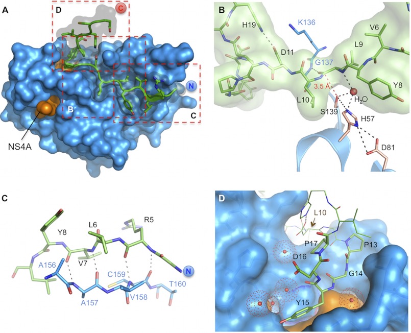
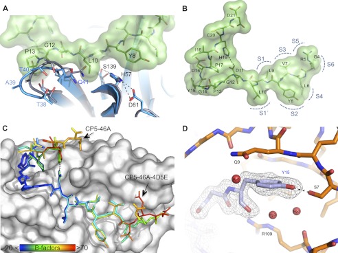
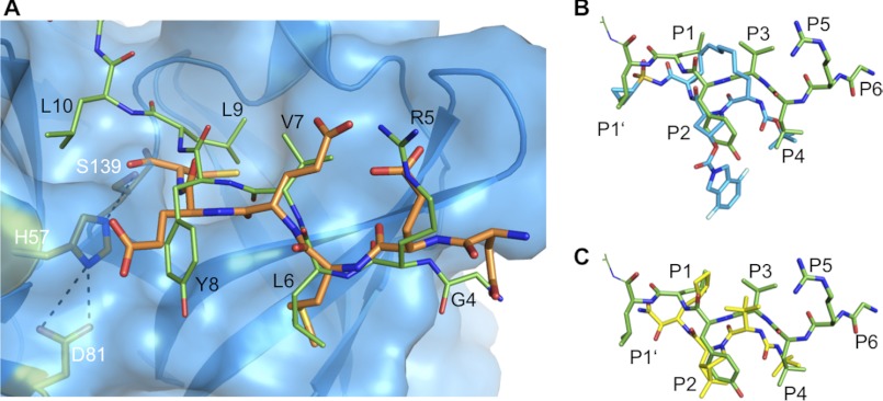
Hepatitis C virus (HCV) NS3-4A protease is essential for viral replication. All current small molecular weight drugs against NS3-4A are substrate peptidomimetics that have a similar binding and resistance profile. We developed inhibitory peptides (IPs) capping the active site and binding via a novel "tyrosine" finger at an alternative NS3-4A site that is of particular interest for further HCV drug development. The peptides are not cleaved due to a combination of geometrical constraints and impairment of the oxyanion hole function. Selection and optimization through combinatorial phagemid display, protein crystallography, and further modifications resulted in a 32-amino acid peptide with a K(i) of 0.53 nm. Inhibition of viral replication in cell culture was demonstrated by fusion to a cell-penetrating peptide. Negligible susceptibility to known (A156V and R155K) resistance mutations of the NS3-4A protease was observed. This work shows for the first time that antiviral peptides can target an intracellular site and reveals a novel druggable site on the HCV protease.
Figures
Similar articles
-
Peptidomimetic escape mechanisms arise via genetic diversity in the ligand-binding site of the hepatitis C virus NS3/4A serine protease.Gastroenterology. 2012 Mar;142(3):654-63. doi: 10.1053/j.gastro.2011.11.035. Epub 2011 Dec 7. Gastroenterology. 2012. PMID: 22155364 Free PMC article.
-
Molecular docking investigation of the binding interactions of macrocyclic inhibitors with HCV NS3 protease and its mutants (R155K, D168A and A156V).Protein J. 2014 Feb;33(1):32-47. doi: 10.1007/s10930-013-9538-6. Protein J. 2014. PMID: 24374429
-
Molecular modeling study on the resistance mechanism of HCV NS3/4A serine protease mutants R155K, A156V and D168A to TMC435.Antiviral Res. 2012 Jan;93(1):126-37. doi: 10.1016/j.antiviral.2011.11.007. Epub 2011 Nov 22. Antiviral Res. 2012. PMID: 22127068
-
Nonstructural protein 3-4A: the Swiss army knife of hepatitis C virus.J Viral Hepat. 2011 May;18(5):305-15. doi: 10.1111/j.1365-2893.2011.01451.x. Epub 2011 Mar 23. J Viral Hepat. 2011. PMID: 21470343 Review.
-
Discovery and development of VX-950, a novel, covalent, and reversible inhibitor of hepatitis C virus NS3.4A serine protease.Infect Disord Drug Targets. 2006 Mar;6(1):3-16. doi: 10.2174/187152606776056706. Infect Disord Drug Targets. 2006. PMID: 16787300 Review.
Cited by
-
Programmable solid-state condensates for spatiotemporal control of mammalian gene expression.Nat Chem Biol. 2025 Sep;21(9):1457-1466. doi: 10.1038/s41589-025-01860-0. Epub 2025 Mar 14. Nat Chem Biol. 2025. PMID: 40087540 Free PMC article.
-
Leveraging the therapeutic, biological, and self-assembling potential of peptides for the treatment of viral infections.J Control Release. 2022 Aug;348:1028-1049. doi: 10.1016/j.jconrel.2022.06.037. Epub 2022 Jul 6. J Control Release. 2022. PMID: 35752254 Free PMC article. Review.
-
Quinazolinone-Peptido-Nitrophenyl-Derivatives as Potential Inhibitors of SARS-CoV-2 Main Protease.Viruses. 2023 Jan 19;15(2):287. doi: 10.3390/v15020287. Viruses. 2023. PMID: 36851501 Free PMC article.
-
Controlled Protein Activities with Viral Proteases, Antiviral Peptides, and Antiviral Drugs.ACS Chem Biol. 2023 May 19;18(5):1228-1236. doi: 10.1021/acschembio.3c00138. Epub 2023 May 4. ACS Chem Biol. 2023. PMID: 37140437 Free PMC article.
-
A Chemically Disrupted Proximity System for Controlling Dynamic Cellular Processes.J Am Chem Soc. 2019 Feb 27;141(8):3352-3355. doi: 10.1021/jacs.8b12382. Epub 2019 Feb 14. J Am Chem Soc. 2019. PMID: 30735038 Free PMC article.
References
-
- Shepard C. W., Finelli L., Alter M. J. (2005) Global epidemiology of hepatitis C virus infection. Lancet Infect. Dis. 5, 558–567 - PubMed
-
- Bartenschlager R., Frese M., Pietschmann T. (2004) Novel insights into hepatitis C virus replication and persistence. Adv. Virus Res. 63, 71–180 - PubMed
-
- Bartenschlager R., Lohmann V. (2000) Replication of the hepatitis C virus. Baillieres Best Pract. Res. Clin. Gastroenterol. 14, 241–254 - PubMed
MeSH terms
Substances
LinkOut - more resources
Full Text Sources
Other Literature Sources
Molecular Biology Databases
