Review
. 2012 Oct;13(10):693-704.
doi: 10.1038/nrg3246. Epub 2012 Sep 11.

The telomere syndromes

Affiliations
Review

The telomere syndromes

Mary Armanios et al. Nat Rev Genet. 2012 Oct.

Erratum in

  • Nat Rev Genet. 2013 Mar;14(3):235

Abstract

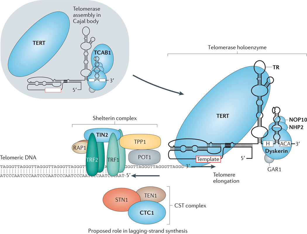
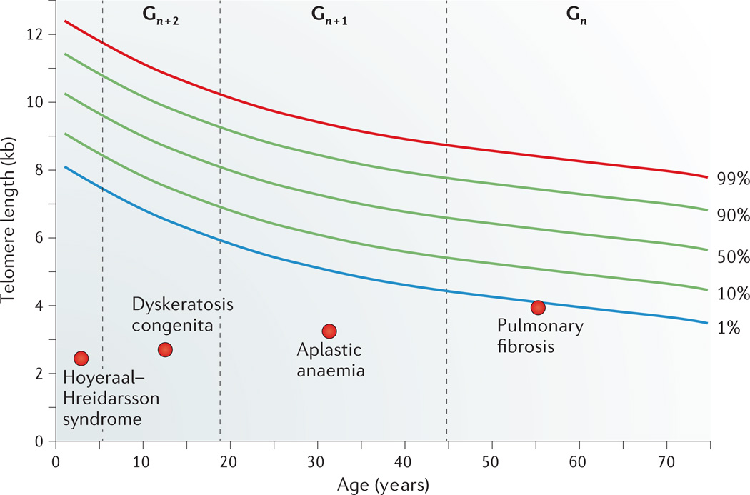
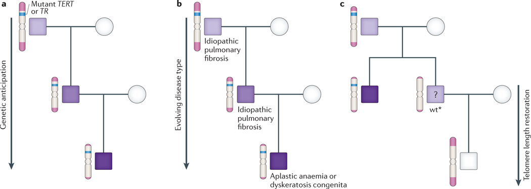
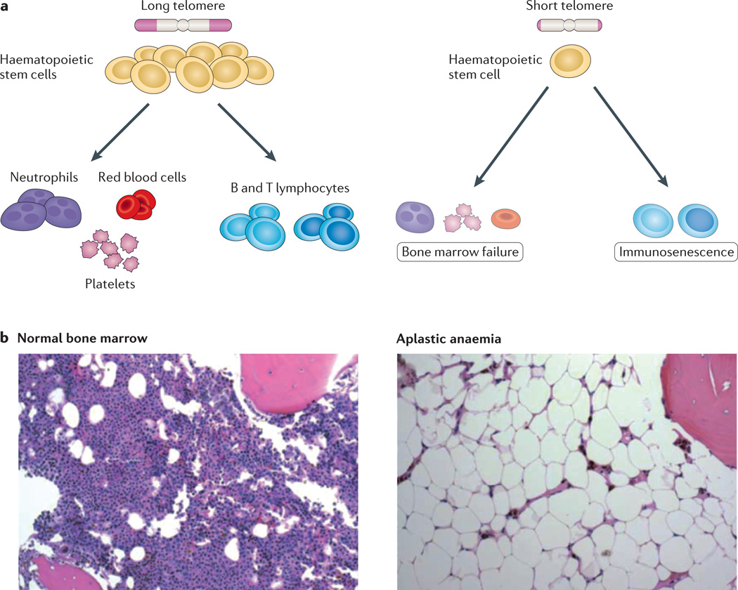
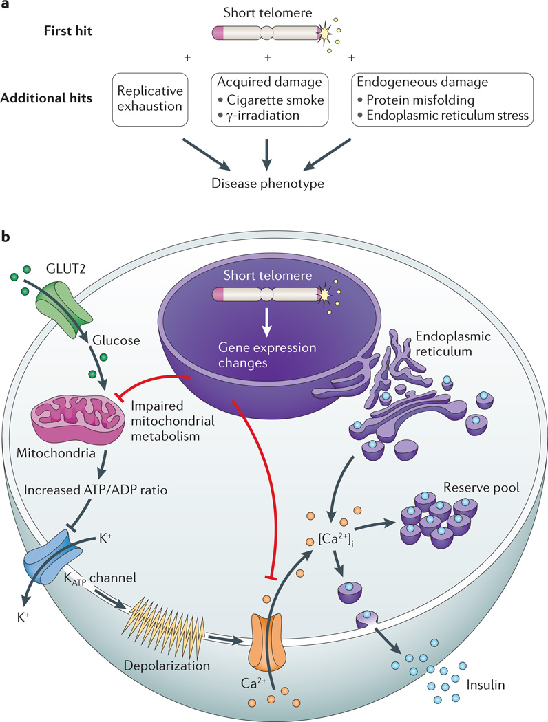
There has been mounting evidence of a causal role for telomere dysfunction in a number of degenerative disorders. Their manifestations encompass common disease states such as idiopathic pulmonary fibrosis and bone marrow failure. Although these disorders seem to be clinically diverse, collectively they comprise a single syndrome spectrum defined by the short telomere defect. Here we review the manifestations and unique genetics of telomere syndromes. We also discuss their underlying molecular mechanisms and significance for understanding common age-related disease processes.


Figures

Figure 1
Figure 1. Telomerase and telomere components involved in human monogenic telomere syndromes
Components for which mutations have been identified in telomere syndromes are indicated in bold type and shaded in blue. Shelterin complex components are made up of six component proteins — telomere repeat-binding factor 1 (TRF1), TRF2, repressor/activator protein 1 (RAP1), TRF1-interacting nuclear protein 2 (TIN2), TIN2-interacting protein 1 (TPP1) and protection of telomeres 1 (POT1) — which are essential for telomere protection and for regulating telomere elongation. The telomerase enzyme complex is comprised of TERT (the reverse transcriptase) and TR (the essential RNA component that contains a template for telomere repeat addition). TR contains a 3′H/ACA box motif that binds the dyskerin protein, which is part of a larger dyskerin complex that also consists of NHP2, NOP10 and GAR1. Note that for simplicity, one dyskerin complex is shown per TR molecule, although two copies are now thought to bind each TR. Telomerase Cajal body protein 1 (TCAB1) binds a Cajal body localization motif in TR and has a role in TR trafficking and biogenesis. In the Cajal body, TR and TERT assemble into a functional holoenzyme complex. The CST complex has three components — conserved telomere protection component 1 (CTC1), suppressor of cdc thirteen 1 (STN1) and telomeric pathway with STN1 (TEN1) — which are thought to function in part in telomere lagging-strand synthesis. Figure adapted, with permission, from Ref. © (2009) Annual Reviews.
Figure 2
Figure 2. Age-dependent manifestations of telomere syndromes
A schematic drawing that illustrates the typical range of telomere lengths by age in, for example, peripheral blood lymphocytes. At every age, telomere length displays a normal distribution that is defined by the percentile lines labelled on the right. Telomere length in individuals with four different clinical presentations across the age range is indicated. The dashed lines represent a typical age range in which these disorders may first manifest, and ‘Gn’, ‘Gn + 1’ and ‘Gn + 2’ designate three successive generations manifesting with earlier-onset and evolving disease type owing to progressive telomere shortening.
Figure 3
Figure 3. Unique genetics of autosomal-dominant telomere syndromes
a | Schema of a typical autosomal-dominant family with an inherited mutation in TERT (the reverse transcriptase) or TR (the telomerase RNA) showing earlier-onset disease with each generation, as illustrated by the darkening shades of purple. b | Disease type evolves in autosomal-dominant families from lung-predominant, which commonly manifests as idiopathic pulmonary fibrosis (IPF), to bone-marrow-failure-predominant, which presents as aplastic anaemia or dyskeratosis congenita. c | In mouse and human families, progeny of telomerase mutation carriers inherit the short telomeres even when they do not carry the mutant telomerase gene and are designated wt*. Mice with the wt* genotype have mild telomere-mediated phenotypes, but it remains unclear whether this is the situation for human cases (as represented by the question mark). The telomere length in human families is restored in progeny of these individuals. Figure adapted, with permission, from Ref. © (2012) Elsevier.
Figure 4
Figure 4. Short telomeres cause haematopoietic stem cell failure
a | Simplified schema of the haematopoiesis hierarchy with intact telomere length (left panel) and in the presence of telomere dysfunction (right panel). Telomere dysfunction causes both quantitative and qualitative defects in haematopoietic stem cells, which cause a decrease in mature blood forms. Defects in lymphopoiesis also cause immune defects. b | Histopathology of normal bone marrow biopsy shows intact marrow cellularity and haematopoietic cellular elements (left panel). In the right panel, a photomicrograph of a bone marrow biopsy from an individual with aplastic anaemia shows acellular marrow and replacement of the marrow parenchyma by fat. Panel b reproduced, with permission, from Ref. © (2009) Annual Reviews.
Figure 5
Figure 5. Mechanisms of telomere-mediated disease in slow-turnover tissues
a | Telomere length lowers the threshold to endogenous and exogenous damage, which is hypothesized to precipitate disease in slow-turnover tissues. b | A schematic of an insulin-producing β-cell showing the mechanism of a telomere-mediated insulin exocytosis defect. Glucose passively enters β-cells through the glucose transporter type 2 (GLUT2; also known as SLC2A2). Following glycolysis, ATP is generated by oxidative phosphorylation in the mitochondria. The net cytosolic change of the ATP/ADP ratio leads to closure of ATP-dependent K+ channels and opening of Ca2+ channels. The influx of extracellular Ca2+ results in an increase in the concentration of free, intracellular Ca2+ ([Ca2+]i), which triggers the release of insulin from both reserve and back-up pools. Short telomeres cause gene expression changes in pancreatic islets, which are associated with global metabolic dysregulation, mitochondrial dysfunction and concurrent defects in glucose-dependent and glucose-independent Ca2+ handling.

References

    1. Blackburn EH. Telomeres and telomerase: the means to the end (Nobel lecture) Angew. Chem. Int. Edn Engl. 2010;49:7405–7421. - PubMed
    1. Harley CB, Futcher AB, Greider CW. Telomeres shorten during ageing of human fibroblasts. Nature. 1990;345:458–460. - PubMed
    1. Allsopp RC, et al. Telomere length predicts replicative capacity of human fibroblasts. Proc. Natl Acad. Sci. USA. 1992;89:10114–10118.. References and showed that telomere length shortens in human cells with age and limits their in vitro replicative potential.

    1. Greider CW, Blackburn EH. Identification of a specific telomere terminal transferase activity in Tetrahymena extracts. Cell. 1985;43:405–413. - PubMed
    1. Greider CW, Blackburn EH. The telomere terminal transferase of Tetrahymena is a ribonucleoprotein enzyme with two kinds of primer specificity. Cell. 1987;51:887–898. - PubMed

Publication types