

Angiofibrotic response to vascular endothelial growth factor inhibition in diabetic retinal detachment: report no. 1
- PMID: 22965588
- PMCID: PMC3984910
- DOI: 10.1001/archophthalmol.2012.1611
Angiofibrotic response to vascular endothelial growth factor inhibition in diabetic retinal detachment: report no. 1
Abstract
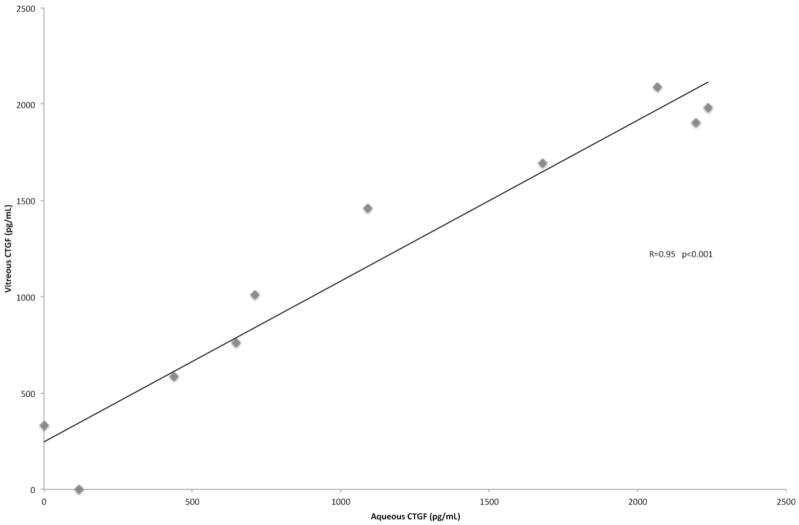
OBJECTIVES To assess the effect of bevacizumab injection on connective tissue growth factor (CTGF) and vascular endothelial growth factor (VEGF) in the ocular fluids of patients with diabetic traction retinal detachment, and to determine whether intraoperative and postoperative complications are decreased in eyes given adjunctive preoperative bevacizumab injection. METHODS Twenty eyes of 19 patients were randomized to receive intravitreal bevacizumab or sham injection 3 to 7 days before vitrectomy for severe proliferative diabetic retinopathy. We collected aqueous samples before injection and at the time of vitrectomy and extracted undiluted vitreous samples. RESULTS Five eyes had decreased vascularization of membranes from preinjection to the time of vitrectomy (all in the bevacizumab treatment arm). Median visual acuities were 20/400 in control eyes at baseline and postoperative month 3 (POM3) and 8/200 in the bevacizumab-treated group at baseline and 20/100 at POM3 (P= .30 between control and bevacizumab-treated groups at POM3). All retinas were attached at POM3. Vitreous levels of VEGF were significantly lower in the bevacizumab group than in the control group (P= .03). Vitreous levels of CTGF were slightly lower in the bevacizumab group compared with the control group, but this difference was not statistically significant (P= .38). Levels of CTGF in the aqueous were strongly correlated with CTGF levels in the vitreous of controls (Spearman correlation coefficient, 0.95 [P< .001]). CONCLUSIONS Intravitreal bevacizumab injection reduces vitreous levels of VEGF and produces a clinically observable alteration in diabetic fibrovascular membranes. Ocular fluid levels of CTGF are not significantly affected within the week after VEGF inhibition. Retinal reattachment rates and visual acuity are not significantly altered by preoperative intravitreal bevacizumab injection at POM3. TRIAL REGISTRATION clinicaltrials.gov Identifier: NCT01270542.
Figures
References
-
- Eliott D, Lee MS, Abrams GW. Proliferative Diabetic Retinopathy: Principles and Techniques of Surgical Treatment. In: Ryan SJ, editor. Retina. 4. St. Louis: Elsevier Mosby; 2006. pp. 2413–2449.
-
- Miller JW, Adamis AP, Aiello LP. Vascular endothelial growth factor in ocular neovascularization and proliferative diabetic retinopathy. Diabetes Metab Rev. 1997 Mar;13(1):37–50. - PubMed
-
- Aiello LP, Avery RL, Arrigg PG, et al. Vascular endothelial growth factor in ocular fluid of patients with diabetic retinopathy and other retinal disorders. N Engl J Med. 1994 Dec 1;331(22):1480–1487. - PubMed
-
- Adamis AP, Miller JW, Bernal MT, et al. Increased vascular endothelial growth factor levels in the vitreous of eyes with proliferative diabetic retinopathy. Am J Ophthalmol. 1994 Oct 15;118(4):445–450. - PubMed
-
- Frank RN, Amin RH, Eliott D, Puklin JE, Abrams GW. Basic fibroblast growth factor and vascular endothelial growth factor are present in epiretinal and choroidal neovascular membranes. Am J Ophthalmol. 1996 Sep;122(3):393–403. - PubMed
Publication types
MeSH terms
Substances
Associated data
Grants and funding
LinkOut - more resources
Full Text Sources
Other Literature Sources
Medical
Miscellaneous
