

Factors associated with changes in visual acuity and central subfield thickness at 1 year after treatment for diabetic macular edema with ranibizumab
- PMID: 22965591
- PMCID: PMC3543147
- DOI: 10.1001/archophthalmol.2012.1107
Factors associated with changes in visual acuity and central subfield thickness at 1 year after treatment for diabetic macular edema with ranibizumab
Abstract
Objective: To identify factors that predict the success or failure of treatment with intravitreal ranibizumab for patients with diabetic macular edema.
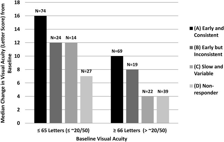
Methods: A total of 37 baseline demographic, systemic, ocular, optical coherence tomographic, and fundus photographic variables were assessed for association with change in visual acuity or central subfield thickness between baseline and 1 year in 361 eyes that were randomly assigned to intravitreal ranibizumab with prompt or deferred laser treatment within a trial of ranibizumab, triamcinolone acetonide, and laser treatment for center-involved diabetic macular edema. A categorical variable describing follow-up anatomic responses to therapy was added to the visual acuity outcome model.
Results: After adjusting for baseline visual acuity, a larger visual acuity treatment benefit was associated with younger age (P< .001), less severe diabetic retinopathy on clinical examination (P= .003), and absence of surface wrinkling retinopathy (P< .001). The reduction in central subfield thickness during the first treatment year also predicted better visual acuity outcomes (P< .001). After adjusting for baseline central subfield thickness, the presence of hard exudates was associated with more favorable improvement on optical coherence tomographic scan (P= .004). Because only 11 eyes experienced vision loss and 6 eyes experienced an increase in central subfield thickness, factors for poor outcomes could not be evaluated.
Conclusions: A review of baseline factors and anatomic responses during the first year of ranibizumab therapy for association with visual acuity outcome did not identify any features that would preclude ranibizumab treatment. However, baseline central subfield thickness is the strongest predictor of anatomic outcome, and reduction in central subfield thickness during the first treatment year is associated with better visual acuity outcomes.
Figures
References
-
- Moss S, Klein R, Klein B. The 14-year incidence of visual loss in a diabetic population. Ophthalmology. 1998;105(6):998–1003. - PubMed
-
- Centers for Disease Control and Prevention. [Accessed December 14,2011];Obesity and overweight for professionals, data and statistics: Adult obesity. http://www.cdc.gov/obesity/data/adult.html.
-
- Early Treatment Diabetic Retinopathy Study Research Group. Photocoagulation for diabetic macular edema. Early Treatment Diabetic Retinopathy Study report number 1. Arch Ophthalmol. 1985;103(12):1796–806. - PubMed
-
- Antonetti DA, Barber AJ, Hollinger LA, Wolpert EB, Gardner TW. Vascular endothelial growth factor induces rapid phosphorylation of tight junction proteins occludin and zonula occluden 1. A potential mechanism for vascular permeability in diabetic retinopaty and tumors. J Bio Chem. 1999;274(33):23463–7. - PubMed
-
- Michaelides M, Kaines A, Hamilton RD, et al. A prospective randomized trial of intravitreal bevacizumab or laser therapy in the management of diabetic macular edema (BOLT study) 12-month data: report 2. Ophthalmology. 2010;117(6):1078–86 e2. - PubMed
Publication types
MeSH terms
Substances
Grants and funding
LinkOut - more resources
Full Text Sources
Other Literature Sources
Medical
