. 2012 Oct;158A(10):2393-406.
doi: 10.1002/ajmg.a.35561. Epub 2012 Sep 10.

Beyond Gómez-López-Hernández syndrome: recurring phenotypic themes in rhombencephalosynapsis

Affiliations

Beyond Gómez-López-Hernández syndrome: recurring phenotypic themes in rhombencephalosynapsis

Hannah M Tully et al. Am J Med Genet A. 2012 Oct.

Abstract

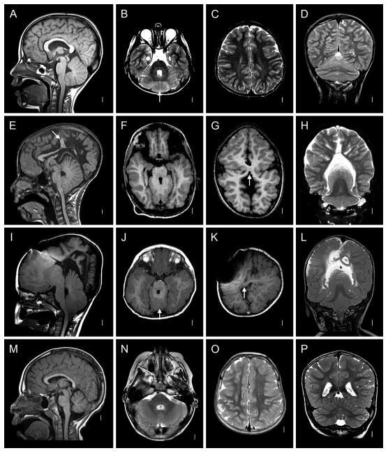
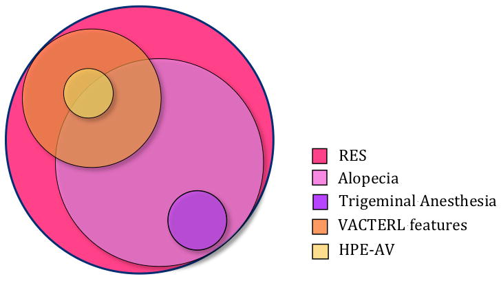
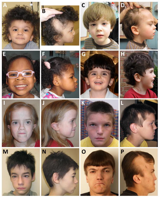
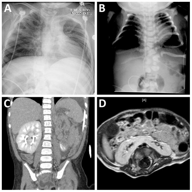
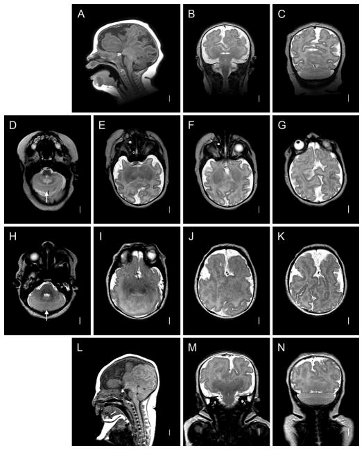
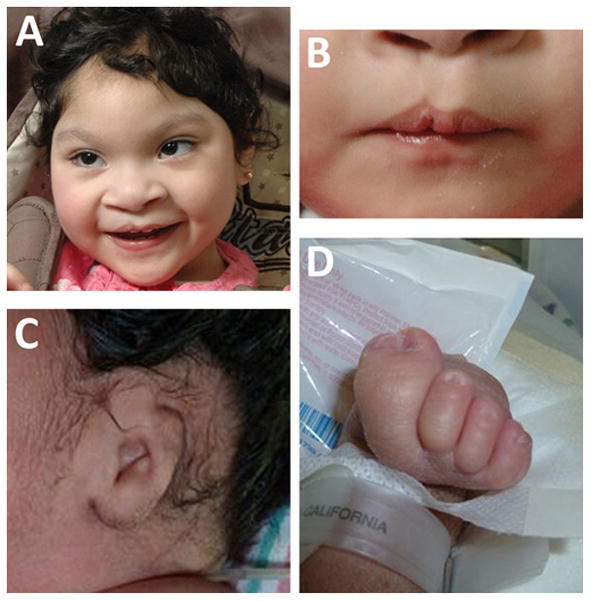
Rhombencephalosynapsis (RES) is an uncommon cerebellar malformation characterized by fusion of the hemispheres without an intervening vermis. Frequently described in association with Gómez-López-Hernández syndrome, RES also occurs in conjunction with VACTERL features and with holoprosencephaly (HPE). We sought to determine the full phenotypic spectrum of RES in a large cohort of patients. Information was obtained through database review, patient questionnaire, radiographic, and morphologic assessment, and statistical analysis. We assessed 53 patients. Thirty-three had alopecia, 3 had trigeminal anesthesia, 14 had VACTERL features, and 2 had HPE with aventriculy. Specific craniofacial features were seen throughout the cohort, but were more common in patients with alopecia. We noted substantial overlap between groups. We conclude that although some distinct subgroups can be delineated, the overlapping features seen in our cohort suggest an underlying spectrum of RES-associated malformations rather than a collection of discrete syndromes.


Figures

Figure 1
Figure 1. Radiographic Features of RES
A–D: Patient 48. Midline sagittal T1-weighted image through the cerebellum demonstrates hemispheric rather than vermian configuration (A). Axial T2 through the cerebellum shows fusion of white matter across the midline and a keyhole-shaped 4th ventricle (B). Axial T2 through though the cerebral hemispheres shows normal anatomy (C). Coronal T2 showing continuity of cerebellar folia across the midline without an intervening vermis (D). E–H: Patient 46. Midline sagittal T1-weighted image demonstrating a towering cerebellum with hemispheric architecture. Note the absence of a visible aqueduct. This patient had severe congenital hydrocephalus. Many of the supratentorial abnormalities (arrow) are likely a consequence of distortion from hydrocephalus and subsequent decompression (E). Axial T1 through the cerebellum demonstrating similar findings as the patient above (F). Axial T1 through the cerebral hemispheres shows an area of white matter continuity suspicious for mild HPE, but likely representing post-hydrocephalus distortion(G). Coronal T2 showing a towering cerebellum with upward displacement through the tentorial notch (H). I–L: Patient 14. I, Mid-sagittal and axial T1 demonstrating similar findings to Patient 46. This patient also had severe congenital hydrocephalus (J). Axial T1 showing area suspicious for HPE (arrow), though post-hydrocephalic distortion makes this difficult to confirm (K). Coronal T2 demonstrating similar findings to Patient 46 (L). M–P: normal brain.
Figure 2
Figure 2
Areas of overlap among patients with RES.
Figure 3
Figure 3. Faces of Patients with RES
A–B: Patient 39. C–D: Patient 28. E–F: Patient 26. G–H: Patient 17. I–J, Patient 7. K–L: Patient 9. M–N: Patient 46. O–P: Patient 44.
Figure 4
Figure 4. VACTERL Features
A: Patient 33. Radiograph demonstrating multiple fused ribs associated with vertebral segmentation defects. B: Patient 35. Radiograph demonstrating multiple thoracic and lumbar vertebral segmentation defects and deformed ribs. C: Patient 21. Coronal CT showing absent kidney on the left with partially duplicated kidney on the right (cross-fused renal ectopia). D: Patient 36. Axial T1-weighted image of the abdomen showing horseshoe kidney.
Figure 5
Figure 5. Holoprosencephaly-Aventriculy
A–G: Patient 15. Mid-sagittal view demonstrating dramatically infolded brain, absent 3rd ventricle and aqueduct, and mass-like fusion of the midbrain (A). Coronal T2 demonstrating gray matter continuity across the midline (HPE) and absent lateral ventricles (B). Coronal T2 demonstrating posterior predominance of HPE and continuity of cerebellar folia across the midline (C). Axial T2 demonstrating RES(D). Axial T2s showing posterior-predominant fusion, infolding and aventriculy (E–G). H–N: Patient 35. Multiple axial, coronal and mid-sagittal sections showing striking similarity of brain malformation to that of Patient 15.
Figure 6
Figure 6. Additional features in patients with HPE-AV
A and B: Patient 15. Broad nose, upslanting palpebral fissures and forme fruste cleft lip (A). Close-up of lip (B). C–D: Patient 35. Microtia (C). Partially duplicated, fused right hallux (D).

Similar articles

Cited by

  • Midbrain and hindbrain malformations: advances in clinical diagnosis, imaging, and genetics.
    Doherty D, Millen KJ, Barkovich AJ. Doherty D, et al. Lancet Neurol. 2013 Apr;12(4):381-93. doi: 10.1016/S1474-4422(13)70024-3. Epub 2013 Mar 18. Lancet Neurol. 2013. PMID: 23518331 Free PMC article. Review.
  • Rhombencephalosynapsis: Fused cerebellum, confused geneticists.
    Aldinger KA, Dempsey JC, Tully HM, Grout ME, Mehaffey MG, Dobyns WB, Doherty D. Aldinger KA, et al. Am J Med Genet C Semin Med Genet. 2018 Dec;178(4):432-439. doi: 10.1002/ajmg.c.31666. Am J Med Genet C Semin Med Genet. 2018. PMID: 30580482 Free PMC article. Review.
  • Diagnosis of rhomboencephalosynapsis by MRI in a 5-year-old child.
    Gómez NT, Mancilla WP, Mejía CHR, Leal JCA. Gómez NT, et al. Radiol Case Rep. 2020 May 1;15(7):867-870. doi: 10.1016/j.radcr.2020.04.015. eCollection 2020 Jul. Radiol Case Rep. 2020. PMID: 32382369 Free PMC article.
  • Infantile hydrocephalus: a review of epidemiology, classification and causes.
    Tully HM, Dobyns WB. Tully HM, et al. Eur J Med Genet. 2014 Aug;57(8):359-68. doi: 10.1016/j.ejmg.2014.06.002. Epub 2014 Jun 13. Eur J Med Genet. 2014. PMID: 24932902 Free PMC article. Review.
  • MN1 C-terminal truncation syndrome is a novel neurodevelopmental and craniofacial disorder with partial rhombencephalosynapsis.
    Mak CCY, Doherty D, Lin AE, Vegas N, Cho MT, Viot G, Dimartino C, Weisfeld-Adams JD, Lessel D, Joss S, Li C, Gonzaga-Jauregui C, Zarate YA, Ehmke N, Horn D, Troyer C, Kant SG, Lee Y, Ishak GE, Leung G, Barone Pritchard A, Yang S, Bend EG, Filippini F, Roadhouse C, Lebrun N, Mehaffey MG, Martin PM, Apple B, Millan F, Puk O, Hoffer MJV, Henderson LB, McGowan R, Wentzensen IM, Pei S, Zahir FR, Yu M, Gibson WT, Seman A, Steeves M, Murrell JR, Luettgen S, Francisco E, Strom TM, Amlie-Wolf L, Kaindl AM, Wilson WG, Halbach S, Basel-Salmon L, Lev-El N, Denecke J, Vissers LELM, Radtke K, Chelly J, Zackai E, Friedman JM, Bamshad MJ, Nickerson DA; University of Washington Center for Mendelian Genomics; Reid RR, Devriendt K, Chae JH, Stolerman E, McDougall C, Powis Z, Bienvenu T, Tan TY, Orenstein N, Dobyns WB, Shieh JT, Choi M, Waggoner D, Gripp KW, Parker MJ, Stoler J, Lyonnet S, Cormier-Daire V, Viskochil D, Hoffman TL, Amiel J, Chung BHY, Gordon CT. Mak CCY, et al. Brain. 2020 Jan 1;143(1):55-68. doi: 10.1093/brain/awz379. Brain. 2020. PMID: 31834374 Free PMC article.

References

    1. Armstrong DK, Burrows D. Congenital triangular alopecia. Pediatr Dermatol. 1996;13:394–396. - PubMed
    1. Assouly P, Happle R. A hairy paradox: congenital triangular alopecia with a central hair tuft. Dermatology. 2010;221:107–109. - PubMed
    1. Aydingoz U, Cila A, Aktan G. Rhombencephalosynapsis associated with hand anomalies. Br J Radiol. 1997;70:764–766. - PubMed
    1. Bargman H. Congenital triangular alopecia. J Am Acad Dermatol. 1988;18:390. - PubMed
    1. Barkovich AJ, Quint DJ. Middle interhemispheric fusion: an unusual variant of holoprosencephaly. Am J Neuroradiol. 1993;14:431–440. - PMC - PubMed

Publication types

MeSH terms

Supplementary concepts