. 2013 Jan;51(1):50-8.
doi: 10.1002/dvg.22347. Epub 2012 Nov 3.

Generation of a germ cell-specific mouse transgenic CHERRY reporter, Sohlh1-mCherryFlag

Affiliations

Generation of a germ cell-specific mouse transgenic CHERRY reporter, Sohlh1-mCherryFlag

Hitomi Suzuki et al. Genesis. 2013 Jan.

Abstract

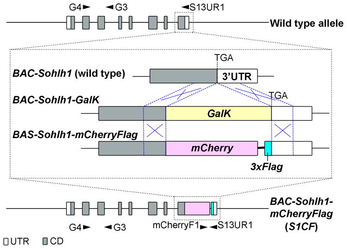
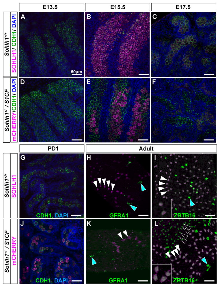
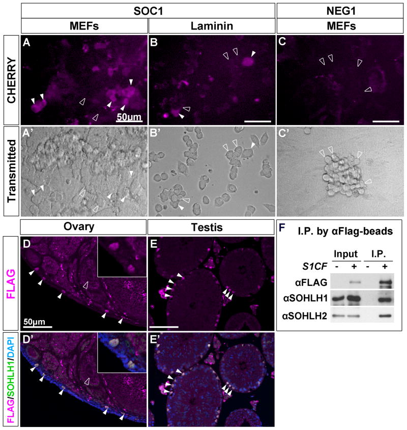
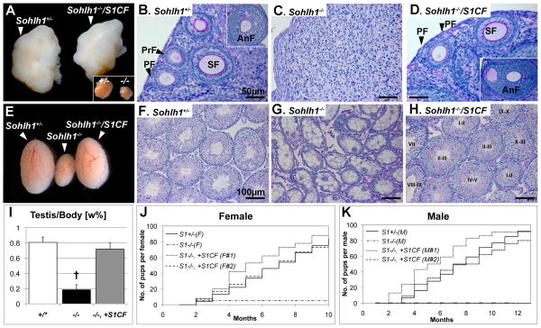
Visualization of differentiating germ cells is critical to understanding the formation of primordial follicles in the ovary, and the commitment of spermatogonial stem cells to differentiation. We engineered and generated a BAC transgenic mouse line, Sohlh1-mCherryFlag (S1CF), under the direction of the native Sohlh1 promoter. Sohlh1 is a germ cell-specific gene that encodes the basic helix-loop-helix (bHLH) transcriptional regulator that is essential in oogenesis and spermatogenesis. Sohlh1 expression is unique, and is limited to perinatal and early follicle oocytes and differentiating spermatogonia. The Sohlh1-mCherryFlag transgene was engineered to fuse SOHLH1 to the red fluorescent protein CHERRY with 3-tandem-FLAG tags. S1CF animals fluoresce specifically in the oocytes of perinatal ovaries and small follicles in adult ovaries, as well as in spermatogonia, a pattern that is similar to endogenous SOHLH1. Moreover, S1CF rescued germ cell loss and infertility in both male and female Sohlh1(-/-) animals. The FLAG-tag on S1CF was effective for immunostaining and immunoprecipitation. The Sohlh1-mCherryFlag transgenic mouse provides a unique model to study early germ cell differentiation, as well as in vivo imaging and purification of differentiating germ cells.


Figures

Figure 1
Figure 1. BAC-Sohlh1-mCherryFlag engineering
(A) BAC-Sohlh1-mCherryFlag (S1CF) construction. For BAC-Sohlh1-mCherryFlag (S1CF) engineering, the GalK sequence (yellow box) was first inserted just before the stop codon on the Sohlh1 exon 8, using a homologous recombination system in bacteria, followed by the replacement of mCherry-3xFlag (Magenta and turquoise boxes). Homologous regions used for recombination are indicated in blue break lines. PCR primer locations are shown with black arrowheads. Gray boxes represent the Sohlh1 coding region (CD) and white boxes represent the untranslated region (UTR). The following primer sets were used for PCR: G4 and G3 for wild type (633bp), mCherryF1 and S13UR1 for S1CF (313bp). In matings with the Sohlh1+/−, we also used HPRT2 and G3 primers previously described to detect the mutant allele (220bp) (Pangas et al., 2006).
Figure 2
Figure 2. CHERRY expression mimics endogenous SOHLH1 expression in female gonads
Endogenous SOHLH1 (A–C) and CHERRY (D–I) expression in female gonads at E13.5 (A and D), E15.5 (B and E), E17.5 (C and F), postnatal 1 day (PD1; G and G′), 3 weeks old (H and H′) and 7 weeks old (I and I′). (A–F) Gonads were immunostained with anti-CDH1 (green) (A–F) and anti-SOHLH1 (magenta) (A–C), and counter stained with DAPI (blue). Native CHERRY fluorescence is shown in magenta. Genotypes are indicated. (G–I) Ovaries were counter stained with DAPI (gray in G′–I′). Native CHERRY fluorescence is shown in magenta (not immunostained). The endogenous SOHLH1 localization in wild type PD1 ovary is shown in insets of G and G′. P, primordial follicle; Pr, primary follicle; S, secondary follicle. White arrowheads indicate oocytes expressing CHERRY and open arrowheads indicate oocytes that do not express CHERRY. Scale bars represent 50μm. Endogenous SOHLH1 expression commences between E14.5–15.5 in a subset of germ cells and expands to all germ cells by the time of birth. Endogenous SOHLH1 protein can be detected in germ cell cysts, primordial follicles and primary follicles but not in secondary follicles. Endogenous SOHLH1 was dominantly localized to the cytoplasm in most oocytes. CHERRY mimics endogenous SOHLH1 expression and developmental localization, however, the onset of CHERRY expression was slightly later than that of endogenous SOHLH1. We did not observe this delay when CHERRY was expressed on the background of Sohlh1 deficient gonads, arguing that some negative regulation by endogenous SOHLH1 may exist.
Figure 3
Figure 3. CHERRY expression mimics endogenous SOHLH1 expression in male gonads
SOHLH1 (A–C and G–I) and CHERRY (D–F and J–L) expression in male gonads at E13.5 (A and D), E15.5 (B and E), E17.5 (C and F), PD1 (PD1; G and J) and 8 weeks old adult (H, I, K and L). Gonads were immunostained with anti-CDH1 (green) (A–G and J), anti-SOHLH1 (magenta) (D–F and G–I), anti-GFRA1 (green) (H and K) and ZBTB16 (green) (I and L), and counter stained with DAPI (blue) (A–G and J). Genotypes are indicated. CHERRY signals are shown in magenta (D–F and J–L). Cyan arrowheads indicate GFRA1-positive or ZBTB16 positive As undifferentiating spermatogonia that do not express SOHLH1 or CHERRY. White arrowheads indicate SOHLH1 or CHERRY positive spermatogonia. Open arrowheads indicate CHERRY negative differentiating spermatogonia that would be expected to express SOHLH1. Scale bars represent 50μm. Endogenous SOHLH1 expression commences at around E15.5 and decreases in the perinatal testis until PD3, when SOHLH1 can be detected again in spermatogonia. Endogenous SOHLH1 protein was detected in both undifferentiated (H) and differentiating (I) sprematogonia, but was not observed in most of the GFRA1-positive undifferentiated sprematogonial population. CHERRY closely mimics this endogenous SOHLH1 expression throughout the development of male germ cells.
Figure 4
Figure 4. S1CF is useful for multiple analysis
(A–C) Spermatogonial stem/progenitor cells derived from Sohlh1+/−/S1CF (SOC1; A, B) or Sohlh1+/− (NEG1; C) were cultured on MEFs (A, C) or laminin (B). On MEFs the cultured cells proliferated to form large 3-dimensional clusters of interconnected cells while on laminin the cultured cells exhibited a flatter morphology. CHERRY fluorescence was observed in a subset of the SOC1 spermatogonial population (A, B) but not in NEG1 (C). White arrowheads indicate spermatogonia expressing CHERRY and open arrowheads indicate spermatogonia that do not express CHERRY. Scale bars represent 50μm. (D and E) Section immunofluorescence of adult ovary (D) and testis (E) harboring the S1CF transgene. Specimens were immunostained with anti-SOHLH1 (green) and anti-FLAG (magenta), and counter stained with DAPI (blue). White arrowheads indicate oocytes or spermatogonia stained with SOHLH1 and FLAG. The open arrowhead in A, A′ and A″ indicates an oocyte in the secondary follicle stage that expresses neither SOHLH1 nor FLAG. Scale bars represent 50μm. (F) Immunoprecipitation–Western blot analyses of proteins from PD7 testes extracts of wild type and S1CF transgenic mice using anti-FLAG antibody conjugated beads. Each antibody used for western blot analysis is indicated at the left side of panel. Input: pre-immunoprecipitation testes lysate (1%). S1CF was immunoprecipitated only from S1CF-positive lysate (I.P.). Endogenous SOHLH1 and SOHLH2 were co-immunoprecipitated with S1CF, indicating that SOHLH1 homodimerizes and heterodimerizes in vivo in the testis.
Figure 5
Figure 5. S1CF transgene rescues the Sohlh1−/− phenotype in both males and females
Sohlh1−/−/S1CF adult anatomy, histology, and cumulative number of pups from females (A–D and J) and males (E–I and K).(A) Ovaries shown under the same magnification for Sohlh1+/− (left), and knockout rescued by S1CF, Sohlh1−/−/S1CF (right). Inset in the right lower corner shows that Sohlh1−/− (−/−) ovaries are one third the size of wild type or Sohlh1+/− (+/−) ovaries as previously reported (Pangas et al., 2006). (B–D) PAS-Hematoxylin staining of adult ovaries. Various stages of follicular development are shown, primordial (PF), primary (PrF), secondary (SF) and antral (AnF) follicles, in Sohlh1+/− (B) and Sohlh1−/−/S1CF (D) ovaries but no follicles are visible in Sohlh1−/− at the same age (C). Scale bars represent 50μm. (E) Gross testes dissected from Sohlh1+/− (left), Sohlh1−/− (middle) and Sohlh1−/−/S1CF (right) mice. Sohlh1−/− testis size was recovered in the presence of S1CF and was equivalent to Sohlh1+/− testes. (F–H) PAS staining of adult testes. Various epithelial stages of seminiferous tubules were observed in Sohlh1+/− (F) and Sohlh1−/−/S1CF (H) testis but not in Sohlh1−/− testes at the same age (G). Scale bars represent 100μm. (I) Comparison of testes weights. Sohlh1−/− (−/−) testes weights recovered with S1CF (−/−, +S1CF), similarly as in Sohlh1+/* (+/*) (Sohlh1+/+ or Sohlh1+/−) (P < 0.1). More than 3 mice were examined for each genotype. Error bars show SE. Student t-test was used to calculate P values. †, P < 0.01 against wild type. (J and K) Comparison of the cumulative number of pups per female (J) or male (K). Genotypes for each line are indicated in the figure. (J) Sohlh1−/− females are infertile, except for occasional, one-time litter observed among a subset of females prior to 8 weeks of age, but Sohlh1−/−/S1CF (n=3) fertility was comparable to Sohlh1+/− females. (K) Sohlh1−/− males were infertile and did not yield any pups, but Sohlh1−/−/S1CF (n=8) males fertility was comparable to Sohlh1+/− males.

Similar articles

Cited by

References

    1. Baibakov B, Gauthier L, Talbot P, Rankin TL, Dean J. Sperm binding to the zona pellucida is not sufficient to induce acrosome exocytosis. Development. 2007;134:933–43. - PubMed
    1. Ballow D, Meistrich ML, Matzuk M, Rajkovic A. Sohlh1 is essential for spermatogonial differentiation. Developmental biology. 2006;294:161–7. - PubMed
    1. Dann CT, Alvarado AL, Molyneux LA, Denard BS, Garbers DL, Porteus MH. Spermatogonial stem cell self-renewal requires OCT4, a factor downregulated during retinoic acid-induced differentiation. Stem Cells. 2008;26:2928–37. - PubMed
    1. Falciatori I, Lillard-Wetherell K, Wu Z, Hamra FK, Garbers DL. Deriving mouse spermatogonial stem cell lines. Methods Mol Biol. 2008;450:181–92. - PubMed
    1. Hoodbhoy T, Aviles M, Baibakov B, Epifano O, Jimenez-Movilla M, Gauthier L, Dean J. ZP2 and ZP3 traffic independently within oocytes prior to assembly into the extracellular zona pellucida. Molecular and cellular biology. 2006;26:7991–8. - PMC - PubMed

Publication types

Substances