. 2012 Dec;87(12):1057-64.
doi: 10.1002/ajh.23317. Epub 2012 Sep 11.

Synergistic anticancer effects of arsenic trioxide with bortezomib in mantle cell lymphoma

Affiliations

Synergistic anticancer effects of arsenic trioxide with bortezomib in mantle cell lymphoma

Hyun Joo Jung et al. Am J Hematol. 2012 Dec.

Abstract

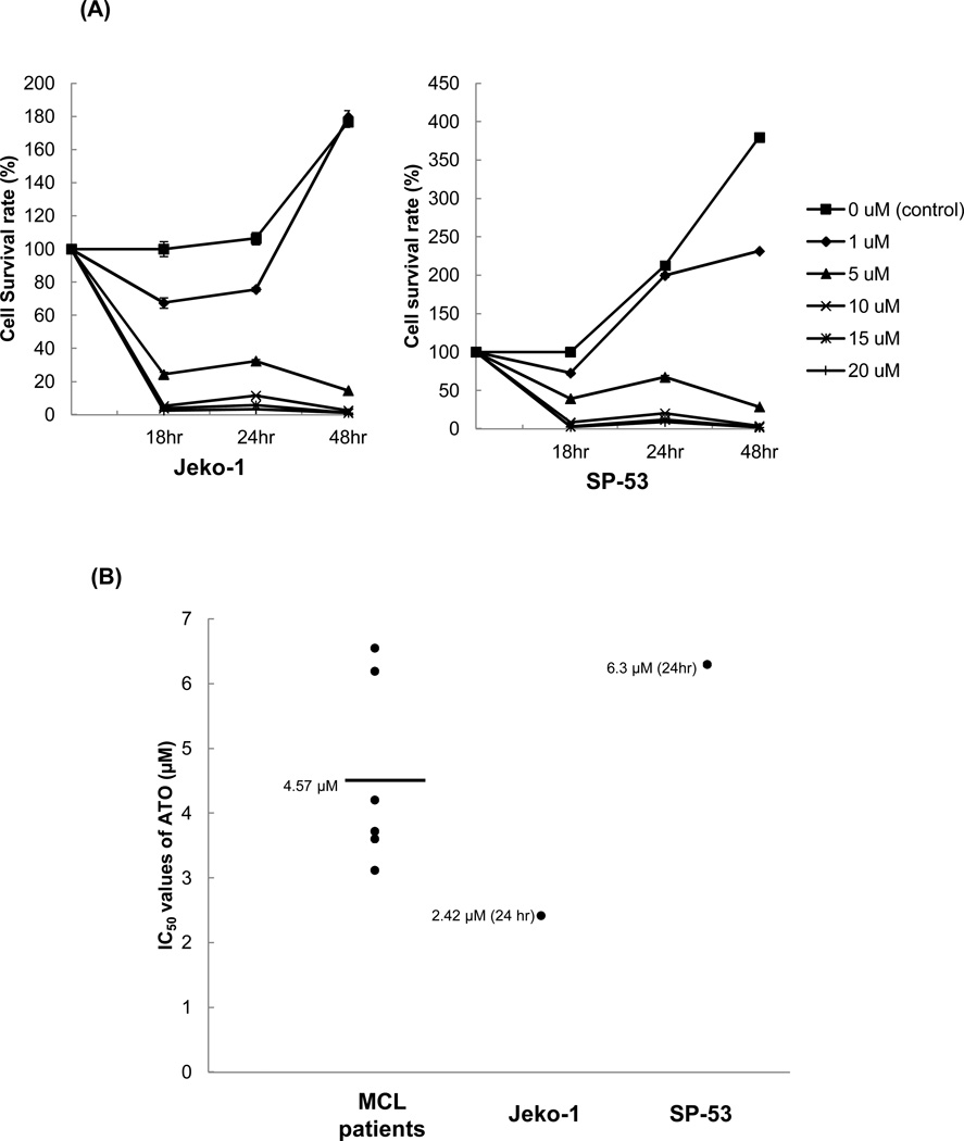
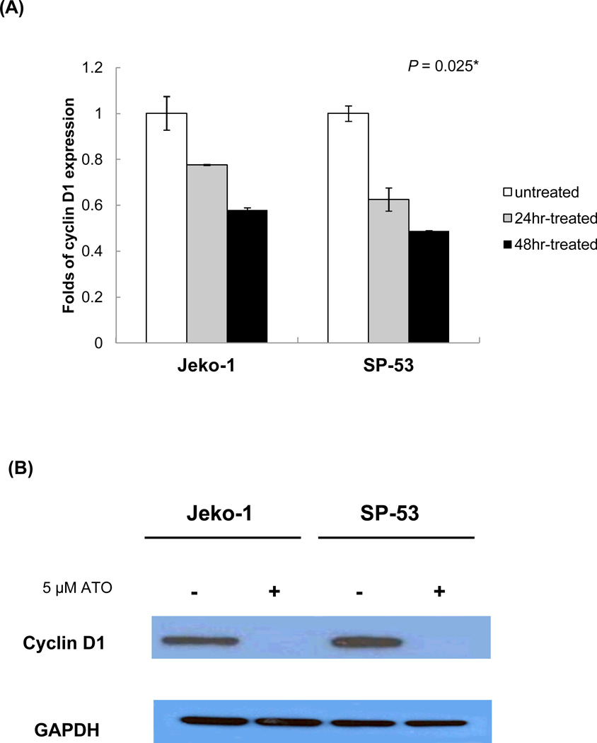
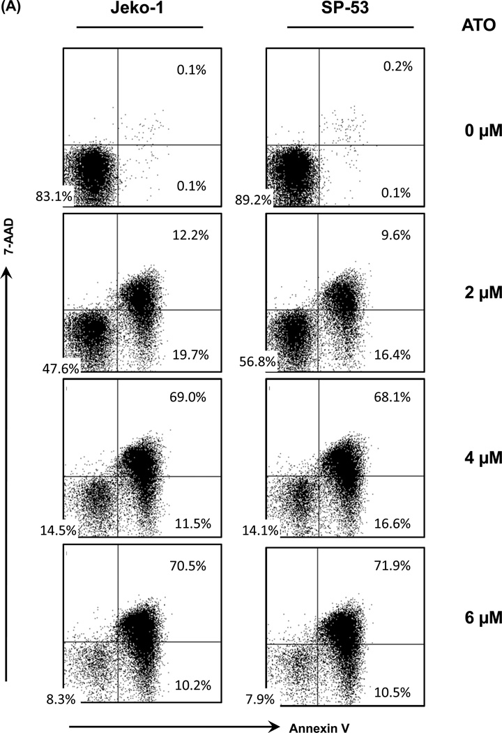
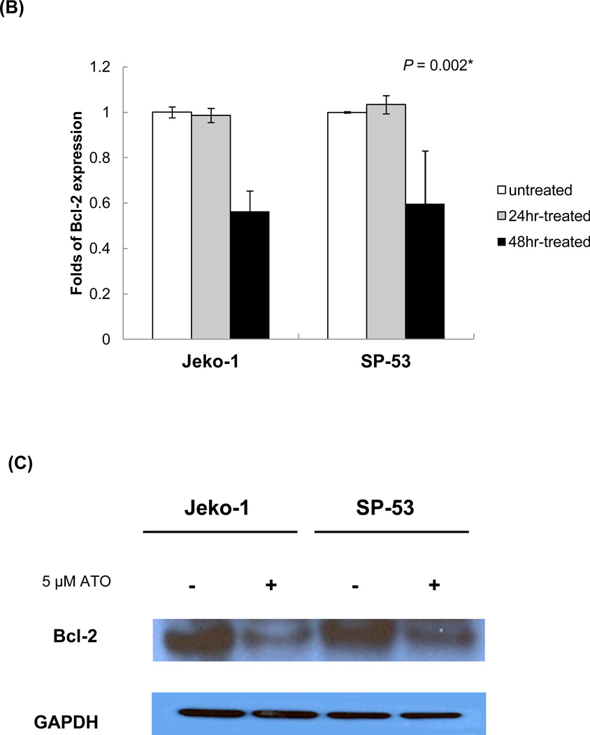
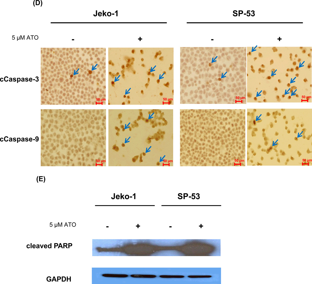
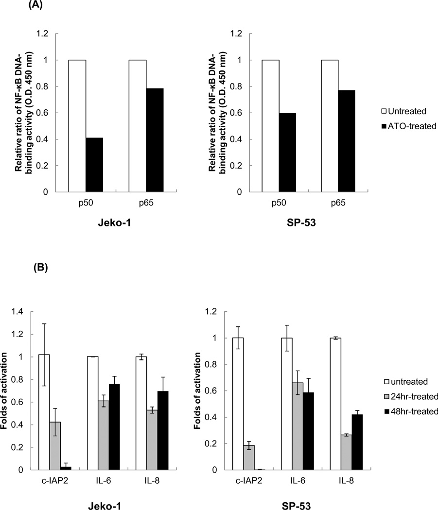
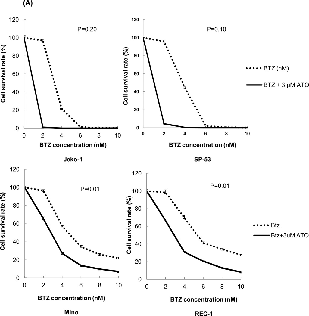
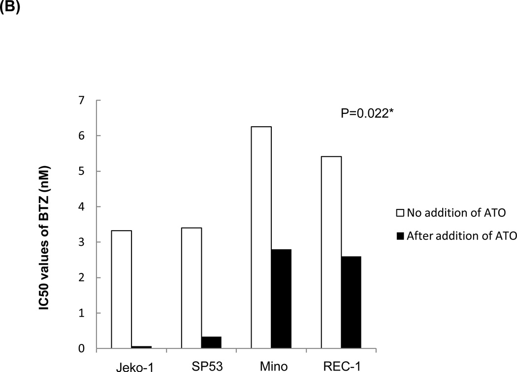
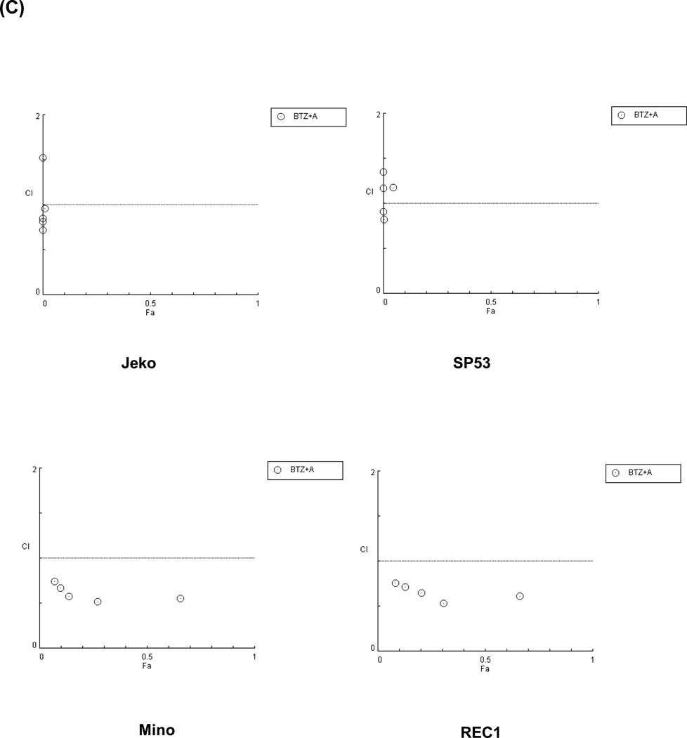
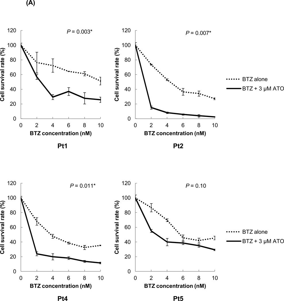
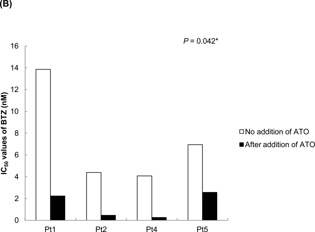
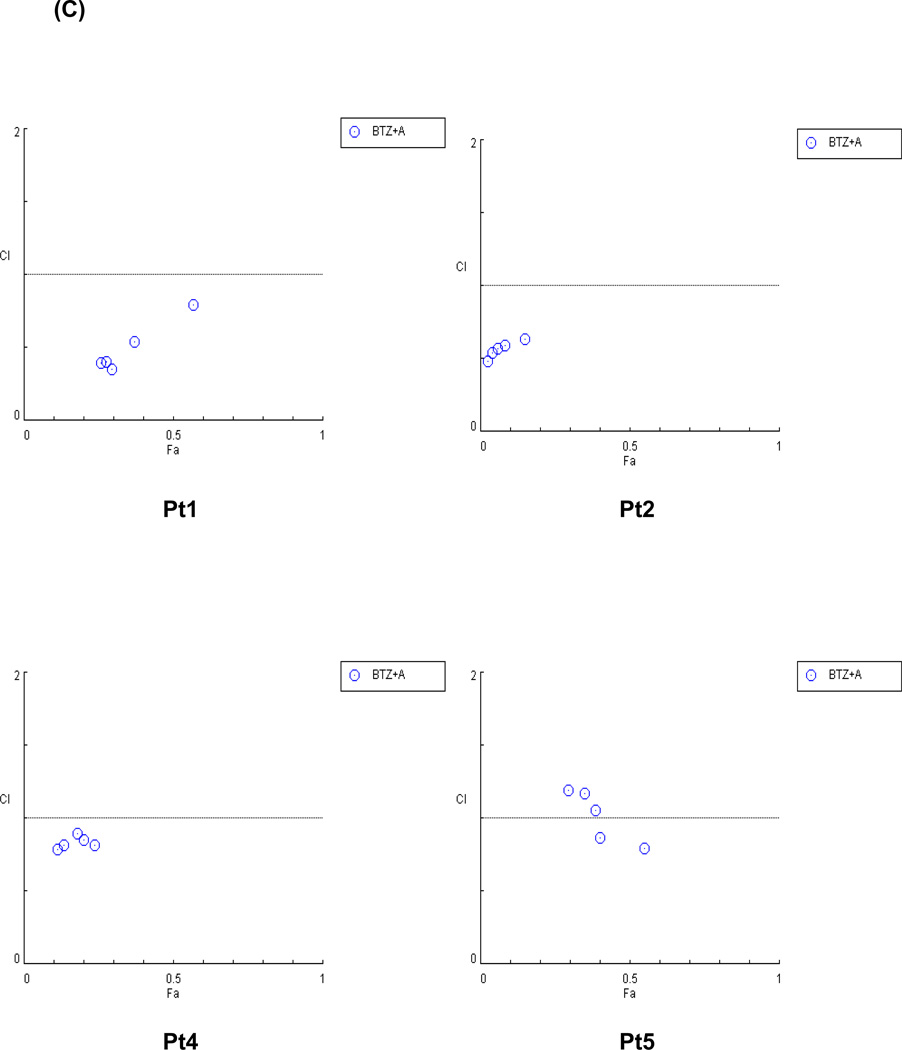
Mantle cell lymphoma (MCL) is a subtype of B-cell Non-Hodgkin's Lymphoma (NHL) and accounts for ~6% of all lymphomas. MCL is highly refractory to most chemotherapy including newer antibody-based therapeutic approaches, and high-grade MCL has one of the worst survival rates among NHLs. Therefore, the development of new therapeutic strategies to overcome drug resistance of MCL is important. In this article, we tested the effects of arsenic trioxide (As(2) O(3) , ATO) in bortezomib-resistant MCL. ATO is reported to induce complete remission in the patients with relapsed or refractory acute promyelocytic leukemia. Their effects in MCL, however, have not been explored. In this report, we show that ATO effectively inhibited the growth of MCL cells in vitro. ATO treatment also reduced cyclin D1 expression which is a genetic hallmark of MCL and NF-kB expression which was reported to have a prosurvival role in some MCL cells. The induction of apoptosis in MCL was partially due to reduced levels of cyclin D1 and increased levels of apoptosis-related molecules. The antiproliferative effects of bortezomib on MCL greatly increased when the cells were also treated with ATO, indicating ATO can sensitize MCL to bortezomib. Similar results were noted in bortezomib-resistant cell lines. In conclusion, ATO may be an alternative drug for use in combined adjuvant therapies for MCL, and further clinical testing should be performed.


Figures

Figure 1
Figure 1. Arsenic trioxide (ATO) affects the growth of MCL cells
(A) ATO inhibited the proliferation of MCL cell lines in a dose- and time-dependent manner. The sensitivities of the MCL cell lines, Jeko-1 and SP-53, to ATO were determined using the CellTiter-Blue® fluorometric cell viability assay (Promega). The cells were cultured for 18, 24, and 48 hrs with different concentrations of ATO. ATO was serially diluted from a maximal dose of 20 εM. Cell viability was presented as a ratio comparing cells given treatment with those not given treatment. The results are shown as the mean ± standard deviation of triplicate experiments. (B) ATO induced the cell death of primary MCL cells. MCL cells from six different patients were cultured for 18 hrs with or without ATO. ATO was serially diluted from a maximal dose of 10 εM. The cytotoxicity of ATO was determined using the CellTiter-Blue® fluorometric assay and was presented as a ratio comparing cells given treatment with those not given treatment. The mean IC50 value of ATO for primary MCL cells was comparable with the IC50 values of the MCL cell lines, Jeko-1 and SP-53. The bar represents the average of IC50 value of ATO for primary MCL cells.
Figure 2
Figure 2. ATO modulates the cyclin D1 expression in MCL
(A) ATO down-regulated the expression of cyclin D1 in MCL. The MCL cell lines, Jeko-1 and SP-53, were incubated with 5 εM ATO for 24 or 48 hrs, after which mRNA was isolated and cDNA prepared. The cDNA was analyzed for the presence of cyclin D1 genes by real time-PCR with GAPDH used as a control. The results are shown as the mean ± standard deviation of triplicate experiments. P values were calculated using the ANOVA test. *, P<0.05. (B) Jeko-1 and SP-53 cells were treated with or without 5 εM ATO for 48 hrs. Immunoblot analyses of cyclin D1 expression were performed using cell lysate proteins from untreated and treated Jeko-1 and SP-53 cells. GAPDH was used as a control. ATO readily suppressed the over-expression of cyclin D1 protein in MCL cells.
Figure 3
Figure 3. ATO induces the apoptosis of MCL cells
(A) ATO induced the dose-dependent apoptosis of MCL cells. Representative flow cytometric profiles show 7-AAD- and Annexin V-stained MCL cell lines (Jeko-1 and SP-53), after incubation with different concentrations of ATO for 48 hrs. ATO was serially diluted from a maximal dose of 6 εM. Cells negative for both Annexin V- and 7-AAD negative cells are viable (lower left quadrant). As apoptosis proceeds, the cells become Annexin V positive and 7-AAD negative (lower right; early apoptosis) and then both Annexin V and 7-AAD positive (upper right quadrant; end stage apoptosis). The results shown are representative of at least two comparable experiments. (B) ATO down-regulated Bcl-2-targeted gene expression in MCL. Jeko-1 and SP-53 cells were treated with 5 εM ATO for 24 or 48 hrs. mRNA was then isolated, and cDNA was prepared for real time-PCR analysis. The cDNA was analyzed for the presence of Bcl-2 target genes, with GAPDH as a control. The results are shown as the mean ± standard deviation of triplicate experiments. P values were calculated using the ANOVA test. *, P<0.05. (C) Immunoblot analyses of Bcl-2, an anti-apoptotic protein, expression were performed using cell lysates from untreated and treated Jeko-1 and SP-53 cells. Jeko-1 and SP-53 cells were treated with 5 εM ATO for 48 hrs. GAPDH was used as a control. ATO suppressed the Bcl-2 protein expression in MCL. (D) ATO activated the cleavage of caspase-3 and -9 in MCL cell lines. Jeko-1 and SP-53 cells were treated with 5 εM ATO for 48 hrs. The cytospin cells were then fixed and immunostained. Representative images with positive immunostaining for the anti-cleaved caspase-3 and -9 antibodies are shown. ATO-treated MCL cells expressed high levels of cleaved caspase-3 and -9 (arrows) compared with untreated cells. At least five sections from each cytospin slides were analyzed. (E) ATO affected the activation of key proteins in the caspase cascade. Immunoblot analyses of cleaved PARP expression were performed using cell lysates from untreated and treated Jeko-1 and SP-53 cells. Jeko-1 and SP-53 cells were treated with 5 εM ATO for 48 hr. GAPDH was used as a control. ATO induced the cleavage of the apoptotic-related protein, PARP, in MCL.
Figure 4
Figure 4. ATO suppress the NF-kB activation in MCL cells
(A) NF-ΚB DNA binding activities were decreased after ATO treatment in MCL cells. Nuclear extracts from Jeko-1 and SP-53 cells that were untreated or treated with ATO (5 εM for 24 hrs) were analyzed using ELISA assays to evaluate the DNA-binding activity of p50 and p65. Bars represent the relative ratio of p50 and p65 DNA-binding activity levels before and after treatment with ATO. (B) Jeko-1 and SP-53 cells were cultured with 5 εM ATO for 24 or 48 hrs. The mRNA was isolated from untreated and ATO-treated MCL cells, and then cDNA was prepared for real time-PCR. The cDNA was analyzed for the presence of genes involved in NF-ΚB-linked transcriptional targets. GAPDH was used as a control. ATO induced the generalized suppression of the expression of the NF-kB target genes, IL-6, IL-8, and c-IAP2, in MCL cells. The columns are mean of duplicate experiments, and bars represent standard deviation.
Figure 5
Figure 5. ATO and bortezomib synergistically induce apoptosis in MCL cell lines
(A) The combination of ATO and bortezomib increased the cytotoxicity of not only bortezomib-sensitive MCL cell lines, but also bortezomib-resistant MCL cell lines. Jeko-1 and SP-53 cells were used as bortezomib-sensitive MCL cell lines, and Mino, and Rec-1 cells were used as the bortezomib-resistant MCL cell lines. The cells were cultured for 24 hrs with bortezomib alone or in combination with 3 εM ATO. Bortezomib was serially diluted as indicated. The survival rates of MCL cells treated with bortezomib alone or the combination of ATO and bortezomib were compared using the CellTiter-Blue® fluorometric cell viability assay. Cell survival rates were presented as a ratio comparing treated and untreated cells. The results are shown as the mean ± standard deviation of triplicate experiments. P values were calculated using unpaired t-test. *, P<0.05. BTZ, bortezomib; ATO, arsenic trioxide. (B) ATO markedly decreased the IC50 values of bortezomib in bortezomib-resistant MCL cells and in bortezomib-sensitive MCL cells. Jeko-1, SP-53, Mino, and Rec-1 cells were cultured for 24 hrs after the addition of bortezomib with or without 3 εM ATO. Cell viability was determined by the CellTiter-Blue® assay. The IC50 values of bortezomib for all tested MCL cells were significantly decreased when in combination with ATO. *, P<0.05 by unpaired t-test. BTZ, bortezomib; ATO, arsenic trioxide. (C) ATO showed synergistic effects with bortezomib to induce cytotoxicity in bortezomib-resistant MCL cells and in bortezomib-sensitive MCL cells. The synergistic cytotoxic effects of bortezomib and ATO were determined using the combination index (CI) based on the data from cell viability assays. CI plots were generated using the CompuSyn software (ComboSyn, NJ, USA) according to the Chou-Talalay method. The combination of ATO and bortezomib is synergistic when CI < 1.0, additive when CI = 1, and antagonistic when CI > 1.0.
Figure 6
Figure 6. ATO sensitizes MCL patient cells to bortezomib
(A) ATO significantly improved the bortezomib sensitivity in primary MCL cells. The chemosensitivities of MCL patient cells to bortezomib were compared with or without ATO using the CellTiter-Blue® fluorometric cell viability assay. The cells were cultured for 16 hrs with bortezomib alone or in combination with 3 εM ATO. The drugs were serially diluted as indicated. Cell survival rates were presented as a ratio comparing treated and untreated cells. The results are shown as the mean ± standard deviation of triplicate experiments. P values were calculated using the unpaired t-test. *, P<0.05. BTZ, bortezomib; ATO, arsenic trioxide. (B) ATO markedly decreased the IC50 values of bortezomib in MCL patient cells. Four different MCL patient samples were cultured for 16 hrs after the addition of bortezomib with or without 3 εM ATO. Cell viability was determined by the CellTiter-Blue® assay. The IC50 values of bortezomib for primary MCL cells were significantly decreased when combined with ATO. *, P<0.05 by unpaired t-test. BTZ, bortezomib; ATO, arsenic trioxide. (C) ATO synergized with bortezomib to induce cytotoxicity in MCL patient samples. The synergistic cytotoxic effects of bortezomib and ATO were determined using the combination index (CI) based on the data from cell viability assays. CI plots were generated using the CompuSyn software (ComboSyn, NJ, USA) according to the Chou-Talalay method. The combination of ATO and bortezomib is synergistic when CI < 1.0, additive when CI = 1, and antagonistic when CI > 1.0.

Similar articles

Cited by

References

    1. Mounter PJ, Lennard AL. Management of non-Hodgkin's lymphomas. Postgraduate Medical Journal. 1999;75:2–6. - PMC - PubMed
    1. Salaverria I, Perez-Galan P, Colomer D, Campo E. Mantle cell lymphoma: from pathology and molecular pathogenesis to new therapeutic perspectives. Haematologica. 2006;91:11–16. - PubMed
    1. Pileri SA, Falini B. Mantle cell lymphoma. Haematologica. 2009;94:1488–1492. - PMC - PubMed
    1. Bonati A, Rizzoli V, Lunghi P. Arsenic trioxide in hematological malignancies:The new discovery of an ancient drug. Curr Pharm Biotechnol. 2006;7:397–405. - PubMed
    1. Douer D, Tallman MS. Arsenic trixoide:New clinical experience with and old medication in hematological melignancies. J. Clin. Oncol. 2005;33:2396–2410. - PubMed

Publication types

MeSH terms