. 2012 Sep 24;209(10):1825-40.
doi: 10.1084/jem.20120774. Epub 2012 Sep 10.

Extrafollicular B cell activation by marginal zone dendritic cells drives T cell-dependent antibody responses

Affiliations

Extrafollicular B cell activation by marginal zone dendritic cells drives T cell-dependent antibody responses

Craig P Chappell et al. J Exp Med. .

Abstract

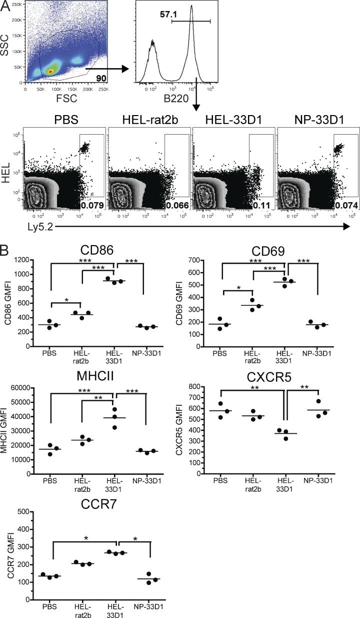
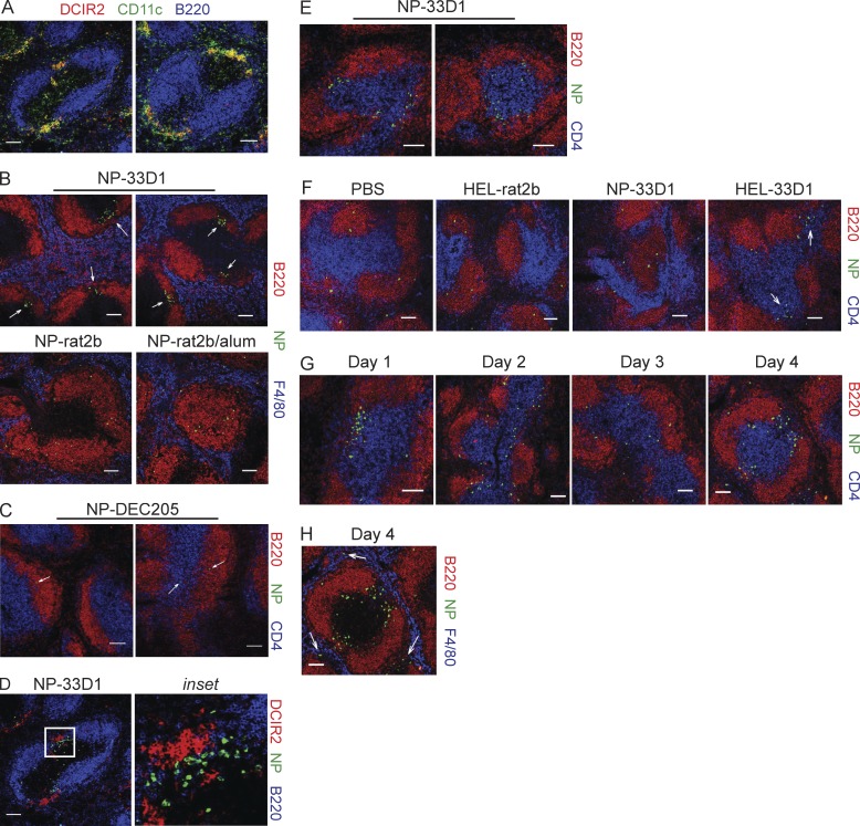
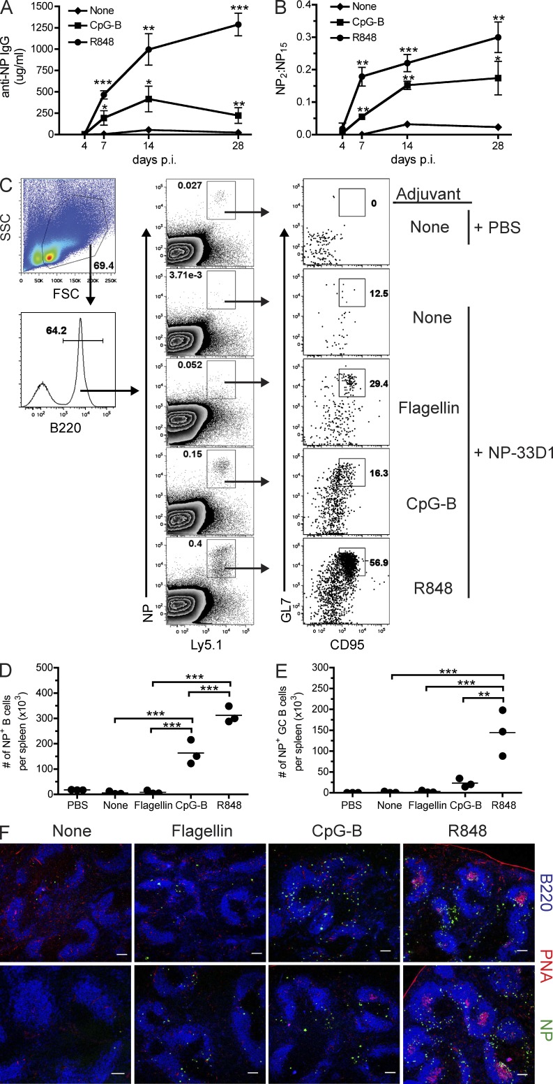
Dendritic cells (DCs) are best known for their ability to activate naive T cells, and emerging evidence suggests that distinct DC subsets induce specialized T cell responses. However, little is known concerning the role of DC subsets in the initiation of B cell responses. We report that antigen (Ag) delivery to DC-inhibitory receptor 2 (DCIR2) found on marginal zone (MZ)-associated CD8α(-) DCs in mice leads to robust class-switched antibody (Ab) responses to a T cell-dependent (TD) Ag. DCIR2(+) DCs induced rapid up-regulation of multiple B cell activation markers and changes in chemokine receptor expression, resulting in accumulation of Ag-specific B cells within extrafollicular splenic bridging channels as early as 24 h after immunization. Ag-specific B cells primed by DCIR2(+) DCs were remarkably efficient at driving naive CD4 T cell proliferation, yet DCIR2-induced responses failed to form germinal centers or undergo affinity maturation of serum Ab unless toll-like receptor (TLR) 7 or TLR9 agonists were included at the time of immunization. These results demonstrate DCIR2(+) DCs have a unique capacity to initiate extrafollicular B cell responses to TD Ag, and thus define a novel division of labor among splenic DC subsets for B cell activation during humoral immune responses.


Figures

Figure 1.
Figure 1.
Ag delivery to DCIR2+ DCs in vivo induces IgG1-restricted Ab responses. (A–D) ELISA analyses of NP-specific serum Ab from B6 mice immunized 10 d earlier with the indicated dose of NP-33D1, NP-DEC205, or NP-rat IgG. Graphs show the mean ± SEM of a representative experiment from three independent experiments using three to four mice/group (using the 10 µg dose). Data were analyzed by two-way ANOVA analysis. (E) ELISPOT analysis showing the number of IgG-secreting NP-specific AFCs in the spleens of C57BL/6 mice on the days indicated after administration of NP-33D1 or NP-rat2b. The mean ± SEM from a single experiment using four mice/group/day is shown. (F) Day 10 anti-NP IgG1 Ab responses in FcγRγ-deficient and C57BL/6 mice immunized with NP-33D1. (G) Day 10 anti-NP IgG1 Ab responses in CD11c-DTR transgenic and C57BL/6 mice treated with 100 ng DT 24 h before immunization with NP-33D1. Data in F and G were analyzed by unpaired Student’s t test and are representative experiments from three and two independent experiments, respectively, and show the mean ± SEM using three to four mice/group. P-value indicators *** and * refer to P < 0.001 and P < 0.05, respectively.
Figure 2.
Figure 2.
DCIR2-induced Ab responses are TD. Groups of C57BL/6 (A–D and F) or BALB/c animals (E) and mice deficient in the indicated gene product were immunized with NP-33D1 and bled 10 d later for serum Ab analysis by ELISA. Graphs depict the mean ± SEM of anti-NP IgG1 serum Ab. Data in A, B, and E are representative data from three independent experiments each. Data in C, D, and F are representative of two independent experiments each. All experiments used three to five mice/group and were analyzed by unpaired Student’s t tests. N.D. = not detected. P-value indicators ***, **, and * refer to P < 0.001, P < 0.01, and P < 0.05, respectively.
Figure 3.
Figure 3.
Ag delivery to DCIR2+ DCs does not induce GC formation, affinity maturation, or long-lived Ab responses and fails to prime for Ag recall. (A) Flow cytometry plots show the frequency of splenic B220+GL7+ GC B cells in C57BL/6 mice immunized 7 d previously with 50 µg NP-CGG plus alum or 10 µg NP-33D1 or NP-rat2b. (B) Total numbers of splenic B220+GL7+ GC B cells from the mice in A. Each dot represents an individual animal with the mean (horizontal bars) indicated. Data were analyzed by one-way ANOVA with Bonferroni’s post-tests and are representative of three independent experiments using three to four mice/group. (C and D) Groups of C57BL/6 mice were immunized as in A, and the relative affinity (C) or quantity (D) of anti-NP IgG Ab over time was determined by ELISA. Mean ± SEM is graphed for a single experiment using four mice/group. (E) C57BL/6 mice were immunized as in A and rechallenged with 10 µg NP-CGG, NP-33D1, or NP-rat2b as indicated 90 d after primary immunization. ELISPOT analysis shows the number of NP-specific IgG AFCs recovered from spleens 4 d after challenge. Data in E were analyzed by two-way ANOVA and are representative data from two independent experiments using four mice/group. P-value indicators *** and ** refer to P < 0.001 and P < 0.01, respectively.
Figure 4.
Figure 4.
Agonists for TLR3, TLR5, or RIG-I do not increase affinity maturation of serum Ab after NP-33D1 immunization. (A) Day 28 anti-NP Ab responses from C57BL/6 mice immunized with NP-33D1 or NP-rat2b with or without 100 µg pU/UC ssRNA or 50 µg polyI:C. Means ± SEM are graphed. (B) Relative affinity ELISA assay showing the ratio of anti-NP IgG quantities that bound NP2 versus NP15 for the groups shown in A. Means ± SEM are graphed. Ab responses in mice immunized with NP-rat2b plus alum are shown as a positive reference for affinity maturation. Data in A and B are representative data from two independent experiments using three to four mice/group. (C and D) C57BL/6 mice were immunized with NP-33D1 with or without 20 µg flagellin and analyzed as A and B. Data are representative of three independent experiments using three mice/group and depict the mean ± SEM. Statistical differences were calculated using two-way ANOVA (A and C) or one-way ANOVA followed by Bonferroni’s post-tests (B and D). P-value indicators ** and * refer to P < 0.01 and P < 0.05, respectively.
Figure 5.
Figure 5.
Ag-specific B cells are rapidly activated in vivo after Ag uptake by DCIR2+ DCs. (A) Flow cytometry plots show the gating strategy for identification of adoptively transferred Ly5.1+ NP-binding B1-8hi B cells from the spleens of C57BL/6 recipients immunized as indicated 24 h previously. Numbers denote frequency of gated cells among total B220+ lymphocytes. (B) Expression of CD69, CD86, MHCII, and CXCR5 among NP-binding Ly5.1+ B cells gated in A for immunized mice (blue dots and solid lines) and PBS-injected controls (red dots and shaded histograms). (C) Geometric mean fluorescence intensities (MFIs) are plotted for the indicated surface molecule among NP-binding Ly5.1+ B cells gated in A. Each dot represents an individual animal with the mean (horizontal bars) indicated. Data shown in A–C are representative data from more than four independent experiments using three to four mice/group. (D) Up-regulation of OX40L expression over time among transferred Ly5.1+ NP-binding B cells from C57BL/6 recipients that received NP-33D1 (solid line), NP-rat2b (dotted line), NP-rat2b plus alum (dashed line), or PBS-injected controls (shaded). Data are representative of two independent experiments using three mice/group. (E) Expression of the indicated activation marker is graphed for gated NP+ (blue line) and NP-negative (red line) populations from mice in A immunized with NP-33D1. Shaded histograms depict NP-binding B cells from a PBS-injected control for reference. (F) The top shows gating strategy for analysis of DC subsets and B cells. The bottom shows AF647 fluorescence among the indicated population 30 min after injection of 10 µg NP-33D1 (red line) or NP-rat2b (blue line) conjugated to Alexa Fluor 647. Data are representative of two independent experiments using two to three mice/group. (G) MFIs of the indicated surface marker are plotted for transferred NP-specific B cells (gated as in A) obtained from C57BL/6 recipients immunized with 10 µg NP-rat2b, NP-DEC205, or NP-33D1. The mean ± SEM from a representative experiment of three independent experiments using three to four mice/group is shown. P-value indicators ***, **, and * refer to P < 0.001, P < 0.01, and P < 0.05, respectively.
Figure 6.
Figure 6.
DC-mediated B cell activation requires specific Ag. (A) Flow cytometry plots show gating of adoptively transferred Ly5.2+ HEL-binding VDJ9κ5 B cells from the spleens of recipient Ly5.1+ congenic C57BL/6 mice immunized as indicated 24 h previously. Numbers denote frequency of gated cells among total B220+ lymphocytes. (B) MFIs of the indicated surface marker 24 h p.i. among HEL-binding Ly5.2+ B cells gated in A. Each dot represents an individual animal with the mean (horizontal bars) indicated. Data in A and B are representative data from three independent experiments using three mice/group. Statistical differences were calculated using one-way ANOVA analysis followed by Bonferroni’s post-tests. P-value indicators ***, **, and * refer to P < 0.001, P < 0.01, and P < 0.05, respectively.
Figure 7.
Figure 7.
DC-mediated B cell activation is TI. (A and B) MFIs of the indicated surface marker are plotted for NP-binding B cells (gated as in Fig. 5 A) obtained from C57BL/6 and MHCII-deficient (A) or TCR-β/δ–deficient (B) hosts 24 h after immunization with NP-33D1 or NP-rat2b as indicated. Each dot represents an individual animal with the mean (horizontal bars) indicated. Data in A and B are representative data from two independent experiments each using three to four mice/group. Data were analyzed by one-way ANOVA analyses followed by Bonferroni’s post-tests. P-value indicators ***, **, and * refer to P < 0.001, P < 0.01, and P < 0.05, respectively. n.s. = not significant.
Figure 8.
Figure 8.
NP-specific B cells rapidly accumulate in MZ bridging channels after Ag delivery to DCIR2+ DCs. 6–8-µm sections were prepared from frozen spleens of B1-8hi or VDJ9κ5 recipient C57BL/6 mice immunized 1–4 d earlier with NP-rat2b, NP-rat2b plus alum, NP-DEC205, or NP-33D1 and stained to determine the location of Ag-specific B cells and/or DCIR2+ DCs. (A) Two examples showing colocalization of DCIR2+ (red) and CD11c+ (green) cells in bridging channels between B cell follicles (blue) in B1-8hi recipients immunized 24 h previously with NP-33D1. (B) NP-binding B cells (green) accumulate at apical poles (arrows) of B220+ B cell follicles (red) 24 h after administration of NP-33D1 (two examples, top row), but not NP-rat2b or NP-rat2b plus alum (bottom row). (C) Two examples indicating NP-binding B cells (green, arrows) do not localize to T cell zones (blue) or bridging channels 24 h p.i. with NP-DEC205. (D) NP-binding B cells (green) found in extrafollicular bridging channels are in close association with DCIR2+ DCs (red) 24 h after immunization with NP-33D1. (E) Two examples showing NP-binding B cells (green) are oriented toward T cell zones (blue) 24 h p.i. with NP-33D1. Data in A–E are representative data from three independent experiments from which more than five sections were analyzed from two or more animals from each group. (F) HEL-binding B cells (green) accumulate in bridging channels (arrows) 24 h p.i. with HEL-33D1, but not NP-33D1. Data in F are representative of two independent experiments from which more than five sections from two animals from each group were analyzed. (G) NP-binding B cells (green) migrate from bridging channels (day 1) to CD4 T cell areas (blue; days 2 and 3) before relocating on day 4 to bridging channels (G) and F4/80+ red pulp (blue; H) after immunization with NP-33D1 (arrows indicate extrafollicular NP-binding B cells). Data for days 2–4 are representative of more than five sections analyzed from two animals for each time point from a single experiment. Bars, 100 µm.
Figure 9.
Figure 9.
Ag-specific B cells primed by DCIR2+ DCs are highly efficient APCs for CD4 T cell proliferation. (A) Schematic showing experimental design. (B) Histograms show dilution of CFSE among gated CD4+Va2+ OT-II T cells after 72 h in culture with NP-specific B cells from spleens of B1-8hi recipient C57BL/6 mice immunized 12 or 36 h previously with NP-33D1 or NP-rat2b. (C) Proliferation indices generated from the data in B are shown. (D) Same as in B, except sorted DCIR2+ DCs from spleens of immunized C57BL/6 mice were used as APCs. (E) Proliferation indices generated from the data in D are shown. Proliferation index is defined as total CFSE fluorescence of the negative control (APC/T cell ratio of 0:1) divided by total CFSE fluorescence of indicated samples. A representative experiment of two independent experiments for each time point is shown for both B and D. (F) Day 12 anti-NP IgG Ab responses in MHCII-deficient and C57BL/6 recipients of OT-II CD4 T cells and B1-8hi B cells after immunization with NP-OVA-33D1 or NP-OVA-rat2b is shown. Each dot represents an individual animal with the mean (horizontal bars) indicated. A representative experiment of two independent experiments using four to five mice/group is shown. P-value indicator *** refers to P < 0.001.
Figure 10.
Figure 10.
TLR7 and TLR9 agonists promote increased Ab responses, GC formation, and affinity maturation of serum Ab. (A) Kinetics of anti-NP IgG Ab responses in C57BL/6 mice that received NP-33D1 alone or together with 20 µg R848 or 50 µg CpG-B. (B) Kinetics of affinity maturation among total anti-NP IgG serum Ab from the animals in A. Statistical values comparing each group with NP-33D1–only control were generated using an unpaired Student’s t test. Data in A and B depict a representative experiment of two independent experiments using three mice/group. Means ± SEM are graphed. (C) C57BL/6 recipients of B1-8hi B cells were immunized as indicated and analyzed on day 7 p.i. Flow cytometry plots show gating strategy (left column) and frequency of NP-binding B cells (middle column) among total B220+ B cells. Right column shows expression of GL7 and Fas among gated NP-binding B cells. Numbers indicate frequency of GC B cells among total NP-binding B cells. Total numbers per spleen of NP-binding B cells (D) and NP-binding GC B cells (E) are plotted for the mice in C. Each dot represents an individual animal with the mean (horizontal bars) indicated. Data in C–E are representative of two independent experiments using three to four mice/group. Statistical values were generated using one-way ANOVA analyses followed by Bonferroni’s post-tests. (F) Immunofluorescence showing GC formation 7 d p.i. in spleen sections derived from mice immunized as in C and stained with B220 (blue), PNA (red), and NP (green). Two examples from each immunization are shown (top and bottom rows). Bars, 100 µm. Data are representative of more than six sections per mouse taken from two mice per condition. P-value indicators ***, **, and * refer to P < 0.001, P < 0.01, and P < 0.05, respectively.

Similar articles

Cited by

References

    1. Akira S., Uematsu S., Takeuchi O. 2006. Pathogen recognition and innate immunity. Cell. 124:783–801 10.1016/j.cell.2006.02.015 - DOI - PubMed
    1. Balázs M., Martin F., Zhou T., Kearney J. 2002. Blood dendritic cells interact with splenic marginal zone B cells to initiate T-independent immune responses. Immunity. 17:341–352 10.1016/S1074-7613(02)00389-8 - DOI - PubMed
    1. Barbalat R., Ewald S.E., Mouchess M.L., Barton G.M. 2011. Nucleic acid recognition by the innate immune system. Annu. Rev. Immunol. 29:185–214 10.1146/annurev-immunol-031210-101340 - DOI - PubMed
    1. Bergtold A., Desai D.D., Gavhane A., Clynes R. 2005. Cell surface recycling of internalized antigen permits dendritic cell priming of B cells. Immunity. 23:503–514 10.1016/j.immuni.2005.09.013 - DOI - PubMed
    1. Berney C., Herren S., Power C.A., Gordon S., Martinez-Pomares L., Kosco-Vilbois M.H. 1999. A member of the dendritic cell family that enters B cell follicles and stimulates primary antibody responses identified by a mannose receptor fusion protein. J. Exp. Med. 190:851–860 10.1084/jem.190.6.851 - DOI - PMC - PubMed

Publication types

MeSH terms