. 2012 Oct;140(4):375-89.
doi: 10.1085/jgp.201110763. Epub 2012 Sep 10.

Plasmin and chymotrypsin have distinct preferences for channel activating cleavage sites in the γ subunit of the human epithelial sodium channel

Affiliations

Plasmin and chymotrypsin have distinct preferences for channel activating cleavage sites in the γ subunit of the human epithelial sodium channel

Silke Haerteis et al. J Gen Physiol. 2012 Oct.

Abstract

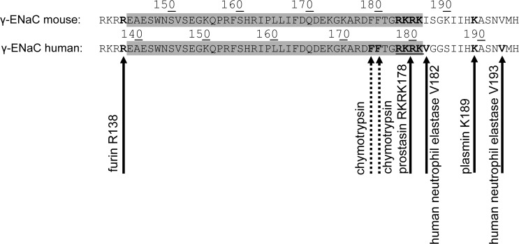
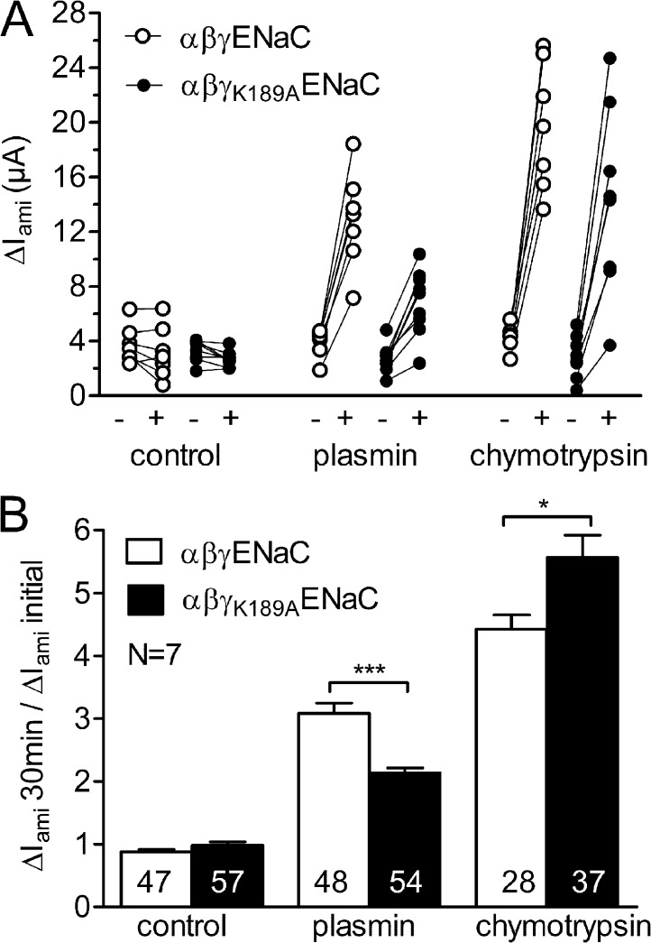
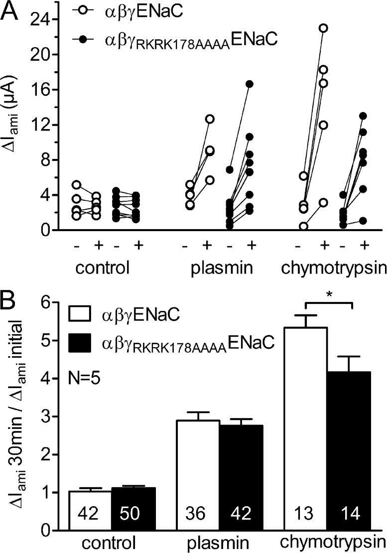
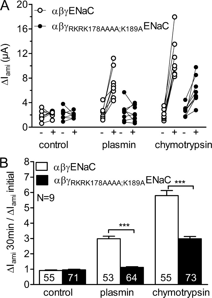
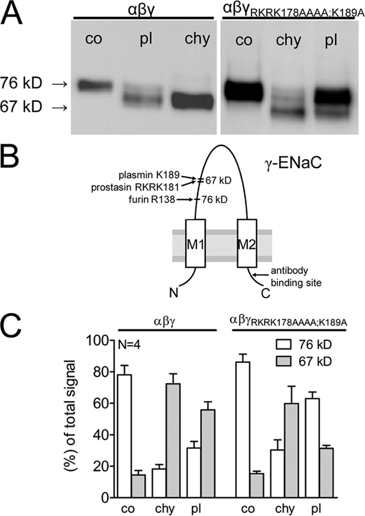
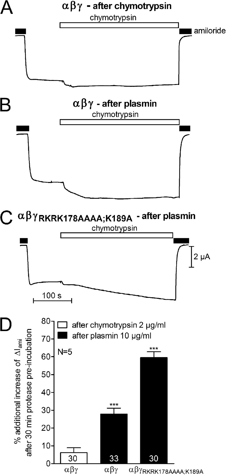
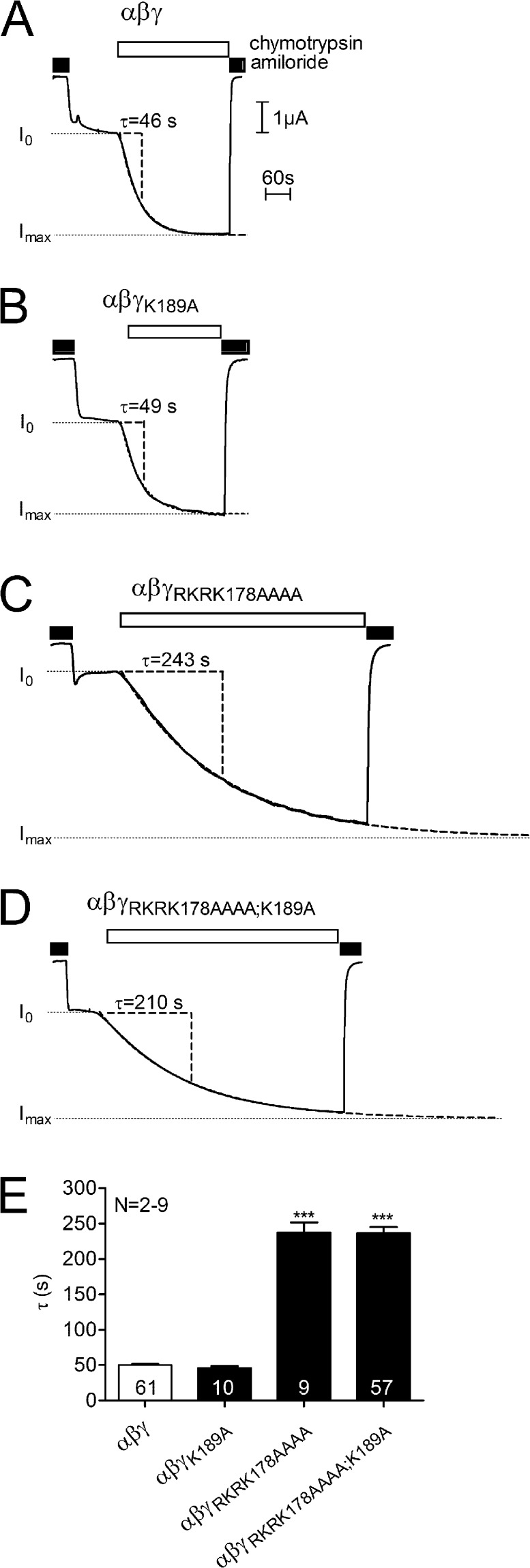
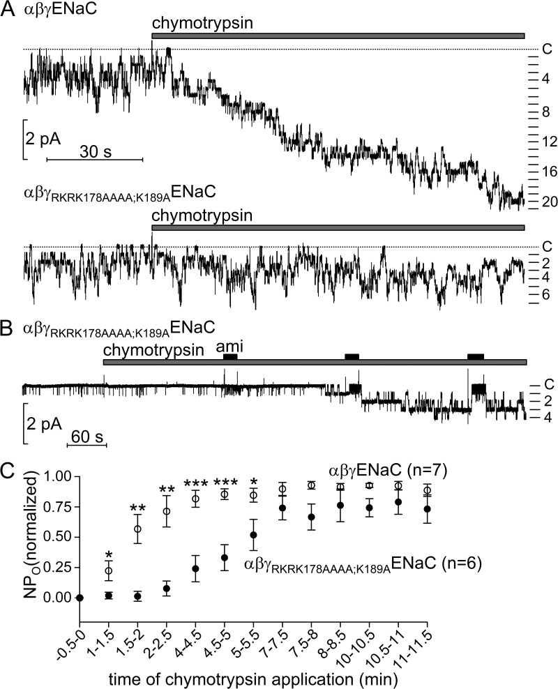
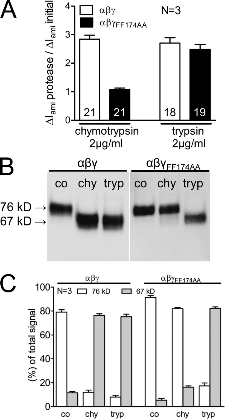
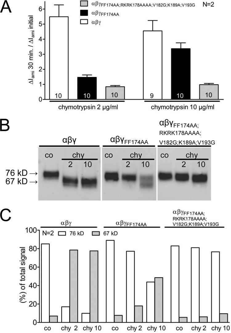
Proteolytic activation of the epithelial sodium channel (ENaC) involves cleavage of its γ subunit in a critical region targeted by several proteases. Our aim was to identify cleavage sites in this region that are functionally important for activation of human ENaC by plasmin and chymotrypsin. Sequence alignment revealed a putative plasmin cleavage site in human γENaC (K189) that corresponds to a plasmin cleavage site (K194) in mouse γENaC. We mutated this site to alanine (K189A) and expressed human wild-type (wt) αβγENaC and αβγ(K189A)ENaC in Xenopus laevis oocytes. The γ(K189A) mutation reduced but did not abolish activation of ENaC whole cell currents by plasmin. Mutating a putative prostasin site (γ(RKRK178AAAA)) had no effect on the stimulatory response to plasmin. In contrast, a double mutation (γ(RKRK178AAAA;K189A)) prevented the stimulatory effect of plasmin. We conclude that in addition to the preferential plasmin cleavage site K189, the putative prostasin cleavage site RKRK178 may serve as an alternative site for proteolytic channel activation by plasmin. Interestingly, the double mutation delayed but did not abolish ENaC activation by chymotrypsin. The time-dependent appearance of cleavage products at the cell surface nicely correlated with the stimulatory effect of chymotrypsin on ENaC currents in oocytes expressing wt or double mutant ENaC. Delayed proteolytic activation of the double mutant channel with a stepwise recruitment of so-called near-silent channels was confirmed in single-channel recordings from outside-out patches. Mutating two phenylalanines (FF174) in the vicinity of the prostasin cleavage site prevented proteolytic activation by chymotrypsin. This indicates that chymotrypsin preferentially cleaves at FF174. The close proximity of FF174 to the prostasin site may explain why mutating the prostasin site impedes channel activation by chymotrypsin. In conclusion, this study supports the concept that different proteases have distinct preferences for certain cleavage sites in γENaC, which may be relevant for tissue-specific proteolytic ENaC activation.


Figures

Figure 1.
Figure 1.
Sequence comparison of mouse γENaC (amino acids 140–200) and human γENaC (amino acids 135–195). The amino acid sequences of mouse γENaC and human γENaC are from the UniProt database (accession nos. Q9WU39 and P51170). The alignment demonstrates the high homology between the two species. The putative cleavage sites for furin (R138), chymotrypsin (FF174), prostasin (RKRK178), human neutrophil elastase (V182 and V193), and plasmin (K189) are indicated in bold and marked by an arrow.
Figure 2.
Figure 2.
The stimulatory effect of plasmin is reduced but not abolished in oocytes expressing ENaC with a mutated putative plasmin site (γK189A). Oocytes expressing αβγ (open symbols) or αβγK189AENaC (closed symbols) were preincubated for 30 min in protease-free solution (control) or in solution containing either 10 µg/ml plasmin or 2 µg/ml chymotrypsin. Amiloride-sensitive whole cell currents (ΔIami) were determined before (−) and after (+) incubation. (A) Individual ΔIami values from a representative experiment using one batch of oocytes. Data points obtained from individual oocytes are connected by a line. (B) Summary of similar experiments as shown in A. Columns represent relative stimulatory effect on ΔIami calculated as the ratio of ΔIami measured after a 30-min preincubation (ΔIami 30 min) to the initial ΔIami (ΔIami initial) measured before incubation. Numbers inside the columns indicate the number of individual oocytes measured. N indicates the number of different batches of oocytes. *, P < 0.05; ***, P < 0.001; unpaired t test.
Figure 3.
Figure 3.
The stimulatory effect of plasmin is preserved in oocytes expressing ENaC with a mutated putative prostasin site (γRKRK178AAAA). Oocytes expressing αβγ (open symbols) or αβγRKRK178AAAAENaC (closed symbols) were preincubated for 30 min in protease-free solution (control) or in solution containing either 10 µg/ml plasmin or 2 µg/ml chymotrypsin. Amiloride-sensitive whole cell currents (ΔIami) were determined before (−) and after (+) incubation. (A) Individual ΔIami values from a representative experiment using one batch of oocytes. Data points obtained from individual oocytes are connected by a line. (B) Summary of similar experiments as shown in A. Columns represent relative stimulatory effect on ΔIami calculated as the ratio of ΔIami measured after a 30-min preincubation (ΔIami 30 min) to the initial ΔIami (ΔIami initial) measured before incubation. Numbers inside the columns indicate the number of individual oocytes measured. N indicates the number of different batches of oocytes. *, P < 0.05; unpaired t test.
Figure 4.
Figure 4.
The stimulatory effect of plasmin on ENaC is abolished in oocytes expressing ENaC with a combined mutation of the plasmin and prostasin sites (αβγRKRK178AAAA;K189A). Oocytes expressing αβγ (open symbols) or αβγRKRK178AAAA;K189AENaC (closed symbols) were preincubated for 30 min in protease-free solution (control) or in solution containing either 10 µg/ml plasmin or 2 µg/ml chymotrypsin. Amiloride-sensitive whole cell currents (ΔIami) were determined before (−) and after (+) incubation. (A) Individual ΔIami values from a representative experiment using one batch of oocytes. Data points obtained from individual oocytes are connected by a line. (B) Summary of similar experiments as shown in A. Columns represent relative stimulatory effect on ΔIami calculated as the ratio of ΔIami measured after a 30-min preincubation (ΔIami 30 min) to the initial ΔIami (ΔIami initial) measured before incubation. Numbers inside the columns indicate the number of individual oocytes measured. N indicates the number of different batches of oocytes. ***, P < 0.001; unpaired t test.
Figure 5.
Figure 5.
Mutating both the plasmin and the prostasin cleavage site (αβγRKRK178AAAA;K189A) reduces proteolytic cleavage of γENaC at the cell surface. Oocytes were treated as described in the legend of Fig. 4. In parallel to the detection of ΔIami (shown in Fig. 4), expression of biotinylated γENaC at the cell surface was analyzed by SDS-PAGE. γENaC was detected with an antibody against the C terminus of human γENaC. Oocytes expressing αβγ or αβγRKRK178AAAA;K189AENaC were preincubated for 30 min in protease-free solution (control [co]) or in solution containing either 10 µg/ml plasmin (pl) or 2 µg/ml chymotrypsin (chy). (A) Representative Western blots from oocytes expressing αβγ or αβγRKRK178AAAA;K189AENaC. (B) Model of the γENaC subunit showing cleavage sites for proteolytic activation and the binding site of the antibody used. (C) Densitometric analysis of similar Western blots as shown in A. For each lane, the signals detected in the regions of 76 kD (open columns) and 67 kD (gray columns) were determined and normalized to the sum of the total signal detected. N indicates the number of different batches of oocytes.
Figure 6.
Figure 6.
Activation with chymotrypsin after activation with plasmin. Representative whole cell current traces from oocytes expressing αβγ (A and B) or αβγRKRK178AAAA;K189A (C) ENaC after a 30-min preincubation in a solution containing 2 µg/ml chymotrypsin (A) or 10 µg/ml plasmin (B and C). 2 µM amiloride and 2 µg/ml chymotrypsin were present in the bath solution, as indicated by closed and open bars, respectively. (D) Average data obtained from five different batches of oocytes. Columns represent additional increase of ΔIami calculated as the ratio of ΔIami after superfusion with chymotrypsin to ΔIami after a 30-min preincubation in chymotrypsin (open column) or plasmin (closed columns). Numbers inside the columns indicate the number of individual oocytes analyzed. N indicates the number of different batches of oocytes. ***, P < 0.001; unpaired t test.
Figure 7.
Figure 7.
Mutating both the plasmin and the prostasin cleavage site delays proteolytic ENaC activation by chymotrypsin. Oocytes expressing αβγ (open symbols) or αβγRKRK178AAAA;K189AENaC (closed symbols) were preincubated for 30 min in protease-free solution (control) or for 5, 30, or 60 min in a solution containing 2 µg/ml chymotrypsin. Amiloride-sensitive whole cell currents (ΔIami) were determined before (−) and after (+) incubation. In parallel to the detection of ΔIami, expression of biotinylated γENaC at the cell surface was analyzed by SDS-PAGE. γENaC was detected with an antibody against the C terminus of human γENaC. (A) Circles represent the ratio of ΔIami measured after a 5-, 30-, or 60-min preincubation (ΔIami min) to the initial ΔIami (ΔIami initial) measured before incubation. Each data point represents the mean ΔIami measured in 22–24 individual oocytes of four different batches. (B and D) γENaC was detected with an antibody against the C terminus of human γENaC. Representative Western blot from one batch of oocytes. In noninjected oocytes, γENaC-specific signals were absent (not depicted). (C and E) Densitometric analysis of three Western blots similar to those shown in B or D. For each lane, the signals detected in the regions of 76 kD (open columns) and 67 kD (gray columns) were determined and normalized to the sum of the total signal detected. N indicates the number of different batches of oocytes.
Figure 8.
Figure 8.
Time constant of proteolytic ENaC activation by chymotrypsin is significantly increased in the αβγRKRK178AAAA and the αβγRKRK178AAAA;K189A mutant channel. Representative whole cell current traces from oocytes expressing αβγ (A), αβγK189A (B), αβγRKRK178AAAA (C), and αβγRKRK178AAAA;K189A (D) ENaC. 2 µM amiloride and 2 µg/ml chymotrypsin were present in the bath solution, as indicated by closed and open bars, respectively. Values for the time constant (τ) of proteolytic current activation were estimated by fitting individual current traces with an exponential function I(t) = I0 + (Imax − I0) · (1 − e(−t/τ)). Fitted curves are shown by broken lines superimposed on the representative current traces. The baseline current level before the application of chymotrypsin (I0) and the extrapolated current maximum (Imax) are indicated by dotted lines. The length of the time constant (τ) is indicated. (E) Average τ obtained from up to nine different batches of oocytes. Numbers inside the columns indicate the number of individual oocytes analyzed. N indicates the number of different batches of oocytes. ***, P < 0.001; unpaired t test.
Figure 9.
Figure 9.
Delayed proteolytic activation of near-silent channels in outside-out patches from oocytes expressing the αβγRKRK178AAAA;K189A mutant channel. (A) Representative single-channel current recordings obtained at a holding potential of −70 mV from an outside-out patch of an oocyte expressing αβγ (top trace) or αβγRKRK178AAAA;K189A (bottom trace) ENaC. 2 µg/ml chymotrypsin was present in the bath solution, as indicated by the gray bar. The current level at which all channels are closed (C) is indicated by a dotted line. (B) The experiment was performed as described in A, obtained from an outside-out patch of an oocyte expressing αβγRKRK178AAAA;K189A mutant ENaC. 2 µM amiloride (ami) and 2 µg/ml chymotrypsin were applied as indicated by closed and gray bars, respectively. (C) Time course of the averaged normalized NPO values after chymotrypsin application calculated from outside-out patch-clamp recordings as shown in A and B. In each individual experiment, NPO values before the application of chymotrypsin were set to zero, and maximal NPO values after chymotrypsin application were set to one. *, P < 0.05; **, P < 0.01; ***, P < 0.001; unpaired t test.
Figure 10.
Figure 10.
Mutating two phenylalanine residues (γFF174) adjacent to the prostasin cleavage site prevents proteolytic activation of ENaC by chymotrypsin. Continuous whole cell current measurements were performed in oocytes expressing αβγ or αβγFF174AAENaC. ΔIami was determined before and after superfusing the oocytes with 2 µg/ml chymotrypsin or 2 µg/ml trypsin (A). For biotinylation experiments, matched oocytes were preincubated for 30 min in protease-free solution (control) or in a solution containing either 2 µg/ml chymotrypsin or 2 µg/ml trypsin (B and C). In parallel to the detection of ΔIami, expression of biotinylated γENaC at the cell surface was analyzed by SDS-PAGE. γENaC was detected with an antibody against the C terminus of human γENaC. (A) Columns represent relative stimulatory effect on ΔIami calculated as the ratio of ΔIami after chymotrypsin/trypsin superfusion (ΔIami protease) to the initial ΔIami (ΔIami initial). (B) γENaC was detected with an antibody against the C terminus of human γENaC. Representative Western blots from oocytes expressing αβγ or αβγFF174AAENaC. In noninjected oocytes, γENaC-specific signals were absent (not depicted). (C) Densitometric analysis of four Western blots similar to those shown in B. For each lane, the signals detected in the regions of 76 kD (open columns) and 67 kD (gray columns) were determined and normalized to the sum of the total signal detected. N indicates the number of different batches of oocytes.
Figure 11.
Figure 11.
High concentrations of chymotrypsin can overcome the effect of the γFF174AA mutation to prevent channel activation. Oocytes expressing αβγ, αβγFF174AA, or αβγFF174AA;RKRK178AAAA;V182G;K189A;V193GENaC were preincubated for 30 min in protease-free solution (control) or in a solution containing 2 or 10 µg/ml chymotrypsin. Amiloride-sensitive whole cell currents (ΔIami) were determined before and after incubation. In parallel to the detection of ΔIami, expression of biotinylated γENaC at the cell surface was analyzed by SDS-PAGE. γENaC was detected with an antibody against the C terminus of human γENaC. (A) Columns represent relative stimulatory effect on ΔIami calculated as the ratio of ΔIami measured after a 30-min preincubation (ΔIami 30 min) to the initial ΔIami (ΔIami initial) measured before incubation. Numbers inside the columns indicate the number of individual oocytes measured. N indicates the number of different batches of oocytes. (B) γENaC was detected with an antibody against the C terminus of human γENaC. Representative Western blots from oocytes expressing αβγ, αβγFF174AA, or αβγFF174AA;RKRK178AAAA;V182G;K189A;V193GENaC. In noninjected oocytes, γENaC-specific signals were absent (not depicted). (C) Densitometric analysis of two Western blots similar to those shown in B. For each lane, the signals detected in the regions of 76 kD (open columns) and 67 kD (gray columns) were determined and normalized to the sum of the total signal detected. N indicates the number of different batches of oocytes.

References

    1. Adebamiro A., Cheng Y., Rao U.S., Danahay H., Bridges R.J. 2007. A segment of γ ENaC mediates elastase activation of Na+ transport. J. Gen. Physiol. 130:611–629 10.1085/jgp.200709781 - DOI - PMC - PubMed
    1. Bruns J.B., Carattino M.D., Sheng S., Maarouf A.B., Weisz O.A., Pilewski J.M., Hughey R.P., Kleyman T.R. 2007. Epithelial Na+ channels are fully activated by furin- and prostasin-dependent release of an inhibitory peptide from the γ-subunit. J. Biol. Chem. 282:6153–6160 10.1074/jbc.M610636200 - DOI - PubMed
    1. Caldwell R.A., Boucher R.C., Stutts M.J. 2004. Serine protease activation of near-silent epithelial Na+ channels. Am. J. Physiol. Cell Physiol. 286:C190–C194 10.1152/ajpcell.00342.2003 - DOI - PubMed
    1. Caldwell R.A., Boucher R.C., Stutts M.J. 2005. Neutrophil elastase activates near-silent epithelial Na+ channels and increases airway epithelial Na+ transport. Am. J. Physiol. Lung Cell. Mol. Physiol. 288:L813–L819 10.1152/ajplung.00435.2004 - DOI - PubMed
    1. Canessa C.M. 2007. Structural biology: unexpected opening. Nature. 449:293–294 10.1038/449293a - DOI - PubMed

Publication types