. 2012 Nov;18(11):1968-83.
doi: 10.1261/rna.033829.112. Epub 2012 Sep 10.

The essential polysome-associated RNA-binding protein RBP42 targets mRNAs involved in Trypanosoma brucei energy metabolism

Affiliations

The essential polysome-associated RNA-binding protein RBP42 targets mRNAs involved in Trypanosoma brucei energy metabolism

Anish Das et al. RNA. 2012 Nov.

Abstract

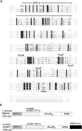
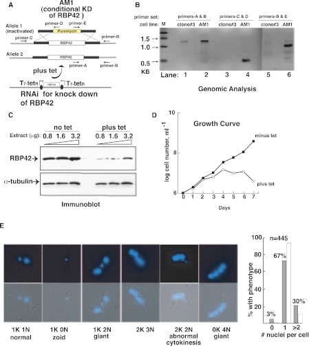
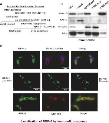
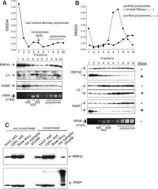
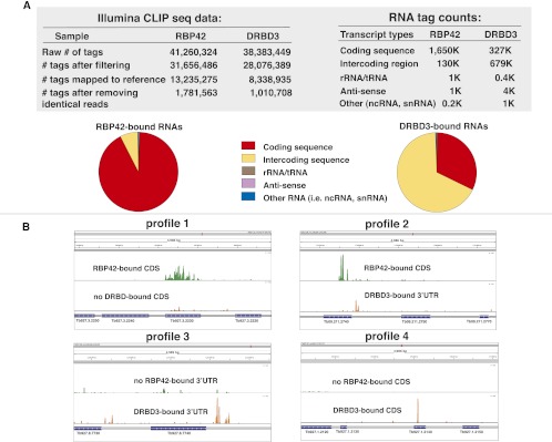
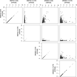
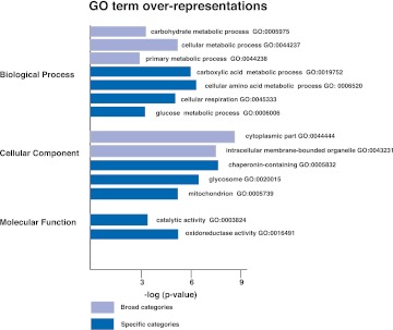
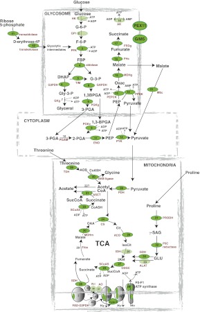
RNA-binding proteins that target mRNA coding regions are emerging as regulators of post-transcriptional processes in eukaryotes. Here we describe a newly identified RNA-binding protein, RBP42, which targets the coding region of mRNAs in the insect form of the African trypanosome, Trypanosoma brucei. RBP42 is an essential protein and associates with polysome-bound mRNAs in the cytoplasm. A global survey of RBP42-bound mRNAs was performed by applying HITS-CLIP technology, which captures protein-RNA interactions in vivo using UV light. Specific RBP42-mRNA interactions, as well as mRNA interactions with a known RNA-binding protein, were purified using specific antibodies. Target RNA sequences were identified and quantified using high-throughput RNA sequencing. Analysis revealed that RBP42 bound mainly within the coding region of mRNAs that encode proteins involved in cellular energy metabolism. Although the mechanism of RBP42's function is unclear at present, we speculate that RBP42 plays a critical role in modulating T. brucei energy metabolism.


Figures

FIGURE 1.
FIGURE 1.
Alignment of T. brucei RBP42 and human G3BP1. (A) Multiple sequence alignments of two trypanosomatid RBP42s and two metazoa G3BP1s. Gene annotations are T. brucei (Tb927.6.4440), Leishmania major (LmjF.30.3090), Homo sapiens (G3BP1,Q13283, Gene identification 10146), and Mus musculus (P97855). The NTF-2 domain is overlined by a gray bar; the RNA recognition motif (RRM) domain is underlined by a white bar; and the three PxxP motifs in T. brucei are overlined with a black line. The single PxxP motif in the human and mouse genes is underlined. Numbering at the ends of lines identifies amino acid location for each organism. (B) Schematic comparison of T. brucei RBP42 and human G3BP1. RBP42 is 79% of the size of G3BP1 (drawing is approximately to scale). The NTF-2 domain (light gray), weakly acidic and acidic regions (dashed-stripe and striped), triple or single PxxP motifs (PxxP), RRM domain (white), and RRG-rich region (black) are shown. RBP42 and G3BP1 are similar but not identical; therefore, the trypanosome protein is named in accordance with the published trypanosomatid RNA-binding protein designation scheme (De Gaudenzi et al. 2005). Within the RRM, the RNP1 signature sequence motif (K/R) G (F/Y)(G/A)FVX(F/Y) is present in T. brucei RBP42 as KGYVFFDF (amino acids 311–319).
FIGURE 2.
FIGURE 2.
Effect of RBP42 depletion on cell viability. (A) RNAi-dependent ablation of RBP42 mRNA in T. brucei strain AM1. This strain contains a single intact RBP42 allele, as well as the RNAi construct. (B) Analysis of genomic DNA to assess the AM1 genotype. PCR reactions utilized primers-A, -B, -C, and -D located as shown in panel A. The 0.6-kb DNA in lane 4 and the 0.9-kb DNA in lane 6 confirm that AM1 contains a puromycin cassette in place of one of the two endogenous RBP42 alleles. The 1.3-kb band in lane 2 shows that the remaining RBP42 allele is intact. (Lanes 1,3,5) Controls showing the presence of intact RBP42 alleles in the parental clone 3 cell line. (C) RBP42 is decreased after ablation of mRNA by tetracycline-dependent RNAi induction, as demonstrated by immunoblot analysis. α-Tubulin levels are shown as a loading control. (D) T. brucei requires RBP42 for cell viability. Tetracycline, which activates the RNAi-dependent ablation of RBP42, was added at day 0. Parasites were directly observed and counted daily using a hemocytometer. Three clones were studied and AM1 chosen as a representative example of the observed phenotype. (E) Cellular morphology and cytokinesis are altered as a result of RBP42 depletion. Examples of the predominant phenotypes are shown as fluorescent micrographs containing DAPI-stained nuclei and kinetoplasts (both in blue) of parasites that represent each phenotype observed on day 4 after RBP42 depletion. Quantification of DAPI-stained nuclei per cell is shown for n = 445 cells in the histogram to the right of micrographs. Open bars, normal phenotypic distribution.
FIGURE 3.
FIGURE 3.
RBP42 is within the S100-precipitated fraction in the cellular cytoplasm. (A) Subcellular fractionation scheme is shown. (B) Immunoblotting of fractionated lysates identifies RBP42 as cytoplasmic and associated with the ribosome-containing subcellular fraction (S100 pellet). Protein detection was performed using the four antibodies listed on the left side of the panel. RPB1, the largest subunit of RNA polymerase II, is a nuclear marker; GRASP and BiP are cytoplasmic markers that fractionate with the S100 supernatant due to the early detergent step in the fractionation (Ho et al. 2006). (C) Immunofluorescent detection of fixed parasites shows cytoplasmic localization of RBP42. Four of 15 Z-stacks are shown. Z-stacks were collected to refine resolution.
FIGURE 4.
FIGURE 4.
RBP42 fractionates predominantly with polysomes. (A) Polysome fractionation profile is shown. Immunoblotting of fractionated material was accomplished using the RBP42, L5, and PABP antibodies listed on the left side of the panel. L5, a component of the 60S ribosomal subunit, is a monosome (80S) and polysome marker; PABP, a cytoplasmic poly(A)-binding protein, is an mRNA marker. Locations of the small subunit (40S), large subunit (60S), intact monosomes (80S), and polysomes in the gradients was accomplished using an ethidium bromide (ETBR)–stained Agarose gel. T. brucei 28S is naturally present as two large fragments, which migrate below 18S rRNA. (B) Purified polysomes obtained from fractions 7–10 in A were further fractionated after mock (solid line) or limited RNase A treatment (dotted line). Immunoblotting was done as shown in A. (C) Immunoblotting of fractionated lysates before or after in vivo protein–RNA crosslinking by UV light. An equal amount of each fraction was present in all lanes to show the relative abundance of RBP42 and PABP in each fraction. Fractions, defined at the top of each lane, were obtained after oligo-d(T) cellulose chromatography.
FIGURE 5.
FIGURE 5.
RBP42 antibody recognizes RBP42 in cell extracts. (A) Cell extracts were depleted using either preimmune serum or RBP42 or DRBD3 antibodies. Immunoblots were probed with the indicated antibody (anti-RBP42 was used in lanes 112, and anti-DRBD3 was used in lanes 1324). None indicates no-antibody-treated samples. (Lanes 1,4,7,10,13,16,19,22) Undiluted samples; (lanes 2,5,8,11,14,17,20,23) 1:5 diluted samples; and (lanes 3,6,9,12,15,18,21,24) 1:25 diluted samples from either starting or immunodepleted extracts. (B) Proteins were captured from crosslinked (+) or not crosslinked (−) parasites using the antibodies indicated above the lanes. Immunocaptured proteins were detected with anti-RBP42 (lanes 18) or anti-DRBD3 (lanes 916) antibodies. None indicates no-antibody-treated samples. (C) Immunoprecipitated protein–RNA complexes were 5′-end radiolabeled on their RNA, separated by denaturating-polyacrylamide gel electrophoresis (SDS-PAGE) and transferred to nitrocellulose membranes. A PhosphorImager scan of protein– 32P-radiolabeled RNA complexes is shown. RBP42 and DRBD3 migrate similarly as the two proteins are similar in molecular mass, and the RNA fragments associated with the proteins are 30–50 nt long.
FIGURE 6.
FIGURE 6.
HITS-CLIP data, RNA read assignments, and quantification. (A) Illumina CLIP sequencing data and RNA tag count data. Pie charts depict the relative abundance of the RNA targets, which are derived from the RNA tag count data. Coding sequences are defined as the annotated open reading frames of mRNAs in version 4 of the T. brucei brucei 927 genome at TriTrypDB and GeneDB (tritrypdb.org/tritrypdb and www.genedb.org). Intercoding sequences contain 5′ and 3′ UTRs, as well as sequences that may contain primary transcripts. rRNA/tRNA, anti-sense, and other RNAs are as defined in the database referenced above. Reads with poor quality and sequence ambiguity, as well as reads generated by PCR amplification, were eliminated prior to mapping. (B) RBP42–mRNA interactions and DRBD3–mRNA interactions (control protein–RNA interactions) within specific genes in the T. brucei genome. (Profile 1) RBP42-bound sequences within the coding region of succinyl-CoA synthetase (α-subunit shown here; Tb927.3.2230). Two upstream genes (Tb927.3.2250 and Tb927.2240) and one downstream gene (Tb927.2220) that are not bound by RBP42 reveal binding specificity, as does the absence of DRBD3 binding to all four genes shown. (Profile 2) RBP42-bound sequences within the CDS and DRBD3-bound sequences within the 3′ UTR of Gim5B (Tb09.211.2740). The absence of the reciprocal binding, as well as the absence of binding to the flanking genes, indicates the binding specificities. (Profile 3) DRBD3-bound sequences within the (predicted) 3′ UTR region of MFS mRNA (major facilitator superfamily protein; Tb927.8.7740). DRBD3-targeting of the MFS mRNA 3′ UTR was initially discovered by Estevez (2008). (Binding specificity is revealed by the low binding of DRBD3 or RBP42 to the CDS of this gene, as well as the low binding of RBP42 to the 3′ UTR of this gene. The small amount of signal indicating DRBD3 interactions with the 3′ UTR of the upstream gene, Tb927.8.7730, was not investigated.) (Profile 4) DRBD3-bound sequences within the coding region of a hypothetical protein mRNA (Tb927.1.2140). Binding specificity is revealed by the absence of RBP42 binding to this gene, as well as the absence of DRBD3 and RBP42 binding to two upstream flanking genes (Tb927.1.2120 and Tb927.1.2130) and the downstream flanking gene (Tb927.1.2150). Scales, 0–1500 (Profile 1), 0–300 (Profile 2), 0–100 (Profile 3), and 0–600 (Profile 4). Green denotes RBP42-bound sequences, and orange denotes DRBD3-bound mRNA sequences.
FIGURE 7.
FIGURE 7.
A comparison of RBP42 and DRBD3 unique reads within the CDS and 3′ UTRs of 835 genes with defined 3′ UTRs. RBP42 and DRBD3 unique reads within the CDS and 3′ UTR of 835 genes were normalized over gene length and read counts to obtain RPKM (reads per kilobase per million mapped reads) values. Gene length for each gene was determined by adding CDS length and 3′ UTR length. The highest RPKM value obtained for RBP42 CDS regions was 7.4; for RBP42 3′ UTR region, 1.4; for DRBD3 CDS, 4.6, and for DRBD3 3′ UTR, 9.9. Pairwise comparisons of RPKM values for each protein over CDs and 3′ UTR regions are shown as a scatterplot matrix. The axes show the RPKM values for the reads mapped to the CDs and 3′ UTR in the RBP42 and DRBD3 experiments.
FIGURE 8.
FIGURE 8.
Gene Ontology (GO) assignments for RBP42-bound mRNAs. Highly significant (P < 0.01) associations of RBP42 targets with GO terms are shown. GO terms are derived from a GO Slim set designed to incorporate trypanosome-specific processes and cellular compartments (Alsford et al. 2011). GO terms that describe broad categories are indicated by light blue bars; GO terms that describe specific categories are indicated by dark blue bars. Statistical analysis of GO term associations was carried out using 137 annotated genes. Since annotated genes lack a comprehensive set of appropriate GO terms, each significant association was verified.
FIGURE 9.
FIGURE 9.
A combination of visual and computer-based analysis reveals that the most significant RBP42-bound mRNAs encode proteins that function in energy production. Metabolism-associated genes with the highest enrichment values (top 10%) for RBP-42–bound RNA tags are depicted as dark green ovals; genes with above-background values are depicted as light green ovals. Enzymes are (1) hexokinase (HK); (2) glucose-6-phosphate isomerase (GPI); (3) phosphofructokinase (PFK); (4) aldolase; (5) triose-phosphate isomerase (TIM); (6) glycerol-3-phosphate dehydrogenase (G3PDH); (7) glycerol kinase-glycosomal (GKg); (8) glyceraldehyde-3-phosphate dehydrogenase (GAPDH); (9) phosphoglycerate kinase (cytosolic, PGKc, and glycosomal [PGKg]); (10) phosphoglycerate mutase (PGAM); (11) enolase (ENO); (12) pyruvate kinase (PYK); (13) phosphoenolpyruvate carboxykinase (PEPCK); (14) pyruvate phosphate dikinase (PPDK); (15) malate dehydrogenase-glycosomal (MDHg); (16) fumarase-cytosolic (FHc); (17) NADH-dependent fumarate reductase (FRDg); (18) adenylate kinase-glycosomal (AK), GIM5-glycosomal membrane protein (GIM5), and PEX11-glycosomal membrane protein (PEX11); (19) malic enzyme-cytoplasmic (MEc); (20) pyruvate dehydrogenase complex (PDH); (21) acetate:succinate CoA transferase (ASCT); (22) unknown enzyme; (23) succinyl CoA synthetase (SCoAS); (24) citrate synthase (CS); (25) aconitase (ACO); (26) isocitrate dehydrogenase (IDH); (27) 2-ketoglutarate dehydrogenase (ODGC); (28) succinate dehydrogenase (SDH); (29) fumarase-mitochondrial (FHm); (30) malate dehydrogenase-mitochondrial (MDHm); (31) proline dehydrogenase (PRODH); (32) pyrroline-5-carboxylate reductase (P5C reductase); (33) L-alanine aminotransferase (ALAT); (34) glutamate dehydrogenase (GDH); (35) glycerol-3-phosphate oxidase (FAD-G3PDH); (36) rotenone-insensitive NADH dehydrogenase (R–I); (37) alternative oxidase (AO); and (38) F0/F1-ATP synthase (β-, γ-subunits and subunit 9) in complexes I–IV of the respiratory chain (in complex I, subunit 20-kDa subunit is indicated; in complex II, no subunits are indicated; in complex III, the Rieske iron-sulfur protein is indicated; in complex IV, the cytochrome oxidase subunits IV, V, and VII–IX are indicated; cytochrome C [Cyto C]); (39) threonine-3-dehydrogenase (TDH); (40) 2-amino-3-ketobutyrate CoA ligase (ABK ligase); (41) transaldolase; and (42) transketolase.

Similar articles

Cited by

References

    1. Acosta-Serrano A, Vassella E, Liniger M, Kunz Renggli C, Brun R, Roditi I, Englund PT 2001. The surface coat of procyclic Trypanosoma brucei: Programmed expression and proteolytic cleavage of procyclin in the tsetse fly. Proc Natl Acad Sci 98: 1513–1518 - PMC - PubMed
    1. Alsford S, Turner DJ, Obado SO, Sanchez-Flores A, Glover L, Berriman M, Hertz-Fowler C, Horn D 2011. High-throughput phenotyping using parallel sequencing of RNA interference targets in the African trypanosome. Genome Res 21: 915–924 - PMC - PubMed
    1. Angenstein F, Evans AM, Settlage RE, Moran ST, Ling SC, Klintsova AY, Shabanowitz J, Hunt DF, Greenough WT 2002. A receptor for activated C kinase is part of messenger ribonucleoprotein complexes associated with polyA-mRNAs in neurons. J Neurosci 22: 8827–8837 - PMC - PubMed
    1. Atlas R, Behar L, Elliott E, Ginzburg I 2004. The insulin-like growth factor mRNA binding-protein IMP-1 and the Ras-regulatory protein G3BP associate with tau mRNA and HuD protein in differentiated P19 neuronal cells. J Neurochem 89: 613–626 - PubMed
    1. Atlas R, Behar L, Sapoznik S, Ginzburg I 2007. Dynamic association with polysomes during P19 neuronal differentiation and an untranslated-region-dependent translation regulation of the tau mRNA by the tau mRNA-associated proteins IMP1, HuD, and G3BP1. J Neurosci Res 85: 173–183 - PubMed

Publication types

MeSH terms

LinkOut - more resources